



Biliary Atresia
Summary
- Biliary atresia is a condition where the outcome is decided by how quickly the diagnosis is made. It is the most common cause of neonatal jaundice requiring surgery and the most common indication for liver transplantation in children [1].
- Untreated it progresses to cirrhosis and hepatic failure. The Kasai procedure must be performed before the age of 3 months to avoid irreversible liver damage, and after that age transplantation is generally needed instead [1].
- The clinical trigger is simple: progressive jaundice persisting beyond 2 weeks after birth suggests atresia [1].
Definition
Biliary atresia is obliteration of the biliary tree presenting in infancy. It can involve the extrahepatic biliary tree, the intrahepatic tree, or both [1].
Pathophysiology
The obstruction to bile flow produces a progressive fibrotic process in the liver. Liver biopsy shows periportal fibrosis, bile plugging and eventual cirrhosis [1], and without relief of the obstruction the child develops continued cirrhosis and eventual hepatic failure [1].
That progression is why the age threshold exists: after roughly 3 months the liver damage is irreversible and restoring bile drainage no longer rescues the organ [1].
Proposed mechanisms and the pattern of obliteration in Schwartz's account
Biliary atresia is a fibroproliferative obliteration of the biliary tree progressing to fibrosis, cirrhosis and end-stage failure, with an incidence of 1 in 8000 to 1 in 18,000; Ladd and Gross called it an "arrest of development during the solid stage of bile duct formation", later theories invoke defective hepatogenesis, prenatal vasculogenesis, immune dysregulation, infection and toxins, cfc1 mutations governing left–right axis determination occur in the biliary atresia–splenic malformation syndrome, maternal microchimerism is commoner in the livers of affected boys and may drive an autoimmune attack, and animal studies implicate perinatal reovirus or rotavirus provoking interferon-γ-mediated periportal inflammation [2]. Obliteration involves the common duct, cystic duct, one or both hepatic ducts and the gallbladder in varying combinations; histology shows parenchymal inflammation, fibrous deposition at the portal plates on trichrome staining of frozen sections and non-specific ductular proliferation; about 25% have coincident malformations, often with polysplenia, including malrotation, preduodenal portal vein and intrahepatic vena cava [2].
Clinical features
Progressive jaundice persisting more than 2 weeks after birth suggests biliary atresia [1].
The distinction that matters at the cot side is between physiological jaundice of the newborn, which is an unconjugated hyperbilirubinaemia from immature glucuronyl transferase, and biliary atresia, which is obstructive and therefore conjugated, the two are separated on the Liver Anatomy and Physiology page [1].
Physiological versus pathological jaundice in Schwartz's account
- Jaundice occurs in the first week in 60% of term and 80% of preterm infants because of high haemoglobin load and immature glucuronyl transferase, appearing on day 2–3 and resolving within 5–7 days; persistence beyond 2 weeks is pathological by definition and the work-up covers obstruction (biliary atresia, choledochal cyst, inspissated bile syndrome), haemolysis (ABO and Rh incompatibility, spherocytosis), metabolic disease (α1-antitrypsin deficiency, galactosaemia, pyruvate kinase deficiency) and congenital infection (syphilis, rubella) [2].
- Infants with biliary atresia are jaundiced at or soon after birth, are often missed because physiological jaundice is so common, pass acholic pale grey stools, fail to thrive and, untreated, develop splenomegaly and oesophageal varices [2].
- Inspissated bile syndrome is persistent obstructive jaundice with a normal tree from viscous bile in infants on parenteral nutrition, with haemolysis or cystic fibrosis, and neonatal hepatitis is acquired biliary inflammation without obliteration, usually viral and self-limiting, for which cholangiography is both diagnostic and therapeutic [2].
Etiology
Biliary atresia is the commonest surgical cause of neonatal jaundice [1]. The other neonatal cause of obstructive jaundice a surgeon must exclude is a choledochal cyst, covered on the Biliary Strictures and Cholangiocarcinoma page.
Diagnosis
Diagnosis is by liver biopsy, which shows periportal fibrosis, bile plugging and eventual cirrhosis [1].
Ultrasound and HIDA scanning can reveal the atretic biliary tree [1].
The diagnostic sequence in Schwartz's account
No single test suffices: bilirubin is fractionated, TORCH and viral hepatitis titres are sent, ultrasound looks for choledochal cyst, an absent gallbladder is highly suggestive but a visible gallbladder does not exclude atresia because in about 10% the distal tract is patent with atretic proximal ducts, and the intrahepatic ducts are never dilated; phenobarbital-primed technetium-99m DISIDA scanning excludes atresia if tracer reaches the intestine, and hepatic concentration without excretion with a normal metabolic screen (particularly α1-antitrypsin) makes the presumptive diagnosis; percutaneous liver biopsy may separate atresia from neonatal hepatitis; and when tests point to or cannot exclude atresia, exploration with cholangiography through the gallbladder, open or laparoscopic, defines the anatomy, distal flow into the duodenum and any hypoplasia of the extrahepatic tree seen with α1-antitrypsin deficiency and Alagille's syndrome [2].
Thresholds and severity
Two weeks is the threshold at which persisting jaundice should prompt investigation [1].
Three months is the threshold beyond which the Kasai procedure will not prevent irreversible liver damage, and beyond which liver transplantation is generally required [1].
Treatment and Management
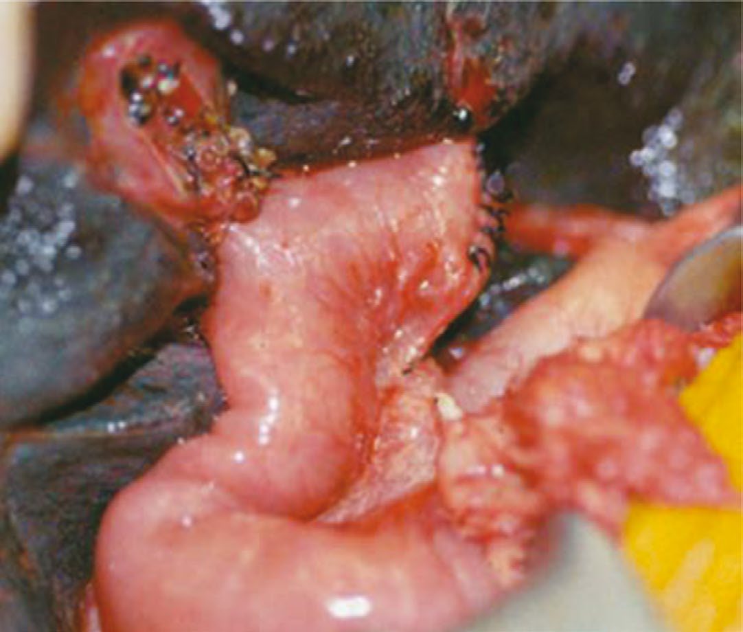
The Kasai procedure (hepaticoportojejunostomy) is attempted first. It involves resecting the atretic extrahepatic bile duct segment and anastomosing a Roux-en-Y limb to the liver [1].
Choleretic agents such as phenobarbital, and steroids, are used to try to increase bile flow after the procedure [1].
Where the child presents after 3 months, transplantation rather than Kasai is generally the route [1]; the transplant itself is covered on the Liver Transplantation page.
Procedural interventions
The operation is a Roux-en-Y hepatoportoenterostomy, anastomosed to the liver at the porta after the atretic extrahepatic duct has been resected [1].
Kasai's rationale, technique and revision in Schwartz's account
Kasai observed that the fibrous tissue at the porta invests microscopically patent ductules communicating with the intrahepatic system, so transecting the portal plate (invariably found cephalad to the portal vein bifurcation) opens these channels into a Roux-en-Y jejunal limb; an intussuscepted antireflux valve is favoured by some but does not alter outcome, a liver biopsy grades fibrosis at operation, and ductule diameter at the plate predicts durable drainage [2]. Bondoc reported 24 revisions among 183 Kasai operations for loss of previously established drainage, 75% regaining drainage and nearly half of those surviving long term with native livers, so revision is reasonable in selected patients whose bile flow was established then lost [2].
Complications
The complication that defines the disease is progression despite surgery: continued cirrhosis and eventual hepatic failure in those for whom the Kasai does not establish adequate drainage [1].
Outcomes
Outcome after the Kasai procedure divides into thirds: one-third get better, one-third go on to liver transplantation, and one-third die [1].
That distribution, and the fact that biliary atresia is the most common indication for paediatric liver transplantation [1], is why the 2-week jaundice rule is treated as a hard referral trigger rather than a period of observation.
Age at surgery, steroids and registry survival in Schwartz's figures
Success falls with age: infants operated before 60 days most often achieve lasting drainage, the outlook is poorer after the 12th week yet surgery remains reasonable because the alternative is certain liver failure and many older infants have done well; bile flow does not imply cure, about a third remain symptom-free and the rest need transplantation, bridging fibrosis at surgery and postoperative cholangitis independently predict failure, cholangitis is the common complication without an effective preventive strategy and unresolved value for long-term prophylactic antibiotics, the Japanese Biliary Atresia Registry of 1381 patients shows 10-year survival of 53% without and 66.7% with transplantation, and the NIH-funded ChiLDREN randomised trial found high-dose postoperative steroids did not significantly improve drainage at 6 months and brought earlier serious adverse events [2].
References
- The ABSITE Review, 2022, Ch. 31 Liver
- Schwartz's Principles of Surgery, 11th ed., Ch. 39, Pediatric Surgery
- Sabiston Textbook of Surgery, 22nd ed., Ch. 117 Pediatric Surgery