



Liver Anatomy and Physiology
Summary
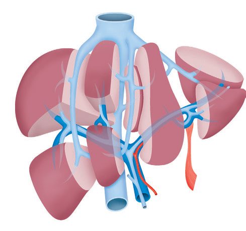
Liver surgery became safe when the internal anatomy was understood rather than the external shape. The liver divides into functional right and left units along a line between the gallbladder fossa and the middle hepatic vein (Cantlie's line) and Couinaud described it as eight segments, each a functional unit with its own branch of hepatic artery, portal vein and bile duct [1]. This page covers the ligaments and the segments, the hilar structures and their variants, the arterial, portal and venous supply segment by segment, the biliary confluence, and the synthetic, metabolic and excretory functions that the operative anatomy exists to preserve. Seventy-five per cent of a normal liver can be safely resected [2].
Definition
Glisson's capsule is the peritoneum that covers the liver; the bare area is the part of the posterosuperior surface not covered by it [2].
The porta hepatis is a pronounced transverse fissure on the visceral surface running between the cephalad end of the fissure for the ligamentum teres and the gallbladder fossa; the neurovascular structures and lymphatics of the hepatoduodenal ligament enter here, and the right and left hepatic ducts emerge [1].
Pathophysiology
Ligaments and surface landmarks
The falciform ligament separates the medial and lateral segments of the left lobe, attaches the liver to the anterior abdominal wall, extends to the umbilicus, and carries the remnant of the umbilical vein [2]. The ligamentum teres carries the obliterated umbilical vein to the undersurface of the liver and extends from the falciform ligament [2]. The triangular ligaments are the lateral and medial extensions of the coronary ligament on the posterior surface, and are made of peritoneum [2].
Cantlie's line (the portal fissure) runs from the middle of the gallbladder fossa to the IVC and separates the right and left lobes [2]. Bailey & Love describes the same division as running between the gallbladder fossa and the middle hepatic vein [1].
The eight segments
| Segment | Position |
|---|---|
| I | Caudate |
| II | Superior left lateral |
| III | Inferior left lateral |
| IV | Left medial (quadrate lobe) |
| V | Inferior right anteromedial |
| VI | Inferior right posterolateral |
| VII | Superior right posterolateral |
| VIII | Superior right anteromedial |
Segments II, III and IV make up the left lobe; V, VI, VII and VIII the right; segment I is the caudate [2]. The portal triad enters segments IV and V, and the gallbladder lies under those same two segments [2].
The hilum
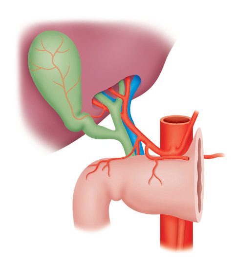
In the most common arrangement the bile duct runs in the free edge of the hepatoduodenal ligament with the hepatic artery medially and the portal vein posteriorly, each dividing into two branches at the hilum [1]. The ABSITE Review gives the same relations for the portal triad (common bile duct lateral, portal vein posterior, proper hepatic artery medial) meeting in the hepatoduodenal ligament [2].
The right hepatic artery crosses the bile duct anteriorly or posteriorly before giving rise to the cystic artery, and multiple branches predominantly from the right hepatic artery supply the bile duct [1], which is why the duct's blood supply is at risk in hilar dissection.
The Pringle manoeuvre is clamping of the porta hepatis. It will not stop hepatic vein bleeding [2].
The boundaries of the foramen of Winslow, the entrance to the lesser sac, are the portal triad anteriorly, the IVC posteriorly, the duodenum inferiorly and the caudate lobe of the liver superiorly [2].
Arterial supply and its variants
The arterial supply is usually derived from the coeliac trunk, which divides into left gastric, common hepatic and splenic arteries; after giving off the gastroduodenal artery the hepatic artery branches at a variable level into right and left hepatic arteries, the larger right branch supplying the right lobe [1].
Two variants account for most of the abnormal anatomy, and each has a predictable location [2]:
- A right hepatic artery arising from the superior mesenteric artery is the commonest hepatic arterial variant, at about 20%; it courses behind the pancreas, posterolateral to the common bile duct. Bailey & Love describes the same vessel running to the liver on the posterior wall of the bile duct after passing behind the uncinate process and head of the pancreas [1].
- A left hepatic artery arising from the left gastric artery, also about 20%, is found medially in the gastrohepatic ligament [2]. Bailey & Love places it running in the lesser omentum from the lesser curve [1].
The middle hepatic artery is most commonly a branch of the left hepatic artery, and most primary and secondary liver tumours are supplied by the hepatic artery [2], the basis of transarterial embolisation.
Portal venous supply
The portal vein forms from the superior mesenteric vein joining the splenic vein and has no valves; the inferior mesenteric vein enters the splenic vein [2]. Bailey & Love places the confluence behind the neck of the pancreas and notes that the left branch has a longer extrahepatic course, approximately 2 cm [1].
The portal vein supplies two-thirds of hepatic blood flow. The left portal vein goes to segments II, III and IV; the right to segments V, VI, VII and VIII [2].
Hepatic venous drainage
Three hepatic veins drain into the IVC, and their territories are the reason segmental resection works [2]:
| Vein | Segments drained |
|---|---|
| Left | II, III and superior IV |
| Middle | V and inferior IV |
| Right | VI, VII and VIII |
The middle hepatic vein merges with the left hepatic vein in 80% of people before entering the IVC; in the other 20% it enters directly [2]. Bailey & Love states the same relation the other way round: the right hepatic vein can be exposed fully outside the parenchyma, while the middle and left usually terminate in a short common trunk [1].
Accessory right hepatic veins drain the medial aspect of the right lobe directly into the IVC, and the inferior phrenic veins also drain directly into it [2]. Bailey & Love notes a variable number of short inferior hepatic veins passing directly from liver to the anterior wall of the IVC, and that the right adrenal gland is adjacent to the retrohepatic IVC and drains into it, usually by a single vein [1].
The caudate lobe is the exception to everything above: it receives separate right and left portal and arterial blood flow, and drains directly into the IVC through its own hepatic veins [2].
The biliary confluence
The left hepatic duct retains a longer transverse extrahepatic portion, travelling under the edge of segment IVB from the umbilical fissure to the biliary bifurcation, which makes it more surgically accessible; it drains segments I, II, III and IV [3].
On the right there are two principal duct systems that join to form a short main right hepatic duct: the right anterior sectoral duct runs vertically and drains segments V and VIII, while the right posterior sectoral duct follows a horizontal course and drains segments VI and VII [3]. In approximately 20% of patients the right posterior sectoral duct courses posterior to the right anterior duct to join the left system [3].
The recognised variations of the confluence are trifurcation at the confluence; either right sectoral duct draining into the common hepatic duct; either right sectoral duct draining into the left hepatic duct; absence of a confluence altogether; and absence of a right hepatic duct with the right posterior sectoral duct draining into the cystic duct [4]. The caudate lobe drains through smaller ducts that typically enter both the right and left systems [3].
At the hilum the biliary bifurcation abuts the base of segment IVB and sits superior to the intrahepatic branches of the portal vein and hepatic arteries [3].
Ligaments, lobes and the caudate in Schwartz's description
- The liver weighs approximately 1500 g and is invested by Glisson's capsule [5].
- The round ligament, the remnant of the obliterated umbilical vein, enters the left hilum at the front edge of the falciform ligament; deep in the plane between the caudate lobe and the left lateral segment lies the ligamentum venosum (Arantius' ligament), the obliterated ductus venosus, covered by the plate of Arantius; the left and right triangular ligaments fix the liver to the diaphragm, the coronary ligaments extend from them anteriorly, and the right coronary ligament also runs from the right undersurface to the peritoneum over the right kidney, anchoring the liver to the retroperitoneum, all of these (round, falciform, triangular and coronary) can be divided in a bloodless plane to mobilise the liver for resection [5].
- The hepatoduodenal ligament (porta hepatis) carries the common bile duct, hepatic artery and portal vein; dorsal to it from the right is the foramen of Winslow (epiploic foramen), which connects to the lesser sac and allows complete inflow control when the ligament is clamped in the Pringle manoeuvre [5].
- Cantlie's line, from the gallbladder fossa to the vena cava, divides the liver into a right lobe of 60–70% of its mass and a left lobe that, with the caudate, makes up the remainder; the caudate lobe lies left and anterior of the vena cava and has three subsegments (the Spiegel lobe, the paracaval portion and the caudate process) and its venous drainage passes directly into the vena cava [5].
- Segment IVA is cephalad beneath the diaphragm from segment VIII to the falciform ligament beside segment II, and IVB caudad beside the gallbladder fossa; "quadrate lobe" is an outdated term for segment IV [5].
- Bismuth's functional anatomy uses the three hepatic veins running in their scissurae to divide the liver into four sectors, the right hepatic vein separating right posterolateral from right anterolateral, the middle vein in the main scissura separating right from left liver, and the left vein separating left posterior from left anterior sectors [5].
Frequencies of the vascular and biliary variants
- Standard arterial anatomy, coeliac axis giving left gastric, splenic and common hepatic arteries, the common hepatic dividing into gastroduodenal and hepatic artery proper, and the latter into right and left hepatic arteries, with the right gastric usually off the hepatic artery proper, is present in only about 76% of people [5].
- In 10–15% a replaced or accessory right hepatic artery arises from the superior mesenteric artery, travels posterior to the portal vein and takes up a right lateral position before entering the parenchyma, recognisable on CT or MRI and by a separate right posterior pulsation in the hilum distinct from the hepatic artery proper lying anteriorly to the left of the duct; in 3–10% a replaced or accessory left hepatic artery from the left gastric runs obliquely in the gastrohepatic ligament anterior to the caudate to enter the hilar plate at the base of the umbilical fissure; and in about 1–2% each both are replaced, or the entire common hepatic artery arises from the superior mesenteric artery, the clue being a strong pulsation to the right of and posterior to the bile duct [5].
- The right hepatic artery passes deep and posterior to the common bile duct about 88% of the time but crosses anterior to it in about 12%, and the cystic artery usually arises from it in Calot's triangle [5].
- Normal portal pressure is 3–5 mmHg, rising to 20–30 mmHg in portal hypertension with decompression most commonly through the coronary (left gastric) vein into oesophageal and gastric varices; the left portal vein has a transverse portion, a 90° turn at the base of the umbilical fissure into an umbilical portion, and usually gives the dominant caudate inflow near that bend, whereas the right portal vein divides higher, close to or inside the hilar plate, and 20–35% of people have aberrant portal anatomy, most often a trifurcation or a branch from the left portal vein to the right anterior lobe [5].
- The superior pancreaticoduodenal vein leaves the main portal vein low and anterolaterally (divided during pancreaticoduodenectomy), and a short posterolateral branch to the caudate process must be identified and ligated during hilar dissection for right hemihepatectomy to avoid avulsion [5].
- The left and middle hepatic veins form a common trunk about 95% of the time while the right vein inserts separately and obliquely; a large inferior accessory right hepatic vein runs in the hepatocaval ligament in 15–20% and can cause torrential bleeding if control is lost during right hepatectomy; and each hepatic vein bisects the portal branches, the right between the right anterior and posterior portal veins, the middle between the right anterior and left portal veins, the left between the segment II and III branches [5].
- The right anterior hepatic duct usually enters the liver above the hilar plate, the right posterior duct dives behind the right portal vein onto the surface of the caudate process, and the left hepatic duct has a longer extrahepatic course before branching behind the left portal vein at the base of the umbilical fissure; Couinaud's 1957 series found a normal bifurcation in only 57%, trifurcation in 12%, the right anterior (16%) or right posterior (4%) duct draining into the common hepatic duct in 20%, the right posterior (5%) or right anterior (1%) duct draining into the left hepatic duct in 6%, absence of a confluence in 3% and the right posterior duct draining into the cystic duct in 2% [5].
- The gallbladder sits on segments IVB and V [5].
Innervation and lymphatics
Parasympathetic supply comes from the anterior hepatic branch of the left vagus and the posterior hepatic branch of the right vagus, and sympathetic supply from the greater thoracic splanchnic nerves and coeliac ganglia, though the denervated transplanted liver functions normally; referred pain to the right shoulder, scapula, flank or back arises from the right phrenic nerve when tumours stretch Glisson's capsule or irritate the diaphragm [5]. Lymph drains from the space of Disse and the periportal clefts of Mall to the cystic duct node in Calot's triangle and the common bile duct, hepatic artery, retropancreatic and coeliac nodes (important in hilar cholangiocarcinoma, which metastasises to nodes frequently) and cephalad to the cardiophrenic nodes, which can be identified on staging CT or MRI [5].
Clinical features
The liver's functions are wider than synthesis alone. Bailey & Love lists them as maintaining core body temperature and heat production; pH balance and correction of lactic acidosis; synthesis of clotting factors; glucose metabolism, glycolysis and gluconeogenesis; urea formation from protein catabolism; bilirubin formation from haemoglobin after breakdown of effete red cells in the spleen; drug and hormone metabolism and excretion; removal of gut endotoxins and foreign antigens; storage of vitamins and minerals including A, D, E, K and B12; immunological function as part of the mononuclear phagocyte system; albumin production for transport of fatty acids, steroids and waste products; angiotensin synthesis; and cytochrome P450 detoxification of contaminants and pollutants, insecticides, food additives and alcohol [1].
The signs of liver dysfunction vary with severity, aetiology and speed of onset, but commonly include jaundice, drowsiness, abdominal pain or swelling, nausea, tremor, vomiting, malaise, confusion and disorientation, bruising, peripheral oedema, and foetor hepaticus, a strong musty smell to the breath [1].
Jaundice becomes apparent when the total bilirubin exceeds 2.5, and is first evident under the tongue [2]. The maximum bilirubin reached is 30, unless there is underlying renal disease, haemolysis, or a bile duct to hepatic vein fistula [2].
Etiology
Two things are not made in the liver, and both matter to a surgeon: von Willebrand factor and factor VIII, which are made in endothelium [2]. Everything else in the clotting cascade is hepatic, which is why the prothrombin time is a synthetic function test.
Ketones are the usual energy source for the liver itself; glucose is converted to glycogen and stored, and excess glucose is converted to fat [2]. Urea is synthesised in the liver [2].
The liver stores a large amount of fat-soluble vitamins, and B12 is the only water-soluble vitamin it stores [2].
Alkaline phosphatase is normally located in the canalicular membrane, whereas nutrient uptake occurs at the sinusoidal membrane [2], which is why a cholestatic picture raises alkaline phosphatase specifically.
Kupffer cells are the liver macrophages [2].
Bile and the enterohepatic circulation
Bile contains bile salts at 85%, with proteins, phospholipids, cholesterol and bilirubin; the final composition is determined by passive reabsorption of water in the gallbladder [2].
Cholesterol is used to make bile salts, which are conjugated to taurine or glycine to improve water solubility [2]. The primary bile acids are cholic and chenodeoxycholic; the secondary bile acids, deoxycholic and lithocholic, are the primary acids dehydroxylated by gut bacteria [2].
Lecithin is the main biliary phospholipid, emulsifying fat and solubilising cholesterol; bile solubilises cholesterol and emulsifies fats to form micelles, which enter enterocytes by fusing with the membrane [2].
Bilirubin
Bilirubin is a breakdown product of haemoglobin (haemoglobin to haem to biliverdin to bilirubin) and is conjugated to glucuronic acid by glucuronyl transferase in the liver, improving water solubility; conjugated bilirubin is then actively secreted into bile [2].
Urobilinogen explains the urine colour. Conjugated bilirubin is broken down by bacteria in the terminal ileum; free bilirubin is reabsorbed, converted to urobilinogen and released in urine as urobilin, giving the yellow colour, and excess urobilinogen turns the urine dark like cola [2].
Bile salts, drug metabolism and paracetamol toxicity
- The liver produces about 1 L of bile daily; cholesterol is converted to the primary bile acids cholic and chenodeoxycholic acid, conjugated to glycine or taurine, and intestinal bacteria deconjugate them and dehydroxylate them to the secondary acids deoxycholic and lithocholic acid [5].
- About 90–95% of bile salts are reabsorbed in the terminal ileum, mainly by active transport, and the remaining 5–10% enter the colon to be converted to secondary salts; those lost in stool are replaced by hepatic synthesis [5].
- The liver synthesises most plasma proteins (albumin, coagulation and fibrinolytic factors and complement) produces glucose in fasting by glycogenolysis and gluconeogenesis from lactate, amino acids and glycerol, and removes excess glucose postprandially by glycogen synthesis, glycolysis and lipogenesis [5].
- Drug metabolism converts lipophilic xenobiotics to hydrophilic products through phase 1 reactions (oxidation, reduction and hydrolysis, largely by the cytochrome P450 hemoproteins) and phase 2 conjugation reactions (glucuronate, acetate, glutathione, glycine, sulphate or methyl groups), mainly in the smooth endoplasmic reticulum; enzyme induction shortens drug action and inhibition prolongs it [5].
- Paracetamol is normally conjugated to harmless glucuronide and sulphate metabolites, but in overdose the pathways are overwhelmed and cytochrome P450 converts some of the drug to a reactive intermediate that glutathione normally detoxifies; once glutathione is depleted the intermediate binds hepatocyte lipid membranes and causes necrosis, which is why treatment replenishes glutathione with sulphydryl compounds such as acetylcysteine [5].
Diagnosis
The pattern of liver enzymes separates hepatitis from obstruction: hepatitis gives very high transaminases with a modest alkaline phosphatase, while obstructive jaundice gives modest transaminases with a very high alkaline phosphatase [2].
Raised unconjugated (indirect) bilirubin, usually with a normal or only mildly raised conjugated fraction, points to prehepatic causes such as haemolysis, or to hepatic deficiencies of uptake or conjugation [2]. Raised conjugated (direct) bilirubin points to secretion defects into the bile ducts, as in hepatitis, or excretion defects into the gastrointestinal tract, obstructive jaundice from gallstones, cancer or benign stricture [2].
Four inherited syndromes divide by which step fails [2]:
| Syndrome | Defect | Bilirubin raised |
|---|---|---|
| Gilbert's | Mild defect in glucuronyl transferase, abnormal conjugation | Unconjugated |
| Crigler-Najjar | Severe glucuronyl transferase deficiency, inability to conjugate; life-threatening | Unconjugated |
| Rotor's | Deficient storage ability | Conjugated |
| Dubin-Johnson | Deficient secretion ability | Conjugated |
Physiologic jaundice of the newborn is the same mechanism as Gilbert's and Crigler-Najjar in miniature, an immature glucuronyl transferase giving a high unconjugated bilirubin [2].
Interpreting the liver panel
- "Liver function tests" is a misnomer, since AST, ALT, alkaline phosphatase, GGT and bilirubin mostly measure cell damage rather than function; synthetic function is better measured by albumin and prothrombin time [5].
- AST is found in liver, heart, skeletal muscle, kidney, brain, pancreas, lung and red cells and is less specific, whereas ALT is predominantly hepatic; enzyme levels correlate poorly with the severity of necrosis and may not be raised in fibrosis or cirrhosis, an AST:ALT ratio above 2:1 is common in alcoholic liver disease, mild elevations occur in fatty liver, chronic viral infection or drug injury, moderate rises in acute viral hepatitis, and levels in the thousands in ischaemia, toxins such as paracetamol and fulminant hepatitis [5].
- The liver makes about 10 g of albumin a day, but its 15–20-day half-life means albumin is not a marker of acute dysfunction and is also affected by nutrition, renal loss, protein-losing enteropathy and hormones; most clotting factors except factor VIII are made exclusively in the liver, the INR standardises the prothrombin time, and because factors II, VII, IX and X are vitamin-K-dependent, the PT is also prolonged by vitamin K deficiency and warfarin [5].
- Normally more than 90% of serum bilirubin is unconjugated; the direct assay measures conjugated and δ-bilirubin (conjugated bilirubin bound to albumin), a raised indirect fraction suggests intrahepatic cholestasis or increased production (haemolysis, haematoma resorption) or defective uptake or conjugation, and because excretion from the hepatocyte is the rate-limiting step, conjugated hyperbilirubinaemia follows disorders of intrahepatic excretion or extrahepatic obstruction and the water-soluble conjugate appears in the urine [5].
- Alkaline phosphatase is expressed by bile duct epithelium and rises with obstruction through increased synthesis; its half-life of about 7 days means levels take several days to normalise after relief of obstruction, and GGT, an early and sensitive but non-specific marker also induced by drugs, alcohol, pancreatic disease, myocardial infarction, renal failure and obstructive lung disease, is interpreted alongside it, a raised GGT with a raised alkaline phosphatase supports a hepatic source [5].
- Jaundice becomes detectable at a bilirubin above 2.5–3 mg/dL; prehepatic causes include the inherited haemolytic anaemias (spherocytosis, elliptocytosis, G6PD deficiency, sickle cell disease, thalassaemia), immune-mediated (Coombs-positive) and non-immune (drugs, mechanical valves, microangiopathy, infection) acquired haemolysis, and impaired albumin transport in malnutrition or burns; Gilbert's syndrome affects 4–7% of the population with transient rises during fasting, stress or illness, Crigler-Najjar is a rare neonatal disorder risking bilirubin encephalopathy, and posthepatic causes range from stones, strictures, cholangiocarcinoma and papillary disorders to extrinsic pancreatic compression and, increasingly, surgical misadventure with clips, retained stones or ischaemic duct injury presenting immediately or years later [5].
Imaging the liver
- Ultrasound is the useful initial test (cheap, radiation-free and excellent for biliary pathology and focal lesions) but misses the dome and subcostal surface, is degraded by obesity and bowel gas, and detected masses usually need CT or MRI; microbubble contrast (<10 μm) improves lesion characterisation through dynamic enhancement patterns and a late liver-specific phase, and transient elastography, in which shear-wave velocity rises with stiffness, has 87% sensitivity and 91% specificity for cirrhosis against biopsy while sampling a larger area [5].
- Intraoperative ultrasound is the gold standard for detecting liver lesions, finds 20–30% more lesions than preoperative imaging, and changes management in almost 50% of planned resections for malignancy [5].
- Because the portal vein supplies about 75% of hepatic blood flow and the hepatic artery 25% while most tumours are arterially supplied, dual- or triple-phase CT images the arterial-dominant phase at 20–30 seconds and the portal phase at 60–70 seconds after contrast: hypervascular tumours enhance in the arterial phase, hypovascular lesions appear hypoattenuating against enhanced parenchyma in the portal phase, and the two phases map the arterial and venous anatomy for operative planning [5].
- CT cholangiography with hepatocyte-excreted contrast depicts small non-dilated peripheral radicals and compares well with ERCP, but fails with grossly dilated ducts or hyperbilirubinaemia [5].
- MRI offers higher soft-tissue contrast and no ionising radiation, with gadolinium agents behaving like CT iodine and liver-specific agents relying on Kupffer-cell uptake (ferumoxide) or hepatocyte biliary secretion (gadoxetate); MRCP shows the level of biliary occlusion without contrast, and MR elastography is promising but costly [5].
- FDG-PET/CT is more sensitive and specific than CT for intra- and extrahepatic colorectal metastases and for surveillance after ablation, though its sensitivity falls after chemotherapy; FDG uptake in hepatocellular carcinoma tracks grade, giving only 50–65% sensitivity, so dual-tracer PET with 11C-acetate for well-differentiated tumours has been introduced, and in cholangiocarcinoma PET helps mainly with regional and distant metastases [5].
Thresholds and severity
Seventy-five per cent of a normal liver can be safely resected [2].
Hepatocytes in the central lobular region (acinar zone III) are the most sensitive to ischaemia [2].
Jaundice appears above a total bilirubin of 2.5 [2].
UK reference ranges for the routine liver blood tests are given by Bailey & Love as bilirubin 5 to 17 μmol/L (0.3 to 1.2 mg/dL), alkaline phosphatase 30 to 140 IU/L, and albumin 35 to 50 g/L (3.5 to 5 g/dL), with the transaminases at 5 to 40 IU/L and gamma-GT 10 to 48 IU/L [1].
Acute liver failure has a definition with a time window in it, and the window is what separates it from cirrhosis. The most widely accepted definition, from the American Association for the Study of Liver Diseases, is evidence of coagulation abnormality, usually an INR above 1.5, together with any degree of mental alteration, in a patient without pre-existing liver disease and with an illness of less than 26 weeks' duration [1].
It is rare in the developed world, with an annual incidence under 10 cases per million and a current mortality of 30 to 40% [1]. In the early stages there may be no objective signs; with severe dysfunction, clinical jaundice is associated with the neurological signs of hepatic encephalopathy, a liver flap, drowsiness, confusion and eventually coma [1].
Acute liver failure, the King's criteria and the future liver remnant
- Acute liver failure, hepatic encephalopathy within 26 weeks of severe liver injury in a patient without prior liver disease or portal hypertension, affects about 2000 patients a year in the United States; intracranial hypertension from cerebral oedema is the commonest cause of death, followed by sepsis and multi-organ failure, and survival has risen from under 20% before transplantation to over 70% [5].
- Viral hepatitis B, A and E predominate in the East, whereas 65% of Western cases are drug- or toxin-induced, paracetamol being the commonest agent in the United States, Australia, the UK and most of Europe (but rare in France and Spain, where its sale is restricted); autoimmune hepatitis, hypoperfusion, pregnancy-related conditions and Wilson's disease account for others, and about 20% remain indeterminate [5].
- In a US multicentre series of 308 patients, 73% were women with a median age of 38, ill for a median 6 days before encephalopathy and 2 days from jaundice to encephalopathy, 40% had a creatinine above 2.0 mg/dL, 14% an arterial pH below 7.30 and 44% a culture-proven infection [5].
- Evaluation includes factor V and VII levels, arterial ammonia, ceruloplasmin, toxicology and paracetamol level, hepatitis and autoimmune panels, HIV and pregnancy tests, and a transjugular biopsy if autoimmune hepatitis or lymphoma is possible; the King's College criteria, with separate paracetamol and non-paracetamol arms, are the most widely applied prognostic system alongside APACHE II, the Clichy criteria and actin-free Gc-globulin, but all have acceptable specificity and low sensitivity and should not replace clinical judgement [5].
- One-year survival after transplantation for acute liver failure reaches 80–90%, yet 10% still die on the waiting list, and liver support devices have shown only transient improvement in encephalopathy and belong in approved trials [5].
- For resection, 25–30% of total liver volume is thought adequate in a normal liver, major complications rising when the estimated future remnant is below 25%; some suggest 40% should remain with underlying liver disease or after chemotherapy for colorectal metastases, a standardised remnant of 20% or less or hypertrophy under 5% after portal vein embolisation predicting more complications, hepatic insufficiency and 90-day mortality [5].
Treatment and Management
The functional anatomy dictates what can be removed together. An extended right hepatectomy takes segments V to VIII plus IV; an extended left hepatectomy takes segments II to IV, with or without the caudate, plus V and VIII [2].
Each segment can be considered a functional unit supplied by its own branch of hepatic artery, portal vein and bile duct [1], which is what makes segment-based resection possible without devascularising the remnant.
Managing acute liver failure
- Patients should be transferred early to a transplant centre; activated charcoal may help within hours of a paracetamol overdose, and N-acetylcysteine should be given as early as possible for suspected paracetamol injury and also for failure of unclear cause, orally as 140 mg/kg then 70 mg/kg every 4 hours for 17 doses or intravenously as 150 mg/kg then 50 mg/kg every 4 hours for 12 doses [5].
- Most drug-induced hepatotoxicity occurs within 6 months of starting a drug, so all prescription, non-prescription, herbal and supplement use over the past year is documented and only essential drugs continued [5].
- In the ICU, surveillance cultures, phosphate monitoring (hypophosphataemia signals regeneration and a better chance of spontaneous recovery, and is corrected intravenously), avoidance of sedation, head-up positioning of at least 30°, frequent neurological examination, intracranial pressure monitoring only when examination is unreliable, head CT to exclude a mass or haemorrhage, blood products only for bleeding or before procedures, renal protection, and continuous rather than intermittent haemodialysis for better haemodynamic and intracranial stability are the principles [5].
- Early identification of patients who will not recover maximises the time to find a graft and avoids transplanting those who would recover [5].
Procedural interventions
The Pringle manoeuvre clamps the porta hepatis and controls inflow, but will not stop bleeding from a hepatic vein [2], the standard reason for continued haemorrhage after the manoeuvre has been applied.
Hilar variation must be identified before division rather than after: there are numerous variations of the hilar structures which are important in the planning and performance of liver operations [1], and both the arterial variants and the biliary confluence variants described above change what is safe to divide.
Brisbane terminology and the steps of hepatectomy
- The Brisbane 2000 terminology of the International Hepato-Pancreato-Biliary Association replaces the older names: right hepatic lobectomy becomes right hepatectomy or hemihepatectomy (V–VIII), left lobectomy becomes left hepatectomy (II–IV), right and left trisegmentectomy become right trisectionectomy or extended right hepatectomy (IV–VIII) and left trisectionectomy or extended left hepatectomy (II, III, IV, V, VIII), left lateral segmentectomy becomes left lateral sectionectomy or bisegmentectomy (II, III), right posterior lobectomy becomes right posterior sectionectomy (VI, VII), and caudate lobectomy becomes segmentectomy I [5].
- Parenchymal transection devices range from blunt fracture and clips through monopolar and bipolar cautery, argon beam, CUSA ultrasonic dissection, water-jet, ultrasonic and tissue-fusion devices to endovascular staplers and topical haemostats; vascular staplers are now used for the hepatic and portal veins and for transection itself, trading cartridge cost against speed, less ischaemia, avoided ICU admission and transfusion, with only one bile leak (1%) in a series of 101 stapled right hemihepatectomies [5].
- Steps common to all open major resections are a right subcostal incision with or without left extension, a fixed retractor, bimanual palpation and ultrasound, division of the round and falciform ligaments to expose the hepatic veins, mobilisation of the relevant triangular and coronary ligaments, opening the gastrohepatic ligament to palpate for replaced arteries, and cholecystectomy [5].
- For right hepatectomy the authors mobilise the liver off the cava "piggyback" ligating the short hepatic veins, divide the right hepatic artery and any replaced branch extrahepatically, ligate the caudate branch off the right portal vein to gain length before dividing it, pass a drain tube between the right and middle hepatic veins and through a notch in the caudate process for a hanging manoeuvre, transect just to the right of the middle hepatic vein under ultrasound guidance (weaving across it causes torrential back-bleeding), take the right hepatic duct late in the transection, divide the right hepatic vein between clamps, check for bile leaks with dilute hydrogen peroxide, confirm left portal inflow and hepatic venous outflow by completion ultrasound, and refix the falciform ligament; bulky right tumours adherent to the diaphragm may be transected by an anterior approach before mobilisation [5].
- For left hepatectomy the left hepatic duct must be divided at the base of the umbilical fissure, not centrally, because the right posterior duct arises from it in about 20% and the right anterior in about 5%, so a central division would transect a right duct in 20–25%; the parenchyma is scored horizontally about 1 cm above the left hilum before turning vertically along Cantlie's line, and the left and middle veins, sharing a trunk about 90% of the time, may be divided after transection if the window behind them is difficult [5].
- Left lateral sectionectomy takes the segment III and then II pedicles at the umbilical fissure, sparing the caudate inflow from the left portal vein, transects flush on the left of the falciform ligament and rarely needs a Pringle because devascularisation precedes transection [5].
Inflow occlusion, preconditioning and remnant augmentation
- Pringle described portal triad clamping a century ago for traumatic haemorrhage; the liver tolerates up to 1 hour of warm ischaemia, intermittent clamping in cycles of about 15 minutes on and 5 minutes off reduces ischaemia-reperfusion injury and enzyme rise compared with continuous occlusion, selective hemihepatic occlusion reduces visceral congestion and total ischaemia and causes less damage than total inflow occlusion in cirrhotic livers, and intraoperative transfusion is an independent risk factor for infection and worse survival [5].
- Ischaemic preconditioning, in Clavien's randomised trial of 100 major resections, a 10-minute clamp, 10-minute reperfusion then 30-minute clamp, produced significantly less liver injury than a 30-minute clamp alone, especially in steatotic livers, through preservation of tissue ATP [5].
- Percutaneous transhepatic portal vein embolisation, first described in the 1980s after the observation that tumour thrombosis of a portal branch caused ipsilateral atrophy and contralateral hypertrophy, is used before a planned right trisectionectomy or extended hepatectomy when the CT-measured future remnant is too small, with surgery about 4 weeks later; Farges found no benefit in normal livers but significantly fewer complications in chronic liver disease, growth still occurs during neoadjuvant chemotherapy (22% versus 26%), complications include bleeding, haemobilia, abscess, incomplete embolisation and bowel obstruction, and ipsilateral arterial or hepatic vein embolisation can be added [5].
- Two-stage hepatectomy clears the left liver by non-anatomic resection, ligates or embolises the right portal vein, and returns for a right or extended right hepatectomy; ALPPS, described in 2012, combines portal vein ligation with in situ parenchymal transection leaving the artery and hepatic vein intact, and remains hotly debated against embolisation [5].
- Cherqui first reported laparoscopic liver resection in 2000; over 9500 cases are now reported, more than half for malignancy, with consensus conferences at Louisville (2008) and Morioka (2014), a learning curve of around 60 cases, less blood loss, morbidity, pain and stay, comparable oncological outcomes for hepatocellular carcinoma and colorectal metastases, cost-effectiveness through a roughly 50% shorter stay, and a hand-port that gives tactile feedback and eases bleeding control for surgeons in transition [5].
Complications
Bleeding and bile leak are the most common problems after hepatic resection [2].
Both trace back to anatomy: bleeding because the Pringle manoeuvre does not control hepatic venous backflow, and bile leak because the biliary confluence varies in ways that are not apparent until a duct has been divided.
Outcomes
The liver's capacity to be resected, 75% of a normal organ [2], rests entirely on the segmental arrangement, since each segment carries its own inflow, outflow and drainage.
Acute liver failure, by contrast, carries a current mortality of 30 to 40% [1], and the 26-week boundary in its definition is what distinguishes it from the chronic disease covered on the Cirrhosis and Portal Hypertension page.
Repeat hepatectomy
Most patients resected for colorectal metastases recur; repeat hepatectomy for limited liver-confined recurrence gave 1-, 3- and 5-year survival of 86%, 51% and 34% in 126 patients, with more than one lesion and size over 5 cm predicting worse survival, and a meta-analysis of 21 studies found a second resection as safe and as beneficial as the first, best reserved for patients without extrahepatic disease and more than a year from the first operation [5]. For hepatocellular carcinoma, second resections have given 2-year overall survival of 90% but disease-free survival of only 31% (against 62% after the first), 5-year survival of 50% with recurrence-free survival of 10%, and in 67 patients 1-, 3- and 5-year survival of 93%, 70% and 56%, with absence of portal invasion, a single primary tumour and a disease-free interval of at least a year as favourable factors [5].
References
- Bailey & Love's Short Practice of Surgery, 28th ed., Ch. 69 The liver
- The ABSITE Review, 2022, Ch. 31 Liver
- Sabiston Textbook of Surgery, 22nd ed., Ch. 88 Biliary System
- Sabiston Textbook of Surgery, 22nd ed., Ch. 89 The Liver
- Schwartz's Principles of Surgery, 11th ed., Ch. 31, Table 31-8