



Head and Neck Cancer
Summary
- Squamous cell carcinoma is the commonest cancer of the oral cavity, pharynx and larynx, and the biggest risk factors are tobacco and alcohol [1].
- The organising principle for treatment is anatomical: oral cavity cancer is a surgical disease, while pharyngeal and laryngeal cancers are treated primarily with radiotherapy, with surgery reserved for advanced disease [1].
- This page covers oral cavity, pharyngeal, nasopharyngeal and laryngeal cancer, the size threshold that changes management, and the neck dissection that accompanies it.
- Salivary gland tumours are on their own page.
Definition
The oral cavity comprises the floor of the mouth, the anterior third of the tongue, the gingiva, the hard palate, the anterior tonsillar pillars and the lips [1].
A verrucous ulcer is a well-differentiated squamous cell carcinoma, often found on the cheek and associated with oral tobacco; it is not aggressive and rarely metastasises [1].
Subsites and boundaries in Schwartz's account
- Squamous cell carcinoma makes up more than 90% of malignancy of the upper aerodigestive mucosa, and treatment varies by subsite, stage, comorbidity and centre, so multidisciplinary review through a joint clinic or tumour board with radiation, surgical and medical oncology, dentistry, speech pathology, radiology and pathology is essential [2].
- The oral cavity runs from the vermilion border to the hard–soft palate junction, circumvallate papillae and anterior tonsillar pillars and has eight subsites, mucosal lip, mandibular alveolus, floor of mouth, anterior two-thirds of tongue, buccal mucosa, retromolar trigone, maxillary alveolus and hard palate; the nasopharynx extends from the posterior septum and choana to the skull base, including the fossa of Rosenmüller and torus tubarius, down to the upper surface of the soft palate; the oropharynx comprises tonsillar region, base of tongue, soft palate and posterolateral walls; the hypopharynx runs from vallecula to the lower cricoid border with pyriform fossa, postcricoid space and posterior wall; and the larynx divides into supraglottis (epiglottis, false cords, medial aryepiglottic folds and upper half of the ventricles), glottis (true cords, commissures and lower half of the ventricles) and subglottis to the superior cricoid border [2].
- A second primary is synchronous within 6 months and metachronous beyond, must satisfy Warren and Gates' criteria (histological confirmation of both, separation by normal mucosa, metastasis excluded), arises at 2–7% a year at a constant lifetime risk, is the second leading cause of death and accounts for a quarter to a third of deaths, is treated as a new index tumour (surgery when feasible after prior radiation, re-irradiation in carefully selected cases without survival advantage over salvage surgery), and carries better survival when it arises in the head and neck than in lung or oesophagus [2].
- Carcinoma of unknown primary is 2–5% of head and neck cancers, rising with HPV-related oropharyngeal disease though thyroid or skin primaries are possible [2].
Pathophysiology
Erythroplakia is considered more premalignant than leukoplakia [1], the reverse of the intuition that the whiter lesion is the more dangerous one.
Nodal drainage differs by subsite, and this determines the field of treatment. Nasopharyngeal and oropharyngeal cancers drain to the posterior cervical nodes; hypopharyngeal cancer drains to the anterior cervical nodes [1].
Lymphatic spread by subsite and the neck levels in Schwartz's account
- Oral cavity cancers metastasise to levels I–III with skip metastases to III–IV; oropharyngeal, laryngeal and hypopharyngeal tumours spread to levels II–IV, oropharyngeal and nasopharyngeal tumours also to retropharyngeal nodes; the supraglottis drains through the thyrohyoid membrane to levels II–IV with frequent bilateral spread, whereas isolated glottic cancer metastasises in only 1–4% and glottic and subglottic tumours drain through the cricothyroid membrane to Delphian, paratracheal and level IV–VI nodes; isolated level V adenopathy in an adult is nasopharyngeal carcinoma until proved otherwise (thyroid and melanoma also reach level V), and nasopharyngeal disease is often bilateral because of its midline position [2].
- The seven levels are Ia (submental, between the anterior bellies of digastric, without laterality), Ib (submandibular), IIa (upper jugular below the accessory nerve), IIb (submuscular recess above it), III (hyoid to cricoid), IV (cricoid to clavicle), Va and Vb (posterior triangle above and below the accessory nerve), VI (hyoid to suprasternal notch between the strap muscles) and VII (upper mediastinal paratracheal) [2].
- Laryngeal tumours spread via the pre-epiglottic and paraglottic spaces and invade the thyroid and cricoid cartilage; the conus elasticus, quadrangular membrane, ventricle, hyoepiglottic ligament and thyrohyoid and cricothyroid membranes act as barriers; cord fixation results from mass effect, paraglottic extension or cricoarytenoid joint or recurrent nerve invasion; and the pharyngobasilar fascia deep to the constrictors is a resectable barrier against the unresectable prevertebral fascia [2].
- HPV-16 oncoproteins E6 and E7 inactivate p53 and Rb, and p16 is the surrogate marker; HPV-positive oropharyngeal tumours occur in younger men with more lifetime sexual partners and oral sex, metastasise distantly no less often but enjoy better locoregional control [2].
- The cheek's layers from within outward are buccal mucosa, pharyngobasilar fascia, buccinator, buccopharyngeal fascia, buccal fat pad, masseter, muscles of expression and SMAS, subcutaneous tissue and skin, explaining early deep invasion of buccal tumours; the floor-of-mouth sling of genioglossus, mylohyoid and hyoglossus is a barrier whose invasion tethers the tongue; and the greater palatine nerve is a route of perineural spread from palatal tumours [2].
Clinical features
The lower lip is the commonest site of oral cavity cancer, because of sun exposure [1].
Each pharyngeal subsite has a characteristic presentation [1]:
- Nasopharyngeal, nose bleeding or obstruction.
- Oropharyngeal, neck mass and sore throat.
- Hypopharyngeal, hoarseness, with early metastases.
Laryngeal cancer presents with hoarseness, aspiration, dyspnoea and dysphagia [1].
Tonsillar carcinoma is asymptomatic until large, and 80% have lymph node metastases at diagnosis [1].
Presentation by subsite in Schwartz's account
- Tongue cancers sit on the lateral and ventral surfaces (midline dorsal lesions are less likely malignant), are the tip of the iceberg on inspection so palpation gauges depth, present early with pain, otalgia, articulation change and dysphagia, and ipsilateral paraesthesia or deviation with fasciculation signal lingual and hypoglossal nerve invasion; trismus indicates masticator-space involvement and advanced disease; oropharyngeal tumours off the palate and tonsil are often invisible, so a muffled "hot potato" voice, dysphagia, weight loss, referred otalgia via the tympanic branches of IX and X or a non-tender neck node (about 50% have metastases at diagnosis, bilaterally with soft palate and base-of-tongue tumours) demand suspicion [2].
- Hypopharyngeal cancer presents with progressive dysphagia to solids then liquids and weight loss, sometimes voice change, otalgia, a neck mass or stridor needing urgent tracheotomy, is missed on routine examination and presents late with the worst survival of any subsite, and cord paralysis signals cricoarytenoid fixation or recurrent nerve invasion [2].
- Laryngeal cancer gives a hot-potato voice in supraglottic and hoarseness in glottic tumours, and subglottic tumours (under 1%) present with cord paralysis or airway compromise [2].
- Sinonasal cancer is usually diagnosed after repeated failed treatment for presumed sinusitis: slowly progressive obstruction, facial pain, headache, epistaxis and numbness, V2 numbness implying pterygopalatine fossa and V3 infratemporal fossa and foramen ovale invasion, and proptosis, epiphora, diplopia and loss of colour vision signalling orbital invasion [2].
- Nasopharyngeal carcinoma, commoner in southern China, Africa, Alaska and Greenland, presents with a level V node, obstruction, epistaxis, unilateral serous otitis media in an adult, otalgia, and cavernous sinus neuropathies (III, IV, V1, V2, VI), with distant metastases in 5% at diagnosis [2].
- Temporal bone tumours (under 0.5%) present with ulceration, granulation or bleeding mistaken for infection, then otorrhoea, otalgia, hearing loss, vertigo and facial palsy, and persistent granulation in the ear must be biopsied and imaged [2].
- Parapharyngeal masses push the lateral pharyngeal wall medially causing sleep apnoea, voice change, dysphagia, cranial neuropathy, Horner's syndrome or vascular compression [2].
Etiology
Tobacco and alcohol are the dominant risk factors across the oral cavity, pharynx and larynx [1]. Tonsillar carcinoma is associated with alcohol, tobacco and male sex, and is most often squamous [1].
Oral cavity cancer is increased in Plummer-Vinson syndrome, glossitis, cervical dysphagia from an oesophageal web, spoon-shaped nails and iron-deficiency anaemia [1].
Nasopharyngeal squamous cell carcinoma is associated with Epstein-Barr virus and with Chinese ethnicity [1].
Two benign lesions are worth separating from cancer. Papilloma is the commonest benign neoplasm of the nose and paranasal sinuses, and also the commonest benign lesion of the larynx [1]. Nasopharyngeal angiofibroma occurs in males under 20, presenting with obstruction or epistaxis, and is extremely vascular [1].
In children, lymphoma is the commonest tumour of the nasopharynx [1].
Risk factors quantified in Schwartz's account
- Smoking triples oral cavity cancer risk and alcohol multiplies it synergistically to 10–15-fold; two packs a day with four drinks a day gives a 35-fold risk; continued smoking after treatment carries a 3–4-fold risk of second primary or recurrence yet only 54% of smokers quit at diagnosis [2].
- Betel quid from the areca catechu tree causes submucous fibrosis and cancers at contact sites (buccal mucosa) and dependent sites (floor of mouth, mandibular alveolus, wet lip); reverse smoking raises hard palate cancer risk 47-fold; marijuana carries an odds ratio of 2.5 with a dose response strongest under 55; ultraviolet light drives lip cancer, over 90% on the more protruding lower lip, while basal cell carcinoma favours the upper lip; transplant recipients and HIV patients have more cancers and HIV patients fare worse; and Plummer–Vinson syndrome, dyskeratosis congenita, Bloom's syndrome and Fanconi anaemia predispose [2].
- Parapharyngeal masses are parotid in nearly half, neurogenic (glomus vagale, carotid body tumour, schwannoma, neurofibroma) in 20–25% and lymphatic in 15%, prestyloid lesions being salivary and poststyloid vascular or neurogenic [2].
Diagnosis
Tonsillectomy is the best way to biopsy a tonsillar carcinoma, with wide resection and margins after that [1].
Nasopharyngeal angiofibroma is approached with angiography rather than biopsy, because of its vascularity [1].
Work-up, staging changes and the unknown primary in Schwartz's account
- All new patients need a full examination with palpation, cranial nerve testing (V2/V3 sensation, VII, XI, XII), flexible nasolaryngoscopy for extent, cord mobility, airway and synchronous tumours, panendoscopy with biopsy, contrast CT of neck and chest, dental review before radiotherapy to prevent osteoradionecrosis, speech pathology assessment and smoking cessation, with PET-CT used in some jurisdictions at the cost of false positives [2].
- The 2017 AJCC 8th edition separates p16-positive oropharyngeal cancer (T1 ≤2 cm, T2 2–4 cm, T3 >4 cm or lingual epiglottis, T4 larynx, extrinsic tongue muscle, medial pterygoid, hard palate or mandible), grades oral cavity T stage by depth of invasion (T1 ≤2 cm and ≤5 mm, T2 ≤2 cm with 5–10 mm or 2–4 cm with ≤10 mm, T3 >4 cm or 10–20 mm, T4a cortical bone, maxillary sinus, skin, bilateral tongue or >20 mm, T4b masticator space, pterygoid plates, skull base or carotid encasement), makes glottic T3 synonymous with cord immobility, and adds extranodal extension to N staging, N3b denoting clinically overt extranodal extension [2].
- For hypopharyngeal tumours a Valsalva during laryngoscopy opens the pyriform sinuses and postcricoid space, FEES assesses penetration and aspiration and modified barium swallow the inferior extent, oesophageal multifocality and aspiration; direct laryngoscopy before laryngectomy also chooses the pharyngeal entry (suprahyoid, lateral or postcricoid pharyngotomy) [2].
- Sinonasal masses need MRI before biopsy to exclude encephalocele and avoid CSF leak, CT for bone and MRI for skull base, orbit and perineural spread, and Ohngren's line from medial canthus to the angle of the mandible separates favourable anteroinferior from unfavourable posterosuperior maxillary tumours; EBV titres track nasopharyngeal treatment response; poststyloid parapharyngeal masses need 24-hour urinary catecholamines because some paragangliomas are functional [2].
- In the unknown primary FNA is preferred to open biopsy to avoid spillage, plane disruption and recurrence, PET-CT (44% sensitivity, 97% specificity in a 246-patient review, detecting tumours over 1 cm) precedes panendoscopy because biopsy causes false positives and directs base-of-tongue sampling, and bilateral tonsillectomy with base-of-tongue biopsies follows if nothing is found [2].
- In adults a neck mass over 2 cm has more than 80% probability of malignancy; a cystic mass may be a branchial cyst or a metastasis from oropharyngeal or papillary thyroid cancer; core biopsy suits suspected lymphoma, and any open biopsy for carcinoma is planned along a neck-dissection incision with frozen section so that a positive result converts to dissection [2].
Thresholds and severity
Four centimetres is the threshold that changes management across the head and neck [1].
For oral cavity cancer, modified radical neck dissection is indicated for tumours above 4 cm, clinically positive nodes, or bone invasion; postoperative radiotherapy for advanced lesions, above 4 cm, positive margins, or nodal or bone involvement [1].
For oropharyngeal and hypopharyngeal cancer, radiotherapy alone is used for tumours under 4 cm with no nodal or bone invasion; combined surgery, MRND and radiotherapy for advanced tumours above 4 cm or with bone or nodal invasion [1].
Survival is lowest for hard palate tumours, because they are hard to resect [1].
Bailey & Love gives oral cavity cancer a chapter of its own, reflecting how much of head and neck practice in the UK sits with maxillofacial and ENT rather than general surgery [4].
The principle that survives across systems is that the larynx is preserved wherever possible. Surgery is not the primary treatment for laryngeal cancer: radiotherapy is used where disease is confined to the vocal cord, and chemoradiotherapy where it extends beyond it [1].
The radiotherapy field follows the midline. Treatment is extended to the ipsilateral neck nodes, and to bilateral neck nodes if the tumour crosses the midline [1].
Where a modified radical neck dissection is performed for laryngeal cancer, the ipsilateral thyroid lobe is taken with it [1].
Risk stratification and unresectability in Schwartz's account
- Ang stratified oropharyngeal cancer into low risk (HPV-positive with ≤10 pack-years, or >10 pack-years but N0–N2a), intermediate (HPV-positive with >10 pack-years and N2b–N3, or HPV-negative with ≤10 pack-years and T2–T3) and high (HPV-negative with ≤10 pack-years and T4, or HPV-negative with >10 pack-years) [2].
- An elective neck dissection is indicated when occult nodal risk exceeds 20%; about 80% of oral cavity cancers present cN0 yet 30% harbour occult disease, more with tongue tumours 4 mm or thicker, and a randomised trial showed elective dissection beats observation regardless of thickness; postoperative radiotherapy is indicated for extranodal extension, perineural or lymphovascular invasion and multiple nodes, and adjuvant chemotherapy adds survival for positive margins or extranodal extension [2].
- Lip cancer nodal metastasis occurs in under 10% (more with upper lip) so observation of the cN0 neck is acceptable, and adjuvant radiotherapy follows margins under 5 mm, nodes, perineural invasion or thickness over 4 mm; negative prognostic factors are perineural invasion, bone invasion, upper lip or commissure involvement, nodes and young age [2].
- Prevertebral fascia invasion, skull base invasion and carotid encasement over 270° preclude surgery with under 20% 5-year survival, carotid sacrifice is not indicated, debulking does not improve survival, but neoadjuvant chemotherapy that renders disease resectable does; sinonasal tumours are unresectable with bilateral optic nerve involvement, carotid invasion or extensive intracranial extension; and for palliation encasement over 180°, prevertebral invasion and skull base invasion define unresectability [2].
- Partial laryngeal procedures demand good pulmonary reserve, formally by pulmonary function tests, or simply the ability to climb two flights of stairs without stopping [2].
Treatment and Management
Oral cavity cancer is treated by wide resection with 1 cm margins [1].
Lip cancer may need flaps if more than a third of the lip is removed, and lesions along the commissure are the most aggressive [1].
Tongue cancer can still be operated on despite jaw invasion, by a commando procedure [1].
Verrucous ulcer is treated by full cheek resection with or without a flap, and needs no lymph node dissection [1].
Maxillary sinus cancer is treated by maxillectomy [1].
Nasopharyngeal carcinoma is treated with radiotherapy as primary therapy, being very sensitive, with chemoradiotherapy for advanced disease and no surgery; paediatric nasopharyngeal lymphoma is treated with chemotherapy [1].
Nasopharyngeal angiofibroma is treated by angiography and embolisation (usually of the internal maxillary artery) followed by resection [1].
Subsite management in Schwartz's account
- Lip cancer is resected with 1 cm margins; oral cavity cancer with 1 cm margins and a supraomohyoid dissection except for T1 tongue lesions under 4 mm deep, with adjuvant radiotherapy for close margins, nodes, T3/T4, perineural or lymphovascular invasion and chemoradiotherapy for positive margins or extracapsular spread [2].
- Floor-of-mouth tumours need bimanual and imaging assessment of the mandible, marginal mandibulectomy when not fixed to cortex and segmental when the inferior alveolar canal or medulla is invaded, en bloc removal with the neck specimen when the sublingual or submandibular spaces are involved, and almost always a pliable fasciocutaneous flap to separate mouth from neck; alveolar tumours need at least marginal mandibulectomy; retromolar trigone tumours need a marginal shave even without cortical invasion and are aggressive, especially with masticator-space or maxillary invasion; buccal tumours may need through-and-through resection with folded free-flap lining; palatal tumours need bone resection even when superficial and infrastructure maxillectomy when larger, and minor salivary tumours (adenoid cystic, mucoepidermoid, adenocarcinoma) arise at the hard–soft palate junction while necrotising sialometaplasia is a self-limiting butterfly-shaped ulcer needing only confirmatory biopsy and torus palatinus a midline bony outgrowth needing none [2].
- Oropharyngeal cancer is treated with single modality for stage I–II and multimodality for III–IV; Parsons' meta-analysis found equal survival but 32% severe complications and 3.5% mortality after surgery against 3.8% and 0.4% after radiotherapy, so concurrent chemoradiotherapy remains standard for T3/T4, N2b–N3 or gross extracapsular disease regardless of HPV status, de-escalation is under study for low- and intermediate-risk HPV-positive disease, and transoral robotic surgery for T1–T2 N0–N2a gives equivalent oncological and functional (gastrostomy, tracheotomy) outcomes cost-effectively but triple-modality treatment follows positive margins or extracapsular spread [2].
- Hypopharyngeal cancer shows no survival difference between surgery-first and chemoradiotherapy-first, concurrent chemoradiotherapy is the laryngeal-preservation modality of choice though salvage carries low cure and high wound complications, primary radiotherapy suits the rare T1 N0, and salvage requires total laryngopharyngectomy with total thyroidectomy and level VI (sometimes VII) dissection en bloc to prevent peristomal recurrence, bilateral level II–IV dissection and reconstruction with radial forearm or anterolateral thigh flap (pectoralis major to prevertebral fascia historically) or gastric pull-up after laryngopharyngo-oesophagectomy [2].
- In the larynx early disease takes single modality and advanced disease two; supraglottic and subglottic tumours receive concurrent chemoradiotherapy for organ preservation unless the patient is already tracheotomy- and gastrostomy-dependent, the organ-preservation trials excluded or under-represented T4 tumours, advanced glottic (T3/T4a) and supraglottic cancers do better with primary surgery, and total laryngectomy with postoperative radiotherapy remains standard for extralaryngeal extension or cartilage destruction, while early glottic and supraglottic lesions do equally well with CO₂ laser transoral resection (best with limited arytenoid or anterior commissure involvement, often repeated, burning no bridges) or radiotherapy [2].
- Sinonasal malignancy, squamous, then adenoid cystic and adenocarcinoma, with SNUC, mucosal melanoma, lymphoma, esthesioneuroblastoma, rhabdomyosarcoma and angiosarcoma, is treated by resection and adjuvant radiotherapy, increasingly endoscopically with lower morbidity and equal outcomes, open surgery being needed for anterior frontal wall, nasal bone, anterior maxillary wall, skin, supraorbital dura, periorbita, infratemporal fossa or palate involvement; regional metastasis is rare (about 15%) so the neck is treated only when involved, vascular tumours are embolised within 24 hours, and chemotherapy applies only to rhabdomyosarcoma (chemotherapy then radiation, surgery for salvage) and SNUC (triple modality) [2].
- Nasopharyngeal carcinoma, well-differentiated or lymphoepithelial squamous cancer, with lymphoma, chordoma, Tornwaldt's cyst, angiofibroma, paraganglioma, plasmacytoma and rhabdomyosarcoma in the differential, is better treated with chemoradiotherapy than radiotherapy alone, recurrence with re-irradiation or increasingly salvage surgery by endoscopic, transpalatal, maxillary-swing or transcervical (early carotid control) approaches [2].
- For the unknown primary an identified primary allows a limited field; if none is found chemoradiotherapy covers nasopharynx to hypopharynx and both necks, neck dissection follows for N2a or worse or persistent nodes, and NCCN prefers up-front neck dissection to radiotherapy for N1 disease because upstaging is common, extranodal extension is not reliably seen on imaging, and some pathological N1 patients without extension then need radiation alone [2].
- Nivolumab produced a 13.3% response against 5.8% for standard therapy in recurrent unresectable disease with fewer adverse events (13.1% versus 35.1%), hypofractionated radiotherapy, chemotherapy, tracheostomy and gastrostomy palliate, and NCCN follow-up is 3-monthly in year 1, 4-monthly in year 2, 6-monthly to year 4 then annually with annual TSH after radiotherapy, nasolaryngoscopy for second primaries, dental care against osteoradionecrosis and physiotherapy for accessory nerve or scapular-flap shoulder dysfunction [2].
Procedural interventions
Modified radical neck dissection is the nodal operation across subsites, indicated by size above 4 cm, clinically positive nodes, or bone invasion [1].
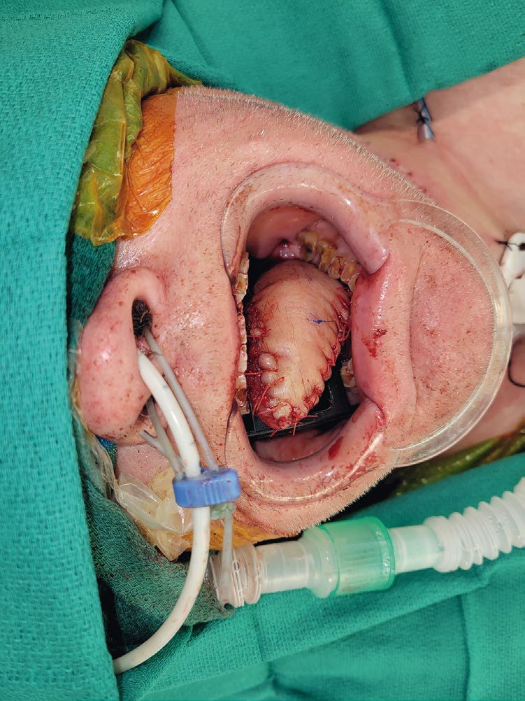
The commando procedure allows resection of tongue cancer with mandibular involvement [1], and lip reconstruction requires flap cover once more than a third of the lip has gone [1], techniques covered on the Grafts and Flaps page.
Neck dissection, partial laryngectomy, reconstruction and rehabilitation in Schwartz's account
- Radical neck dissection sacrificing sternocleidomastoid, internal jugular vein and accessory nerve has been abandoned because selective and modified radical dissection are oncologically equal with better function; selective dissection is standard for cN0 and limited cN1, supraomohyoid (I–III, often with upper IV) for oral cavity, lateral (II–IV) for larynx and hypopharynx, posterolateral (II–V, or partial V) for thyroid, and modified radical dissection remains the choice for advanced nodal disease beyond uncomplicated N1; a planned post-radiotherapy dissection at 6–8 weeks consolidates N2a or worse or a partial response [2].
- Vertical partial laryngectomy through a midline thyrotomy removes the whole true cord and part of the false cord and is reconstructed with strap muscle and perichondrial flaps or a temporoparietal fascial free flap, suiting recurrent T1/T2 tumours of one cord with under 5 mm subglottic extension, a mobile cord and no cricoid or extralaryngeal spread; supraglottic laryngectomy below the hyoid is rarely performed now that laser or TORS suffices; supracricoid laryngectomy with cricohyoidopexy or cricohyoidoepiglottopexy for T3 glottic tumours without pre-epiglottic or cricoarytenoid involvement preserves one cricoarytenoid unit for breathy, coarse phonation with good decannulation and swallowing after an initial high aspiration rate [2].
- Lip defects under a third close primarily, one- to two-thirds use Abbe or Karapandzic flaps (commissure preserved) or Estlander (commissure resected), Bernard–Burow advancement with Burow's triangles when lip tissue is short, Karapandzic up to 80% at the cost of microstomia, and larger defects need free flaps without sphincter function; vermilion realignment and orbicularis reapproximation are the critical steps [2].
- Total glossectomy reconstruction with rectus abdominis or anterolateral thigh flap fills dead space, reaches the premaxilla for articulation and creates posterior bulk for the pharyngeal swallow, 90% being decannulated when the larynx is preserved; soft palate defects use obturators or the modified Gehanno closure; maxillectomy defects take obturators or soft-tissue flaps, with layered fibula or scapular tip for orbitomaxillary defects and orbital floor support to prevent diplopia [2].
- Regional flaps are the pectoralis major (thoracoacromial), submental (facial artery branches, preserved during dissection) and supraclavicular (transverse cervical); free flaps by defect are radial forearm (thin, long pedicle, partial glossectomy, floor of mouth, buccal), anterolateral thigh (hypopharynx, skin, total glossectomy), lateral arm (facial skin colour match), rectus (deep inferior epigastric (total glossectomy, skull base), latissimus (scalp and skull base), fibula (peroneal) segmental mandible and maxilla, bent to a 2.5 mm titanium plate with wedge ostectomies), scapula (circumflex scapular, composite defects), osseocutaneous radial forearm (partial mandible, orbit) and iliac crest (up to 16 cm of bone, high donor morbidity) [2].
- After laryngectomy fewer than 20% achieve fluent oesophageal speech, the electrolarynx bridges, and tracheo-oesophageal puncture with a one-way valve gives functional speech in over 90%, primary and secondary puncture carrying similar complication rates governed more by the extent of pharyngeal reconstruction than prior radiotherapy; speech pathology involvement lowers dysphagia, stricture, weight loss and pneumonia [2].
- Temporal bone tumours are managed by local excision or Mohs for pinna lesions (total auriculectomy for extensive disease, with prosthesis), parotidectomy and supraomohyoid dissection for tumours anterior to the canal and posterolateral dissection behind it, sleeve resection rarely sufficing, lateral temporal bone resection removing canal and middle ear en bloc and subtotal resection adding inner ear and facial nerve, both followed by radiotherapy, with anterolateral thigh flap reconstruction, watertight dural closure, cable grafts or masseteric nerve transfer for the facial nerve and gold weights, lid shortening or tarsorrhaphy to protect the eye [2].
- Parapharyngeal tumours are approached transcervically with submandibular gland excision, transparotid to identify the facial nerve, rarely by mandibulotomy, and never transorally [2].
- Tracheotomy is indicated for prolonged ventilation (early tracheotomy showed no survival benefit in a large randomised trial, only shorter sedation), pulmonary toilet, lost airway reflexes and perioperative obstruction; percutaneous and open techniques are equivalent in meta-analysis of 15 trials with fewer wound infections and better scars percutaneously, cricothyroidotomy is converted early because of cord dysfunction and subglottic stenosis, decannulation follows 24–48 hours of capping, and the stoma closes in 2–3 weeks [2].
Complications
The functional complications drive the treatment choice rather than following it: preserving the larynx is the stated aim in laryngeal cancer, and radiotherapy is chosen over surgery for that reason [1].
For lip and cheek resection, the complication anticipated is cosmetic and functional loss requiring flap reconstruction [1].
Outcomes
Hard palate tumours have the lowest survival of the oral cavity subsites, because resection is difficult [1].
Hypopharyngeal cancer metastasises early [1], and tonsillar cancer has nodal metastases in 80% by diagnosis [1], both consequences of presenting late in a site that stays silent.
Survival figures in Schwartz's account
Early lip cancer has 84–92% 10-year survival, falling to 11–28% with advanced regional or distant disease; cervical metastasis halves 5-year survival; temporal bone cancer has under 40% 5-year survival with nodal spread the strongest predictor; recurrent neck disease after radiotherapy or comprehensive dissection carries very poor survival; and total glossectomy with laryngectomy brings poor quality of life and survival [2].
References
- The ABSITE Review, 2022, Ch. 19 Head and Neck
- Schwartz's Principles of Surgery, 11th ed., Ch. 18, Disorders of the Head and Neck
- Sabiston Textbook of Surgery, 22nd ed., Ch. 69 Plastic Surgical Considerations in Cancer Surgery
- Bailey & Love's Short Practice of Surgery, 28th ed., Ch. 53 Oral cavity cancer