



Grafts and Flaps
Summary
- A graft has no blood supply of its own and must acquire one from the bed; a flap brings its own.
- That single distinction explains most of what follows. A skin graft survives for its first three days by imbibition (osmotic uptake from the bed) with neovascularisation beginning around day 3 [1].
- It follows that poorly vascularised beds will not support a graft, including tendon, bone stripped of periosteum, and irradiated tissue [1].
- For flaps the failure mode is different: the commonest cause of pedicled or free flap necrosis is venous thrombosis, not arterial [2].
Definition
Autografts are the patient's own skin, split-thickness or full-thickness, and are the best option [1]. Homografts (allografts) are cadaveric skin; xenografts are porcine [1].
Split-thickness grafts are 0.12 to 0.15 mm thick and include the epidermis and part of the dermis [1].
Pathophysiology
Graft take happens in two phases. Imbibition, an osmotic process, supplies the graft from day 0 to day 3; neovascularisation begins around day 3 [1].
That two-phase mechanism explains the thickness trade-off. Split-thickness grafts are more likely to survive, because a thinner graft makes imbibition and subsequent revascularisation easier; full-thickness grafts undergo less wound contraction, which is why they are used on the palms and the backs of the hands [1].
The donor site of a split-thickness graft regenerates from hair follicles and skin edges [1], which is why the same donor site can be reharvested and a full-thickness donor site cannot.
Allografts vascularise and are eventually rejected, at which point they must be replaced; xenografts do not vascularise at all [1].
Tissue expansion works by local recruitment, thinning of the dermis and epidermis, and mitosis [2].
Incisions, closure and graft take in Schwartz's account
- Skin rests under tension between points tethered by subcutaneous fibrous tissue, so a linear incision gapes into an ellipse whose long axis lies perpendicular to the lines of greatest tension (Langer's lines from fresh cadavers) while Borges' relaxed skin tension lines follow the furrows produced by pinching relaxed skin, and incisions parallel to them heal least conspicuously; the epidermis and superficial dermis are incised sharply, the deep dermis and subdermal plexus with cautery, the blade kept perpendicular to the surface and the same path traced through fat to avoid devitalised side-tracks, traumatic wounds are converted to controlled surgical wounds by excising damaged edges and reorienting with small local flaps, and old scars are excised through unscarred skin on either side rather than incised, since closing scarred edges invites delayed healing, infection and worse scars [3].
- The dermis is the layer that must be approximated because it carries the blood supply and cells that build matrix; deep dermal sutures are followed by superficial sutures in upper dermis and epidermis, absorbable deep sutures prolong inflammation while non-absorbable ones suit scar-prone patients, step-offs cast shadows that accentuate scars, motion between edges prolongs inflammation and demands more collagen, deep-passing surface sutures leave track marks and come out within a week, shallow dermal or subcuticular sutures avoid them at the cost of harder alignment, and a hypertrophic scar stops growing at 6 months whereas a keloid keeps growing beyond its borders [3].
- Non-healing wounds affect 3–6 million Americans, 85% of them over 65, costing up to $25 billion a year; uncomplicated wounds close within 4–6 weeks and are chronic beyond that, the common thread being persistent pro-inflammatory conditions in which neutrophils and macrophages cyclically scavenge new tissue and degrade matrix; low oxygen tension paradoxically drives capillary budding through hypoxia-inducible factor, fibroblasts become myofibroblasts at about 7 days with contraction peaking around 10 days along an "axis of contraction" at 0.75–1 mm a day, lysyl oxidase cross-links collagen extracellularly, collagenases cleave all three chains into characteristic three-quarter and one-quarter fragments, TGF-β signals through SMAD proteins in remodelling from 3 weeks to beyond a year, and mature scar regains only 80% of uninjured tensile strength as redundant capillaries apoptose and redness fades [3].
- Graft take proceeds through plasmatic imbibition for 24–48 hours (a fibrin film holds the graft while cells survive by diffusion from wound plasma), inosculation after 48 hours (vascular buds from the bed cross the fibrin and align with cut vessels on the graft's deep dermis, the period of greatest risk, lost to shear, seroma, haematoma or bacteria) and revascularisation, complete by 4–5 days and shown by colour and capillary refill; skin substitutes take by the same revascularisation and repopulation [3].
Clinical features
Autografts reduce infection, desiccation, protein loss, pain, water loss, heat loss and red cell loss compared with dermal substitutes [1].
The ranking of cover is consistent: autograft is best, then homograft, then xenograft, then dermal substitutes [1].
Etiology
Skin grafts are contraindicated where the culture is positive for beta-haemolytic streptococcus, or where bacteria exceed 10⁵ [1].
The commonest reason for skin graft loss is seroma or haematoma formation under the graft, which prevents attachment [1]. This is prevented by a pressure dressing applied over the graft [1].
Reasons to delay autografting are infection, insufficient donor sites, a septic or unstable patient, and not wanting to create further donor sites with their attendant blood loss [1].
Diagnosis
The assessment before grafting is of the bed rather than the wound size: a bed of tendon, bare bone or irradiated tissue will not take a graft and needs a flap instead [1].
Thresholds and severity
Homografts last about 4 weeks and are a good temporising material; xenografts last about 2 weeks [1].
Pressure sores are staged, and the stage determines whether a flap is needed [2]:
| Stage | Description | Treatment |
|---|---|---|
| I | Erythema and pain, no skin loss | Keep pressure off |
| II | Partial skin loss with yellow debris, into the dermis | Local treatment, keep pressure off |
| III | Full-thickness skin loss with subcutaneous fat exposed | Sharp debridement; likely needs a myocutaneous flap |
| IV | Involves bony cortex, muscle, adipose tissue, tendon | Myocutaneous flap, for example a gluteal flap |
Meshing a split-thickness graft expands its coverage and allows exudate to escape, and is the standard approach where a large area must be covered from a limited donor site [4].
Meshed grafts are used on the back, flank, trunk, arms and legs, sites where the cosmetic penalty of the mesh pattern is acceptable [1].
The face and palms are treated differently. The face receives topical antibiotics for the first week, with non-meshed full-thickness graft for areas that have not healed; the palm is grafted with full-thickness skin, preserving the specialised palmar aponeurosis, and splinted in extension for 7 days afterwards [1].
The hand is immobilised in extension for 7 days after full-thickness grafting of a deep burn, then physiotherapy begins; wire fixation of joints may be needed if they are unstable or open [1]. Genitals can be covered with meshed split-thickness graft [1].
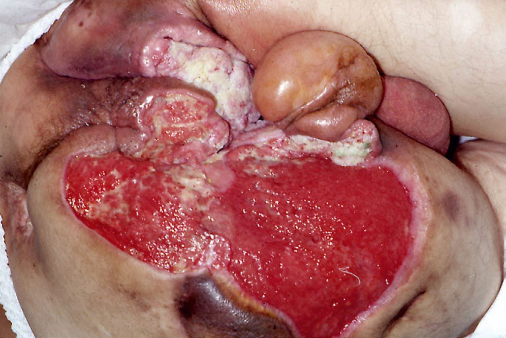
Graft thickness and flap classification in Schwartz's detail
- Thin split grafts contract little primarily (few elastic fibres) but allow much secondary wound contraction (a concern beside mobile structures such as the oral commissure) and take best because epidermis has low metabolic demand, whereas thick split and full-thickness grafts contract more primarily, less secondarily, and are more durable, better pigmented, more resistant to desiccation and better looking but need better beds; meshing at 1:1.5 to 1:6 improves take by draining fluid and conforming to contour but the interstices epithelialise secondarily, giving poorer cosmesis and more contraction, alternatives being micrografting devices and fractional harvesting of full-thickness columns; split donor sites re-epithelialise from basal cells lining the recesses between dermal papillae in about 2 weeks, kept moist, clean and shear-free, with pain eased by adrenaline-lidocaine infiltration, ice, topical local anaesthetic, hydrocolloid or polyurethane dressings with fibrin sealant and airtight film; full-thickness grafts are cleared of all subcutaneous fat, need clean well-vascularised beds free of colonisation, radiation or fibrosis, suit face and hands, and leave a donor site that must be closed primarily; composite grafts of skin with fat, cartilage, perichondrium or a little muscle serve small specialised defects such as the nasal lobule, the helical root of the ear giving matched colour and contour with an inconspicuous primarily closed donor site [3].
- Flaps are classified by tissue (cutaneous, muscle, musculocutaneous, osseous, osteocutaneous, fascial, neural, visceral, jejunum, stomach, colon, omentum), by proximity (local, regional, distant; pedicled when the supply stays attached, free when vessels under about 5 mm are divided and reanastomosed by microvascular technique) and, most importantly, by blood supply: random pattern flaps on unnamed subdermal plexus vessels at a traditional 3:1 length-to-width ratio (transposition including Z-plasty, which lengthens the central limb and redirects tension, and the 60°/120° rhomboid Limberg flap with its Dufourmental modification; semicircular rotation; rectangular and V-Y advancement; interpolation across intact tissue such as the forehead flap for the nose); axial pattern flaps on an artery running along the flap's axis, first Bakamjian's 1971 deltopectoral flap on internal mammary perforators reaching the head and neck, then groin and posterior thigh flaps; musculocutaneous flaps in which a muscle's dominant vessels secondarily perfuse skin through many small vessels, graded by Mathes and Nahai as type I single pedicle (gastrocnemius), II dominant plus minor pedicles that alone cannot sustain it (gracilis), III two dominant pedicles (rectus abdominis), IV segmental pedicles (sartorius) and V one dominant with secondary segmental pedicles that can sustain it (pectoralis major); fasciocutaneous flaps fed through a fascial plexus between rather than through muscles, thinner and without motor loss, Mathes-Nahai types A–C by how the pedicle reaches the fascia (sural perforator flaps for the leg); direct cutaneous flaps; and perforator propeller flaps [3].
- Taylor's angiosomes are cutaneous territories each fed by a single source artery perforating muscle or fascia, usually with two venae comitantes and often a superficial venous system, overlapping like dermatomes and linked at their borders by choke vessels that open or close; the anatomic angiosome is the artery's terminal branching, the dynamic angiosome the tissue stained by intra-arterial fluorescein, and the potential angiosome what a conditioned (delayed) flap can carry (both larger than the anatomic) though no method yet predicts safe harvest limits precisely [3].
- Gustilo grades I–II are open fractures with minimal soft tissue disruption, IIIA severe injury with adequate tissue to repair, IIIB tissue loss needing replacement and IIIC vascular injury needing reconstruction; comminution, bone or soft tissue loss, contamination and devascularisation predict poorly, loss of plantar sensation no longer mandates amputation, and salvage is judged against comorbidity, socioeconomic factors, motivation and rehabilitative potential [3].
Treatment and Management
Flap choice for breast reconstruction has shifted away from muscle-transferring flaps. The DIEP flap is now used more commonly than the TRAM: it transfers the deep inferior epigastric perforators with the overlying fat and skin, with no muscle transferred, anastomosing the inferior epigastric artery and vein to the internal mammary vessels, with the thoracodorsal vessels as an alternative [2]. It produces fewer hernias and less long-term muscle weakness than the TRAM [2].
The TRAM flap transfers a portion of rectus muscle with the flap. A pedicled TRAM relies on the superior epigastric vessels, a free TRAM on the inferior epigastric vessels, and the periumbilical muscle perforators are the most important determinant of TRAM flap viability [2].
Burn scar hypopigmentation and irregularity can be improved with dermabrasion and thin split-thickness grafts [1].
Procedural interventions
A pressure dressing over a fresh graft is not optional, it is the specific measure that prevents the seroma or haematoma that causes most graft failures [1].
Tissue expansion is the technique used where local tissue of matching quality is required but insufficient in quantity, and works by recruitment, dermal and epidermal thinning, and mitosis [2].
Flap technique, expansion and regional reconstruction in Schwartz's account
- Muscle brings bulk, deformability to fill recesses that would harbour fluid and bacteria, projection to correct contour, and motor restoration when its nerve is coapted to a recipient nerve, the latissimus dorsi on the thoracodorsal pedicle for breast reconstruction being the classic case, while fasciocutaneous flaps give thin durable cover for foot and ankle where footwear abrades; propeller flaps are raised on a perforator at the defect's edge with an equal length extending opposite and rotated like a propeller, sparing muscle and often avoiding microsurgery [3].
- Tissue expansion places a silicone elastomer balloon with an accessible filling port under the planned flap and inflates it with serial saline injections over weeks after wound healing; expanded skin shows thickened dermis, richer vasculature and less fat, is true new tissue, and benefits from the delay and the vascular capsule, at the cost of infection, haematoma, seroma, extrusion, implant failure, necrosis, pain, nerve injury and visible temporary deformity; it is most useful for giant congenital naevi, burn scar reconstruction, scalp and breast [3].
- Lower limb trauma follows ATLS, early tetanus and antibiotics, Doppler then angiography for absent pulses, immediate revascularisation, fasciotomy for compartment syndrome (pain on passive stretch in a pale pulseless limb without direct vascular injury, paraesthesia and paralysis being late; recurring after fixation or reperfusion), then debridement, fracture reduction and stabilisation, vascular repair and soft tissue cover ideally by orthopaedic, vascular and plastic teams under one anaesthetic, with negative pressure therapy bridging to reconstruction, soft tissue closed before bone with antibiotic beads or spacer holding the gap and external fixation for segmental loss; cover ranges from split grafts on healthy muscle, temporary substitutes then grafting, local random, axial or perforator flaps from outside the zone of injury, cross-leg flaps only when microsurgery is impossible or in young children, and free flaps (recipient vessels outside the zone, bone stable, latissimus loss hampering crutch use, flow-through designs replacing vessel segments); amputation maximises length, uses the amputated part for grafts or free transfer to the stump, and treats transected nerves by targeted muscle reinnervation, which reduces phantom and residual limb pain and symptomatic neuroma compared with traction neurectomy [3].
- Chest wall: pectoralis major on the thoracoacromial pectoral branch or internal mammary perforators covers sternum, upper chest and neck after sternotomy dehiscence or resection, rectus abdominis on the superior epigastric vessels (or free on the deep inferior epigastric) the lower sternum, latissimus dorsi on thoracodorsal or paraspinal perforators with serratus anterior added for area covers other chest sites, and trapezius on the transverse cervical vessels covers upper back, neck base and shoulder while preserving its superior fibres, acromial attachment and accessory nerve [3].
- Abdominal wall defects, most often fascial dehiscence after laparotomy, need minimal-tension fascial closure to avoid dehiscence, recurrence or compartment syndrome; mesh replaces fascia in clean wounds but is avoided with infected mesh, fistula or perforation, component separation advancing bilateral anterior rectus fascia–rectus–oblique flaps after lateral release closes midline defects up to 10 cm superiorly, 18 cm centrally and 8 cm inferiorly, and larger or full-thickness defects take pedicled tensor fasciae latae (ascending lateral circumflex femoral branch) or anterolateral thigh (descending branch) flaps, sometimes bilateral, or free flaps; perineal reconstruction after surgery and radiation within the rigid pelvic outlet must heal, support pelvic contents, accommodate urinary and bowel function and restore penis or vagina and vulva with local, regional or free flaps [3].
- Pressure sores in stage III–IV are assessed for infection, collections, osteomyelitis (plain films usually suffice, CT or MRI when equivocal) and communication with joint, urethra, colon or spinal canal, debrided without delay including soft, poorly bleeding or purulent bone, and reconstructed (primary closure is rarely possible and split grafts rarely durable) with muscle or musculocutaneous flaps for heavy contamination and complex contours, fasciocutaneous flaps where offloading cannot be guaranteed, suture lines kept off the pressure point and flaps large enough to re-advance: gluteal-based flaps for sacral, gluteal or posterior thigh (descending inferior gluteal branch) for ischial, and tensor fasciae latae, rectus femoris or vastus lateralis for trochanteric ulcers, motor loss being irrelevant in paralysed patients; patients with cord injuries at or above T5 risk autonomic hyperreflexia (intracranial and retinal haemorrhage, seizures, arrhythmia, death), and aftercare demands air-fluidised low-pressure beds for 7–10 days, nutrition, antispasmodics and coordinated care [3].
Complications
TRAM flap complications are flap necrosis, ventral hernia, infection and abdominal wall weakness [2], the last two being what the DIEP was designed to avoid.
Venous thrombosis, not arterial insufficiency, is the commonest cause of flap necrosis in both pedicled and free flaps [2].
Graft loss is most often mechanical rather than biological: a collection under the graft preventing contact with the bed [1].
Protecting the graft in Schwartz's account
Dressings must prevent desiccation and shear through inosculation; tie-over bolsters are traditional but topical negative pressure increases the quantity and quality of split-graft take, especially on irregular or mobile surfaces, and late failure from shear, drying or infection remains possible for up to 2 weeks, so dressings, moisturisers or antibacterials continue until stable [3].
Outcomes
The two rules that determine success are matched to the two failure modes. For grafts, ensure contact with a vascularised bed and prevent a collection beneath it. For flaps, watch the venous outflow.
Thickness sets the remaining trade-off: split-thickness for reliability of take, full-thickness for resistance to contraction where function or appearance demands it [1].
References
- The ABSITE Review, 2022, Ch. 17 Burns
- The ABSITE Review, 2022, Ch. 18 Plastics, Skin, and Soft Tissues
- Schwartz's Principles of Surgery, 11th ed., Ch. 45, Plastic and Reconstructive Surgery, Figs. 45-5 to 45-7
- Bailey & Love's Short Practice of Surgery, 28th ed., Ch. 3 Wound healing and tissue repair
- Sabiston Textbook of Surgery, 22nd ed., Ch. 23 Wound Healing