



Venous Thromboembolism and DVT
Summary
- Venous thromboembolism (VTE) encompasses deep vein thrombosis (DVT) and pulmonary embolism (PE); DVT describes a blood clot in the deep venous system that can detach and embolise to the pulmonary vasculature, causing mechanical obstruction, pulmonary hypertension and acute right ventricular strain [1].
- VTE incidence is approximately 100 per 100,000 people per year, with an estimated 600,000 cases annually in the United States, and death occurs in 6% of DVT and 12% of PE cases within one month of diagnosis [2].
- All hospitalised surgical patients should be risk-stratified for VTE, and prophylaxis, diagnosis (duplex ultrasound for DVT, CT pulmonary angiography for PE) and anticoagulant treatment are central to management [3][4].
Two NICE guidelines cover venous thromboembolism from a surgeon's point of view. NG158 governs diagnosis, management and thrombophilia testing once VTE is suspected or confirmed; NG89 governs prophylaxis, reducing the risk of hospital-acquired DVT or pulmonary embolism [5][6].
NG158 defines "provoked" precisely, and the definition drives the duration of treatment. A provoked DVT or PE is one in a person with a recent (within 3 months) and transient major clinical risk factor for VTE, such as surgery, trauma, significant immobility (bedbound, unable to walk unaided, or likely to spend a substantial proportion of the day in bed or in a chair), pregnancy or puerperium, or in a person having hormonal therapy, meaning the combined oral contraceptive pill or hormone replacement therapy [5]. Active cancer is separately defined as receiving active antimitotic treatment, diagnosed within the past 6 months, recurrent or metastatic, or inoperable, excluding squamous skin cancer and basal cell carcinoma [5].
Definition
- DVT is thrombus formation within the deep venous system of the upper or lower extremities; PE occurs when a thrombus embolises through the venous system to lodge in the pulmonary vasculature [1].
- Massive PE is defined as PE resulting in haemodynamic compromise or where more than 30% of the pulmonary vasculature is compromised [4].
- Phlegmasia alba dolens is a painful, swollen, pale ("white") leg from extensive thrombosis with fluid sequestration and arterial insufficiency due to elevated compartment pressures.
- Phlegmasia cerulea dolens is a painful, swollen, blue leg extending to the buttocks, extensive DVT of the major axial deep venous channels with relative sparing of collateral veins, causing pain, pitting oedema and cyanosis, and is more severe, potentially progressing to venous gangrene [2][7].
Pathophysiology
- Virchow's triad (stasis, endothelial (venous wall) injury, and hypercoagulability) underlies venous thrombosis, in contrast to arterial thrombosis, in which endothelial injury is the key element [7].
- Relative hypercoagulability appears most important in spontaneous ("idiopathic") VTE, whereas stasis and endothelial damage play a greater role in secondary ("provoked") VTE associated with transient risk factors such as immobilisation, surgery and trauma [2].
- DVT most commonly begins in the soleal sinusoids and venae comitantes of the calf before extending proximally into the popliteal, femoral, iliac veins and even the vena cava [8].
- Newly formed thrombi are not firmly attached to the vessel wall and can detach and embolise; once in the pulmonary circulation, emboli are coated with fibrin and platelets, causing mechanical obstruction of pulmonary blood flow [1].
- Anatomical factors can predispose to DVT: May-Thurner syndrome is chronic narrowing of the left common iliac vein at the point where the right common iliac artery crosses over it, predisposing to iliofemoral venous thrombosis [2].
- The left leg is affected roughly twice as often as the right, attributed to compression of the longer left iliac vein by the right iliac artery [7].
Venous structure, thrombus origin and risk in Schwartz's account
- Veins are thin-walled, distensible and collapsible, with a non-thrombogenic endothelium producing nitric oxide and prostacyclin, medial elastic rings and smooth muscle giving capacitance, and bicuspid valves, more numerous distally, absent from the IVC, common iliacs, portal system and cranial sinuses, that close at a retrograde velocity of at least 30 cm/s; the valveless soleal and gastrocnemius sinuses store blood and empty into posterior tibial and peroneal veins with each calf contraction [9].
- Thrombosis begins in areas of stasis such as a soleal sinus or just downstream of a valve cusp in an axial calf vein, isolated proximal DVT without tibial thrombosis being unusual; Virchow's 1862 triad of stasis, endothelial injury and hypercoagulability still holds, hypercoagulability dominating idiopathic ("unprovoked") and stasis and injury the secondary ("provoked") events after immobilisation, surgery or trauma, and multiple risk factors combine exponentially rather than additively [9].
- Compression of the left iliac vein by the crossing right iliac artery (May–Thurner syndrome) or by masses predisposes to iliofemoral thrombosis; VTE is commoner in whites and African Americans than Asians and Native Americans, and single-nucleotide polymorphisms add small risk [9].
Clinical features
- Clinical manifestations of DVT may be entirely absent; when present, features include calf/leg pain, swelling, warmth, erythema, engorged superficial veins, and occasionally mild fever and tachycardia from inflammatory mediator release [3][10].
- Homans' sign (calf pain on dorsiflexion of the foot) is neither sensitive nor specific and should not be relied upon or even performed [3][10].
- The extent of swelling correlates with the level of thrombosis: calf DVT causes minimal swelling, femoral DVT causes ankle and calf swelling, and iliofemoral DVT causes swelling of the entire leg [7].
- In acute PE, the most common symptom is dyspnoea; other features include anxiety, cough, pleuritic or dull chest pain, haemoptysis, and syncope, with examination showing tachycardia, low-grade fever, a loud P2, or poor perfusion; massive PE can cause severe hypotension, unconsciousness, or cardiac arrest [1].
- PE is misdiagnosed in almost 75% of patients, with differentials including acute MI, aortic dissection, septic shock, chest infection, haemothorax, and pneumothorax [4].
- Roughly one-third of symptomatic VTE patients present with PE and two-thirds with DVT [2].
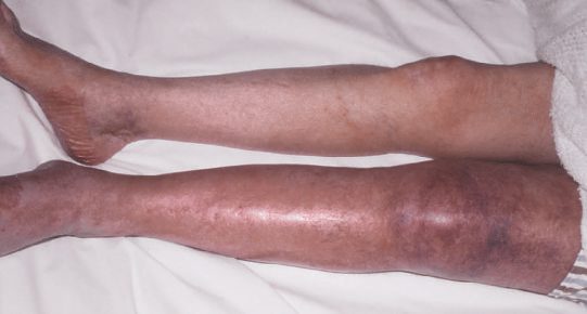
Presentation and the phlegmasias in Schwartz's account
- VTE affects about 100 per 100,000 a year (over 600,000 US cases), 20% within 3 months of surgery, one-third presenting as pulmonary embolism and two-thirds as DVT; death follows 6% of DVT and 12% of PE within a month (not all from the clot), pulmonary hypertension develops in 4% and post-thrombotic syndrome in up to 30%; early DVT may cause no pain or swelling even when extensive in the bed-bound, history and examination are notoriously unreliable and DVT is confirmed in 50% or fewer of clinically suspected cases [9].
- Extensive axial thrombosis sparing collaterals causes phlegmasia cerulea dolens (pain, pitting oedema, cyanosis), and extension into collaterals with massive fluid sequestration causes phlegmasia alba dolens, a pale, painful, grossly swollen limb with arterial insufficiency from raised below-knee compartment pressures; both may progress to venous gangrene and amputation [9].
- Superficial thrombophlebitis usually arises in varicose veins, recurs at variable sites in normal veins as thrombophlebitis migrans (hidden visceral malignancy, blood dyscrasia or collagen disease), or complicates indwelling catheters (38% of PICC lines, 57% of these in the cephalic vein) presenting as redness, warmth, tenderness and a palpable cord, with fever and leukocytosis when suppurative; concomitant DVT accompanies 5–40% of superficial thrombophlebitis, mostly when the great saphenous is involved within 1 cm of the saphenofemoral junction [9].
- Axillary–subclavian thrombosis is primary (venous thoracic outlet syndrome, effort thrombosis, Paget–Schroetter, from repetitive activity damaging the vein between clavicle head and first rib beside the subclavius) in a minority and secondary to catheters or hypercoagulability in most (over 30% of tunnelled subclavian devices thrombose) presenting with arm oedema, tenderness and prominent superficial veins [9].
Etiology
- Acquired VTE risk factors include age over 40, hospitalisation/immobilisation, hormone replacement therapy or oral contraceptive use, pregnancy and the postpartum state, prior VTE, malignancy, major surgery, obesity, nephrotic syndrome, trauma and spinal cord injury, long-haul travel (>6 hours), varicose veins, antiphospholipid syndrome, myeloproliferative disorders, and polycythaemia [2].
- Heritable risk factors include male sex, factor V Leiden mutation, prothrombin 20210A gene variant, and antithrombin, protein C, or protein S deficiency; mixed heritable/acquired causes include hyperhomocysteinaemia and elevated factors VII, VIII, IX and XI [2].
- DVT may arise from vein compression or stasis (immobility, trauma, mass, surgery, paralysis, long-distance travel), inherited hypercoagulability (factor V Leiden, protein C, protein S, antithrombin deficiency), or acquired hypercoagulability (surgery, malignancy, polycythaemia, smoking, hormone replacement therapy, oral contraceptives, dehydration) [10].
- Migratory thrombophlebitis (Trousseau's sign) is classically associated with pancreatic carcinoma [12].
- Without prophylaxis, 30% of patients over age 40 undergoing major surgery will develop DVT, and 0.1–0.2% will die of pulmonary thromboembolism [4].
- About 50% of patients with phlegmasia cerulea dolens have an underlying malignancy [7].
Risk factor catalogue in Schwartz's table
Acquired risks are age over 40, hospitalisation and immobilisation, hormone replacement and oral contraception, pregnancy and the puerperium, prior VTE, malignancy, major surgery, obesity, nephrotic syndrome, trauma and spinal cord injury, long-haul travel over 6 hours, varicose veins, antiphospholipid syndrome, myeloproliferative disease and polycythaemia; inherited risks are male sex, factor V Leiden, prothrombin 20210A, antithrombin, protein C and protein S deficiency, factor XI elevation and dysfibrinogenaemia; mixed heritable–acquired causes are homocysteinaemia, elevated factors VII, VIII, IX and XI, hyperfibrinogenaemia and activated protein C resistance without factor V Leiden, with obesity, hypertension and diabetes also contributing [9].
Diagnosis
- Duplex ultrasound is the investigation of choice for DVT, visualising anatomy, extent of thrombosis, and relying on flow and vein compressibility, though it is operator-dependent and has lower sensitivity for isolated calf DVT [1][10].
- Duplex findings in DVT include cross-sectional vein incompressibility, direct visualisation of thrombus with vein enlargement, abnormal spectral Doppler, and abnormal colour Doppler flow [1].
- D-dimer is often raised postoperatively and is of limited value in that context; a negative d-dimer makes DVT/PE unlikely but the scoring systems and d-dimer are sensitive, not specific, screening tools, so VTE diagnosis should not rest on these alone [1][4].
- The two-level DVT Wells score stratifies patients into "DVT likely" (≥2 points) versus "DVT unlikely" (≤1 point) [3].
- CT or MR venography is more sensitive but reserved for special cases, e.g. suspected iliac vein/IVC thrombus or planning endovenous intervention [4].
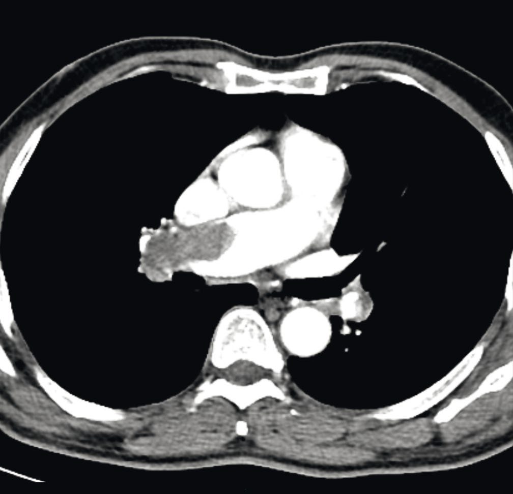
- For PE, CT pulmonary angiography (CTPA) is the gold-standard, first-line test; ventilation/perfusion (V/Q) scanning is used if CTPA is contraindicated (e.g. pregnancy); transthoracic echocardiography (TTE) assesses right ventricular function [1][4].
The UK DVT pathway is built on the two-level Wells score and a 4-hour clock, and the branch that catches people out is what happens when the scan cannot be done in time. If DVT is suspected, use the 2-level DVT Wells score to estimate clinical probability, after a general medical history and physical examination to exclude other causes [5].
DVT likely, Wells 2 points or more [5]:
| Situation | Action |
|---|---|
| Scan obtainable within 4 hours | Proximal leg vein ultrasound with the result available within 4 hours, plus a D-dimer if the scan is negative |
| Scan not obtainable within 4 hours | D-dimer test, then interim therapeutic anticoagulation, then proximal leg vein ultrasound with the result available within 24 hours |
DVT unlikely, Wells 1 point or less [5]: offer a D-dimer with the result available within 4 hours, or, if that is not possible, interim therapeutic anticoagulation while awaiting the result.
Tables reformat the NG158 diagnostic pathway [5]. Note the asymmetry: the D-dimer is ordered after a negative scan in the likely group, but first in the unlikely group.
- The negative-scan, positive-D-dimer branch has its own rule.
- Stop interim therapeutic anticoagulation (but do not stop long-term anticoagulation being used for secondary prevention) and offer a repeat proximal leg vein ultrasound scan 6 to 8 days later [5].
- Where both scan and D-dimer are negative, stop interim anticoagulation, think about alternative diagnoses, and tell the person DVT is unlikely, discussing the signs and symptoms and when to seek further help [5].
- Baseline bloods are taken but must not delay treatment.
- When using interim therapeutic anticoagulation for suspected proximal DVT or PE, carry out full blood count, renal and hepatic function, prothrombin time and APTT, but do not wait for the results before starting anticoagulation, and review and act on them within 24 hours of starting [5].
- Where possible, choose an interim anticoagulant that can be continued if DVT or PE is confirmed [5].
Duplex criteria and the older tests in Schwartz's account
- Duplex (B-mode compression and augmentation with pulsed and colour Doppler) is over 95% sensitive and specific for symptomatic infrainguinal DVT, over 91% and 97% against venography for femoropopliteal thrombus, and 50–93% sensitive for isolated calf thrombus; normal supine flow is phasic (falling with inspiration as the diaphragm descends), augmented by elevation or compression and abolished by Valsalva, and DVT is diagnosed by non-compressibility (the primary criterion, from common femoral to popliteal), absent spontaneous flow, absent colour filling, lost respiratory variation and venous distension, calf thrombus being found mainly by colour flow since calf compression is difficult; bowel gas and habitus limit duplex in the abdomen, where MR and CT venography serve [9].
- Impedance plethysmography (83% sensitive for proximal DVT, poor for calf) and iodine-125 fibrinogen uptake (a 20% rise in one area, 73% sensitive and 71% specific, blind to pelvic thrombus and useless after surgery or with inflammation) are historic; ascending venography via a dorsal foot vein in two projections remains the reference, a normal study virtually excludes DVT (1.3% developed DVT within 3 months in 160 patients), but causes puncture-site thrombosis in 1–9%, pain in 18% with low-osmolar and 44% with conventional contrast, and is now confined to research and pre-intervention planning, while intravascular ultrasound via the common femoral vein outperforms venography for iliac obstruction [9].
- Superficial thrombophlebitis is scanned to exclude DVT and rescanned at 5–7 days when the proximal great saphenous is involved, since 10–20% progress to deep involvement within a week; axillary–subclavian thrombosis may be missed by duplex behind the clavicle or through collaterals, so venography follows discordant findings; mesenteric venous thrombosis is diagnosed by contrast CT or MRI (near 100% sensitive, 98% specific) or ultrasound (93% and 99%) [9].
Scoring and Severity
- The two-level DVT Wells score and two-level PE Wells score categorise patients into likely/unlikely groups to guide further testing [1][3].
- Surgical procedures are stratified by DVT risk: low risk (maxillofacial, neurosurgery, cardiothoracic surgery), medium risk (inguinal hernia repair, abdominal, gynaecological, urological surgery), and high risk (pelvic elective/trauma surgery, total knee/hip replacement) [3].
- Patients with hypotension, right ventricular failure on echocardiography/CT, elevated cardiac biomarkers, and a PE severity index (PESI) class III or higher are considered high risk for PE-related mortality [1].
- The 30-day mortality of acute massive PE is about 50% (40% within the first 2 hours); operative mortality for stable patients undergoing intervention is about 30%, rising to up to 70% for those requiring CPR or mechanical circulatory support preoperatively [4].
Risk stratification and prophylaxis tiers in Schwartz's account
The Rogers score (Patient Safety in Surgery Study: operation type, ASA class 3–5, female sex, work RVU over 17, and two points each for disseminated cancer, chemotherapy within 30 days, sodium over 145, transfusion over 4 units in 72 hours and ventilator dependence, one each for clean/contaminated wound, haematocrit ≤38%, bilirubin over 1 mg/dL, dyspnoea, albumin ≤3.5 and emergency) and the Caprini score (1 point for age 41–60, minor surgery, BMI over 25, swollen legs, varicose veins, pregnancy, recurrent miscarriage, hormones, sepsis, lung disease, infarction, heart failure, inflammatory bowel disease or bed rest; 2 for age 61–74, arthroscopy, malignancy, laparoscopic or major open surgery over 45 minutes, bed over 72 hours, cast or central access; 3 for age ≥75, prior or family VTE, factor V Leiden, prothrombin 20210A, lupus anticoagulant, anticardiolipin, homocysteine, HIT or other thrombophilia; 5 for stroke, arthroplasty, hip, pelvic or leg fracture or spinal cord injury within a month) stratify non-orthopaedic surgical patients: very low risk (<0.5%; Rogers <7, Caprini 0) needs only early ambulation, low risk (~1.5%; Rogers 7–10, Caprini 1–2) mechanical prophylaxis, moderate risk (~3%; Rogers >10, Caprini 3–4) LMWH, low-dose heparin or mechanical prophylaxis, high risk (~6%; Caprini ≥5) LMWH or fondaparinux plus mechanical prophylaxis, with mechanical prophylaxis alone when bleeding risk is high and 4 weeks of extended LMWH after cancer surgery; orthopaedic patients are excluded because their risk is disproportionately higher [9]. Heparin-induced thrombocytopenia is diagnosed by prior heparin exposure with a platelet count under 100,000 or a 50% fall, occurs in 1–5% of heparin-treated patients (antibodies in up to 21% of re-exposed vascular patients, under 2% with LMWH), peaks in the second week and threatens thrombosis in up to 50% over the next 30 days if heparin continues [9].
Treatment and Management
Prophylaxis (compression stockings, calf pumps, pharmacological agents such as low-molecular-weight heparin [LMWH]) is guided by risk assessment performed within 24 hours of admission; compression stockings are avoided in patients with suspected/proven peripheral arterial disease, neuropathy, sensitive/broken skin, severe leg oedema, or leg deformity [3]. Prophylaxis modalities include LMWH, unfractionated heparin (UFH), fondaparinux, and mechanical methods (intermittent pneumatic compression, foot pumps, graduated compression stockings); high-bleeding-risk patients (active GI bleeding, intracranial haemorrhage, liver disease, bleeding disorder, thrombocytopenia, recent head trauma, spinal injury/surgery) require careful risk-benefit assessment before pharmacological prophylaxis [1].
Treating established DVT
- Therapeutic LMWH or fondaparinux is the initial treatment of choice for confirmed DVT; UFH infusion (titrated to APTT) is preferred in severe renal impairment (eGFR <30) or high bleeding risk [4].
- Long-term anticoagulation is with a vitamin K antagonist (warfarin) or a direct oral anticoagulant for at least 3–6 months; duration recommendations are 3 months for a first-time calf DVT or a provoked DVT/PE (e.g. postoperative), versus lifetime anticoagulation for a second calf DVT, unprovoked proximal DVT/PE, active cancer (until cured), or a hypercoagulable state [4][7].
- The American College of Chest Physicians recommends 3 months of antithrombotic therapy after a provoked DVT [2].
- Catheter-directed thrombolysis may be considered for selected iliofemoral DVT; absolute contraindications to thrombolysis include prior ischaemic or haemorrhagic stroke within 3 months, head trauma within 3 months, neurosurgery within 6 months, known intracranial neoplasm, internal bleeding within 6 weeks, active bleeding disorder, traumatic CPR within 3 weeks, or suspected aortic dissection [2][4].
- Schwartz's regimens: unfractionated heparin binds antithrombin through an 18-saccharide sequence, boosting it a thousandfold against IIa and Xa (less IXa, XIa, XIIa) and also acting via tissue factor pathway inhibitor and heparin cofactor II; weight-based bolus of 80 units/kg then 18 units/kg/h, half-life 45–90 minutes, aPTT checked 6-hourly to 1.5–2.5 times control (anti-Xa 0.3–0.7 IU/mL), or subcutaneously as 17,500 units then 250 units/kg twice daily adjusted to aPTT, or fixed unmonitored 333 units/kg then 250 units/kg twice daily; major bleeding occurs in about 5% (1% medical, 8% surgical), reversed by protamine (1 mg neutralises 90–115 units, no more than 50 mg per 10 minutes; hypotension, pulmonary oedema, anaphylaxis, more likely after NPH insulin or with fish allergy), and prolonged high doses cause osteopenia [9].
- LMWH (depolymerised porcine heparin, pentasaccharide-mediated anti-Xa with less anti-IIa) has over 90% bioavailability and a 4–6 hour half-life, needs no monitoring except in renal failure, children, weight over 120 kg and pregnancy (anti-Xa assay, preparation-specific), is only about 60% reversed by protamine, reduces thrombosis, bleeding and mortality against heparin in meta-analyses, cuts HIT to under 2% (but cross-reacts once HIT is established) and allows outpatient treatment, nadroparin versus intravenous heparin gave recurrence 6.9% vs 8.6%, major bleeding 0.5% vs 2.0% and 67% fewer hospital days [9].
- Fondaparinux (synthetic pentasaccharide, pure anti-Xa, 17-hour half-life) is given once daily at 5, 7.5 or 10 mg for under 50, 50–100 and over 100 kg, matching enoxaparin and heparin with 3.8–5% recurrence and 2–2.6% major bleeding; parenteral direct thrombin inhibitors (hirudin, argatroban, bivalirudin) are reserved for suspected or confirmed HIT, argatroban 2 µg/kg/min without bolus, half-life 39–51 minutes, aPTT 1.5–3 times, hepatic clearance, no antidote, continued at least 7 days or until platelets recover with 5 days' warfarin overlap; dabigatran (prodrug, 12–17 hour half-life, renal clearance, no monitoring, avoided at BMI ≥40 or over 120 kg, dyspepsia, reversed by idarucizumab, contraindicated with mechanical valves) is FDA-approved for VTE since 2014; rivaroxaban (7–17 hours; 15 mg twice daily for 21 days then 20 mg daily; avoid creatinine clearance under 30), apixaban (5–9 hours; 10 mg daily for 7 days then 5 mg twice daily; avoid clearance under 15) and edoxaban (10–14 hours; 60 mg daily, 30 mg if clearance 15–50 or weight ≤60 kg) inactivate free and clot-bound Xa, are avoided in obesity, hepatic failure and pregnancy, and are partially reversed only by prothrombin complex concentrate [9].
- Warfarin blocks γ-carboxylation of II, VII, IX, X and proteins C and S, reaches steady state in 4–5 days (factor X and II half-lives 36 and 72 hours), is started at 5–10 mg on day one (less in the elderly, malnourished, hepatic, cardiac or freshly operated), monitored by INR = (patient PT/normal PT)^ISI to 2.0–3.0, overlapped with at least 5 days of parenteral therapy until INR ≥2 for 24 hours, and reversed by dose omission, vitamin K, plasma, prothrombin complex or factor VIIa; warfarin skin necrosis and limb gangrene strike women 4:1 in breast, buttock and thigh in the first days, sometimes with protein C or S deficiency or cancer, and re-challenge uses 2 mg under therapeutic heparin with a 1–2 week escalation [9].
- Duration follows the ACCP: 3 months for a first provoked or surgical DVT; 3 months for a first unprovoked DVT with extended therapy considered for proximal clot, low bleeding risk and stable monitoring; symptomatic unprovoked distal DVT 3 months, asymptomatic distal DVT serial imaging at 2 weeks; second unprovoked DVT extended vitamin K antagonist; and DVT with cancer extended LMWH (up to 6 months lowers recurrence more than warfarin), 4–6 weeks recurs more than 3–6 months, unprovoked DVT recurs in up to 40% at 10 years, extended therapy cuts recurrence 75 to over 90%, and conventional-intensity warfarin beats low-intensity (INR 1.5–2.0) at 0.7 vs 1.9 events per 100 person-years without extra bleeding [9].
- Superficial thrombophlebitis more than 1 cm from the junction gets compression and indomethacin (LMWH and NSAIDs both reduce extension; fondaparinux cut VTE from 1.3% to 0.2%, number needed to treat 88), suppurative phlebitis needs antibiotics, catheter removal and sometimes vein excision, and thrombus within 1 cm of the saphenofemoral junction is treated equally well by 6 weeks' anticoagulation or saphenous ligation; axillary–subclavian thrombosis is anticoagulated, and symptomatic primary cases receive catheter-directed tPA via a basilic approach followed by first rib resection, scalenectomy, venous reconstruction or angioplasty of residual stenosis, with the same anticoagulant course as unlysed patients; mesenteric venous thrombosis (5–15% of acute mesenteric ischaemia, mortality near 50%, in hypercoagulable, malignant or cirrhotic patients and rarely after laparoscopy, peritonism in under half) is treated by fluids, heparin and bowel rest, converting to 3–6 months or indefinite oral anticoagulation, with laparotomy, resection and 24–48 hour second look for peritonitis [9].
Pulmonary embolism
- For suspected PE with high clinical suspicion, heparin should be given without waiting for CT confirmation unless contraindicated [14].
- Haemodynamically unstable PE requires definitive thrombolysis (if no contraindications), with supportive measures, sitting up, 100% oxygen, possible intubation, vasopressors (norepinephrine/Levophed) with cautious fluids to avoid worsening right heart failure, ICU care, and consideration of fibrinolytics (streptokinase, urokinase, rTPA), catheter-directed therapy, surgical embolectomy, or ECMO as a bridge to embolectomy [1][4].
- Most PE cases are haemodynamically stable and managed like DVT, with anticoagulation transitioned to long-term therapy for about 6 months; routine fibrinolytics are reserved for unstable patients deteriorating despite anticoagulation, as the risk generally outweighs benefit in stable disease [1].
- Anticoagulation should continue in patients with a vena cava filter, as duration is determined by the underlying VTE, not the filter [2].
NICE guidance on diagnosis and anticoagulation
- Duration of anticoagulation is 3 months, with cancer the single exception.
- Offer anticoagulation for at least 3 months to people with confirmed proximal DVT or PE [5].
- At 3 months (3 to 6 months for people with active cancer) assess and discuss the benefits and risks of continuing, stopping or changing the anticoagulant [5]. Consider stopping anticoagulation at that point after a provoked DVT or PE if the provoking factor is no longer present and the clinical course has been uncomplicated, giving written information on symptoms and signs to look out for, direct contact details of a healthcare professional or team with expertise in thrombosis, and information about out-of-hours services [5].
IVC filters are restricted to two situations, and removal is mandated. Do not offer an inferior vena caval filter to people with proximal DVT or PE unless it is part of a prospective clinical study, or anticoagulation is contraindicated, or a PE has occurred during anticoagulation treatment [5]. Where anticoagulation is contraindicated, consider a filter, and remove it when anticoagulation is no longer contraindicated and has been established [5].
Catheter-directed thrombolysis has four conditions, all of which must be met. Consider it for people with symptomatic iliofemoral DVT who have symptoms lasting less than 14 days, good functional status, a life expectancy of 1 year or more, and a low risk of bleeding [5].
- Two testing recommendations are prohibitions, and both are commonly breached.
- For cancer: in people with unprovoked DVT or PE not known to have cancer, review the medical history and baseline blood results and offer a physical examination, but do not offer further investigations for cancer unless they have relevant clinical symptoms or signs [5].
- For thrombophilia [5]:
| Situation | NICE position |
|---|---|
| Person continuing anticoagulation | Do not offer hereditary thrombophilia testing |
| Provoked DVT or PE | Do not offer thrombophilia testing |
| Unprovoked DVT or PE, anticoagulation planned to stop | Consider testing for antiphospholipid antibodies |
| Unprovoked DVT or PE with a first-degree relative who has had DVT or PE, anticoagulation planned to stop | Consider testing for hereditary thrombophilia |
| First-degree relatives of a person with DVT or PE and thrombophilia | Do not routinely offer thrombophilia testing |
Table reformats the NG158 thrombophilia testing recommendations [5]. Both "consider" rows carry the same caveat: these tests can be affected by anticoagulants and specialist advice may be needed [5].
Surgeries
- IVC filters (percutaneously inserted via jugular or femoral vein into the infrarenal IVC, below the renal veins) are indicated for contraindication to anticoagulation, PE occurring despite adequate anticoagulation, recurrent PE, free-floating iliofemoral/deep femoral DVT (controversial), and recent pulmonary embolectomy [7][10].
- Routine IVC filter placement in proximal DVT has not been shown to prolong early or late survival, though it decreases PE rate (hazard ratio 0.22), at the cost of an increased rate of recurrent DVT (hazard ratio 1.87).
- The rate of fatal filter-related complications is <0.12%, and FDA guidance recommends removal within 29–54 days of implantation once no longer needed [2].
- PE occurring with a filter already in place likely arises from the SVC/upper extremities, IVC above the filter, or gonadal veins [7].
Thrombectomy and thrombolysis
- Emergent surgical (or catheter) thrombectomy is indicated for phlegmasia cerulea dolens with a threatened extremity (loss of sensation or motor function) [7].
- Thrombolysis or surgical thrombectomy is reserved for severe thrombosis with venous gangrene [10].
- Thrombolytic agents include urokinase (expensive), streptokinase (cheap but with systemic effects, anaphylaxis risk, and antibody resistance limiting repeat use), and recombinant tPA (powerful clot affinity, lower systemic effects, half-life under 6 minutes), typically administered via arterial catheter with simultaneous heparin, with regular clinical and coagulation monitoring given a rising complication rate after 24–36 hours of infusion [10].
- Schwartz's lytic agents, streptokinase (antigenic, fever in 1–4%), urokinase (massive PE only), alteplase (infarction, stroke, massive PE, used off-label for DVT), reteplase and tenecteplase (infarction only), all convert plasminogen to plasmin; systemic lysis in a Cochrane review of 12 trials gave more lysis (RR 0.24–0.37) and less post-thrombotic syndrome (RR 0.66) but more bleeding (RR 1.73), so catheter-directed therapy via popliteal, contralateral femoral or jugular access with multi-side-hole catheters, often pharmacomechanical with the AngioJet, replaced it: a 209-patient trial of iliofemoral DVT within 21 days improved 6-month iliac patency (65.9% vs 47.4%) and cut post-thrombotic syndrome by an absolute 15% at 2 years (number needed to treat 7), while ATTRACT (nearly 700 patients, 43% femoropopliteal) found equal overall post-thrombotic syndrome (47% vs 48%) but less moderate-to-severe disease (18% vs 24%) at the price of more bleeding (4.5% vs 1.7%, major 1.7% vs 0.3%), supporting lysis only for young patients with acute iliofemoral DVT; absolute contraindications are stroke or head trauma within 3 months, neurosurgery within 6 months, intracranial neoplasm, internal bleeding within 6 weeks, bleeding disorder, traumatic CPR within 3 weeks or suspected dissection [9].
- Operative thrombectomy for iliofemoral DVT worsening on anticoagulation or phlegmasia begins with calf fasciotomy if phlegmasia is present, then a longitudinal common femoral venotomy, balloon catheter passes into the IVC, distal clot expressed by an elastic wrap from foot to thigh, ligation if old femoral thrombus cannot be cleared, transperitoneal infrarenal caval control for caval extension, completion venography with angioplasty and stenting of iliac stenosis, a saphenous-to-superficial-femoral arteriovenous fistula to keep the segment open, heparin for days, warfarin for at least 6 months and stockings for a year; PE complicates up to 20% and death under 1%, early failure 16%, and valvular competence was 80% at 5 and 56% at 10 years with over 90% having minimal post-thrombotic symptoms [9].
- IVC filters (since the Kimray–Greenfield of 1973, now percutaneous via femoral, jugular or peripheral veins) are indicated for lower limb VTE with absolute contraindication to anticoagulation, bleeding on anticoagulation, recurrence despite adequate anticoagulation or severe pulmonary hypertension, with anticoagulation continued when possible; routine filters added to anticoagulation reduced PE (HR 0.22) but increased recurrent DVT (HR 1.87) without survival gain, fatal complications are under 0.12% but insertion-site thrombosis, misplacement, caval thrombosis, fracture, migration and erosion occur, so the FDA advised removal once unnecessary in 2010 and within 29–54 days in 2014; retrievable filters (all approved as permanent, often left in place) suit young trauma patients, high-risk operations and transient thrombophilia, and prophylactic filters in 132 high-risk trauma patients gave 0% symptomatic PE when well positioned but 6.3% with three deaths when tilted or malpositioned, 3.1% insertion-site DVT and 97.1% caval patency at 3 years, though the ACCP does not recommend filters for primary prophylaxis [9].
Catheter-associated and suppurative thrombophlebitis
For venous thrombosis associated with a central line, the line should be removed if not needed, followed by heparin; if the access is important, systemic heparin or tPA down the line may be tried [7]. Suppurative thrombophlebitis (pus within the vein, usually following peripheral IV cannulation, most commonly Staphylococcus aureus) requires resection of the entire affected vein if purulence or sepsis persists despite antibiotics [12].
Pulmonary embolectomy
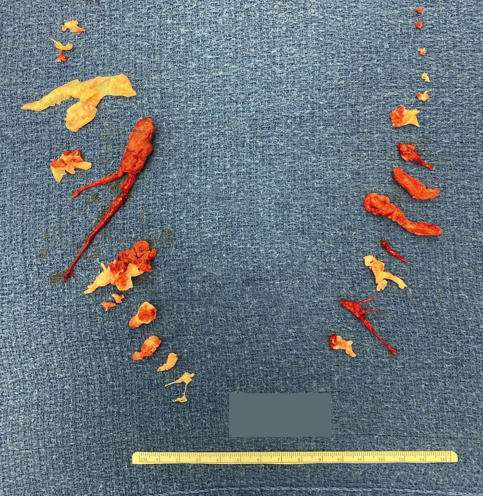
Schwartz reserves open embolectomy through a posterolateral thoracotomy on bypass for pre-terminal massive PE after failed or contraindicated lysis, with 20–40% mortality; catheter fragmentation followed by lysis succeeded in 7 of 10 patients with 20% mortality, and suction embolectomy extracted clot in 76% with 70% 30-day survival [9].
Complications
- VTE is associated with increased postoperative morbidity and mortality [1].
- Death occurs in 6% of DVT and 12% of PE cases within one month of diagnosis [2].
- Post-VTE sequelae include pulmonary hypertension (about 4% incidence) and post-thrombotic syndrome (up to 30% incidence) [2].
- Extensive DVT can progress to phlegmasia alba dolens or phlegmasia cerulea dolens, both of which can be complicated by venous gangrene and the need for amputation [2].
- IVC filter complications include air embolism, arrhythmias, pneumothorax/haemothorax, IVC obstruction, renal vein thrombosis, and filter migration, erosion, or fracture [10].
- Severe chronic venous insufficiency is often secondary to extensive or recurrent lower limb DVT (post-phlebitic limb), presenting with leg/ankle oedema, varicose eczema, pigmentation, lipodermatosclerosis, and venous ulceration [10].
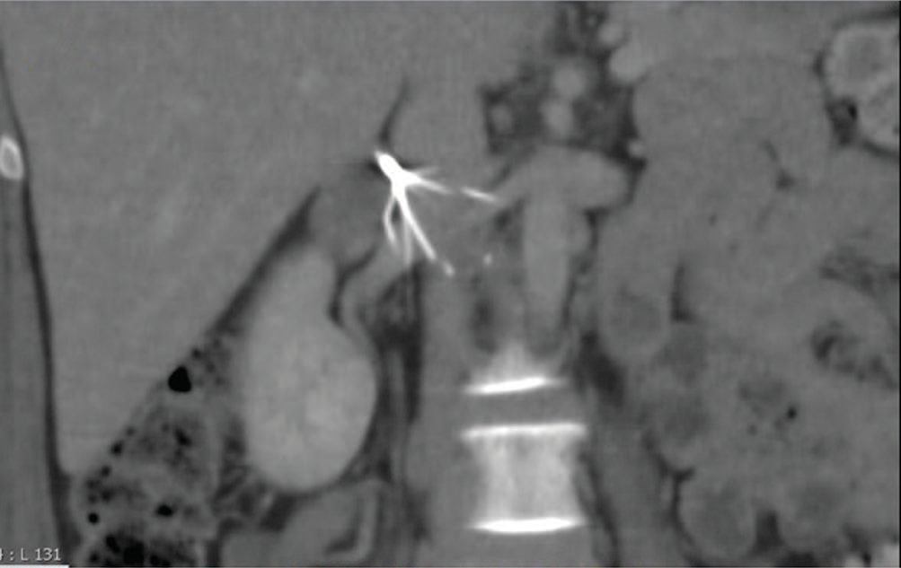
Prophylaxis efficacy and failure in Schwartz's account
About a third of the 150,000–200,000 annual US VTE deaths follow surgery; low-dose heparin and LMWH cut symptomatic and asymptomatic VTE by 60–70%, lower-dose LMWH bleeds less than low-dose heparin, which bleeds less than higher prophylactic LMWH doses, LMWH adds once-daily dosing and fewer heparin antibodies, aspirin alone is inadequate, fondaparinux matched dalteparin after high-risk abdominal surgery and beat pneumatic compression alone at the cost of more bleeding (1.6% vs 0.2%), and prophylaxis continues to discharge or for 4–6 weeks in high-risk cancer patients [9].
Prognosis
- The incidence of VTE is approximately 100 per 100,000 people per year, with about 20% of diagnoses made within 3 months of a surgical procedure [2].
- Without prophylaxis, up to 30% of at-risk surgical patients over 40 develop DVT and 0.1–0.2% die of pulmonary thromboembolism [4].
- The 30-day mortality of acute massive PE is about 50%, with 40% of deaths occurring within the first 2 hours [4].
- A negative d-dimer test makes VTE unlikely, but patients should still be counselled on PE warning signs [3].
- Phlegmasia cerulea dolens carries a risk of venous gangrene and amputation, and about half of these patients have an underlying malignancy [2][7].
References
- Sabiston Textbook of Surgery, 22nd ed., Ch. 26 Surgical Complications: Overview and Grading
- Schwartz's Principles of Surgery: ABSITE and Board Review, Ch. 24 Venous and Lymphatic Disease
- Bailey & Love's Short Practice of Surgery, 28th ed., Ch. 24 Postoperative care
- Oxford Handbook of Clinical Surgery, 5th ed., Ch. 2, Deep vein thrombosis and pulmonary embolism
- NICE Guideline NG158: Venous thromboembolic diseases — diagnosis, management and thrombophilia testing (2020, last updated August 2023), 1.1.1; 1.1.2; 1.1.3; 1.1.4; 1.1.6; 1.1.7; 1.1.8; 1.3.3; 1.3.4; 1.3.5; 1.4.1; 1.4.2; 1.6.1; 1.7.1; 1.7.2; 1.8.1; 1.8.2; 1.9.1; 1.9.1 to 1.9.5; 1.9.2; 1.9.3; 1.9.4; 1.9.5; Recommendations; Terms used in this guideline www.nice.org.uk
- NICE Guideline NG89: Venous thromboembolism in over 16s — reducing the risk of hospital-acquired deep vein thrombosis or pulmonary embolism (2018, last updated August 2019), Recommendations www.nice.org.uk
- The ABSITE Review, 2022, Deep venous thrombosis
- Browse's Introduction to the Symptoms and Signs of Surgical Disease, 6th ed., Ch. 10 The arteries, veins and lymphatics
- Schwartz's Principles of Surgery, 11th ed., Ch. 24, Venous and Lymphatic Disease
- Oxford Handbook of Clinical Surgery, 5th ed., Ch. 19 Peripheral vascular disease, Deep venous thrombosis
- Bailey & Love's Short Practice of Surgery, 28th ed., Ch. 62
- The ABSITE Review, 2022, section on venous disease
- Sabiston Textbook of Surgery, 22nd ed., Ch. 114
- The ABSITE Review, 2022, Ch. 2 Hematology