



Peripheral Arterial Disease
Summary
- Peripheral arterial disease (PAD) is an obliterative, predominantly atherosclerotic process affecting the extremity arteries, most commonly the lower limbs, producing a spectrum from asymptomatic disease through intermittent claudication (IC) to chronic limb-threatening ischaemia (CLTI) and acute limb ischaemia [1].
- It affects an estimated 8.5–12 million people in the United States and carries a 5-year mortality of 32%, higher than many cancers [1].
- Diagnosis rests on history, pulse examination and the ankle–brachial index (ABI), and management combines cardiovascular risk-factor modification with revascularisation (endovascular or surgical) for lifestyle-limiting or limb-threatening disease [2].
UK practice is governed by NICE CG147, Peripheral arterial disease: diagnosis and management, published in 2012 with a 2018 update on diagnosis in diabetes and a 2020 update, and by its quality standard QS52 [3]. Its six sections cover information requirements, secondary prevention, diagnosis, imaging for revascularisation, and separate management sections for intermittent claudication and critical limb ischaemia.
- CG147 opens with an information requirement rather than a clinical one, and the required content is specified: the causes of symptoms and the severity of the disease; the risks of limb loss and cardiovascular events; the key modifiable risk factors of smoking, diabetes control, hyperlipidaemia, diet, body weight and exercise; how to manage pain; all relevant treatment options with risks and benefits; and how to access support for dealing with depression and anxiety [3].
- Secondary prevention of cardiovascular disease is offered to everyone with peripheral arterial disease, covering tobacco dependence, diet, weight and exercise, lipid modification and statin therapy, diabetes and blood pressure [3].
- Three drug recommendations are carried by cross-referenced technology appraisals: clopidogrel for preventing occlusive vascular events, rivaroxaban plus aspirin for people with symptomatic disease at high risk of ischaemic events, and semaglutide alongside diet and physical activity for people with established cardiovascular disease and a BMI of at least 27 kg/m² [3].
Definition
- PAD is defined by stenosis or occlusion of the peripheral arterial system, predominantly caused by atherosclerosis and/or thromboembolic disease, producing symptoms and signs relating to the organ supplied, in the leg, claudication, rest pain and gangrene [2].
- Patients with ischaemic rest pain and/or tissue loss (ulceration/gangrene) persisting for more than two weeks, with objective evidence of arterial insufficiency, are defined as having chronic limb-threatening ischaemia (CLTI) [1][2].
- The European consensus definition of critical limb ischaemia is rest pain unrelieved by analgesia for >2 weeks and/or tissue loss in the presence of an ankle systolic pressure <50 mmHg or toe systolic pressure <30 mmHg [4].
Pathophysiology
- Atherosclerosis is a dynamic process resulting from chronic inflammation of the arterial wall: endothelial activation by proinflammatory stimuli leads to leukocyte adhesion and migration into the intima, where monocytes differentiate into macrophages that take up lipoproteins to form foam cells and fatty streaks [1].
- Vascular smooth muscle cells proliferate and secrete collagen to form a fibrous cap; ongoing apoptosis of foam cells, smooth muscle cells and macrophages produces a necrotic lipid core, and plaque rupture or superficial erosion exposes thrombogenic tissue factor, triggering coagulation and thrombosis [1].
- Calcification increases with age, diabetes mellitus (DM) and chronic kidney disease (CKD) [1].
- Arterial remodelling occurs in compensation and encroachment stages: luminal diameter is preserved by arterial enlargement until a plaque occupies 40% of the cross-sectional area, after which the lumen progressively narrows [1].
- Atherosclerotic plaque develops in predictable distributions at bifurcations and areas of fixation/angulation, most commonly the distal superficial femoral artery (SFA) at the adductor hiatus (Hunter's canal), where the artery is compressed by the adductor magnus tendon [1][5].
- Collateral vessels develop from distributing branches in response to stenosis and may maintain limb viability; symptoms worsen when disease progression outpaces collateral formation or collateral flow is reduced by decreased cardiac output or increased blood viscosity [1].
- Anaerobic muscle metabolism during exercise, when arterial inflow cannot meet oxygen demand, produces the cramp-like pain of intermittent claudication [2].
Epidemiology and the vascular examination in Schwartz's account
- Claudication affects 1.8% of people under 60, 3.7% of those 60–70 and 5.2% over 70; ultrasound of 784 people aged 56–77 showed plaque in 64%, yet only 19% of those with peripheral disease are symptomatic, 6.9% of 3171 adults aged 45–75 had an ABI under 0.95 with only 22% symptomatic, cardiovascular and cerebrovascular disease was three to four times commoner even in the asymptomatic, and 68% of peripheral disease (42% of that with ABI under 0.75) was unknown to the primary physician [6].
- Claudication is a marker of extensive atherosclerosis managed mainly by risk-factor modification and drugs, only 5% needing intervention for disabling pain, with 5-year mortality approaching 30%; about 30% of vascular patients are diabetic and the history covers stroke, coronary disease, prior interventions, diabetes, hypertension, smoking and lipids [6].
- The femoral pulse lies midway between anterior superior iliac spine and pubic tubercle, the popliteal is felt bimanually with the knee flexed to 45° and thumbs on the tibial tuberosity compressing the artery against the tibia, the posterior tibial 2 cm behind the medial malleolus and the dorsalis pedis 1 cm lateral to the extensor hallucis longus tendon, graded 4+ to 1+ or 2+/1+/0; the aorta is felt as an expansile epigastric pulse (it divides at the umbilicus), a carotid bruit correlates better with coronary than carotid disease, and elevation pallor, dependent rubor, nail change and hair loss mark chronic ischaemia [6].
- Aortoiliac occlusion collateralises through SMA to IMA via the superior haemorrhoidal to the middle and inferior haemorrhoidals and internal iliac, lumbar arteries to superior gluteal and internal iliac, lumbar to lateral and deep circumflex arteries and the common femoral, and Winslow's pathway from subclavian through superior and inferior epigastric arteries to the external iliac; the distal SFA in the adductor canal is a favourite site of occlusion because the hiatus prevents compensatory dilatation, and slow occlusion there may be asymptomatic through profunda collaterals reconstituting the popliteal [6].
Clinical features
- Intermittent claudication is a cramp-like muscle pain that is brought on by walking, absent on taking the first step, and relieved by rest within about 5 minutes; the claudication distance varies only slightly day to day and shortens with increased workload (hills, speed, weight) or reduced oxygen delivery (anaemia, cardiorespiratory disease) [2].
- The affected muscle group is classically one anatomical level below the level of disease: superficial femoral artery disease (70% of cases) causes calf claudication, aortoiliac disease (30%) causes thigh or buttock claudication, and Leriche's syndrome is buttock claudication combined with impotence from aortoiliac occlusion [1][2].
- As disease progresses, rest pain develops, typically affecting the forefoot, worsened by elevation/lying flat (loss of gravitational perfusion) and relieved by dependency; patients often sleep with the foot hanging out of bed [1][2].
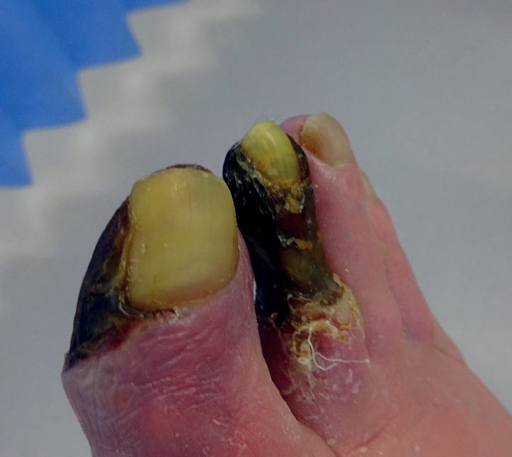
- Ulceration presents as painful erosions between the toes or shallow non-healing ulcers on the dorsum of the foot, shins or malleoli; frank gangrene is blackened and mummified, and may become wet with superadded infection [2].
- Chronic ischaemic limbs (unlike acutely ischaemic ones) tend to equilibrate with ambient temperature and retain sensation and movement; elevation reveals pallor that becomes dependent rubor ("sunset foot") on lowering the leg, and capillary refill may be prolonged to 10 seconds [2].
- Examination signs include diminished/absent pulses, arterial bruits (indicating turbulent flow/stenosis, though unreliable), hair loss, shiny atrophic skin, and slow capillary refill [2][5].
- Differential diagnoses of claudication-type pain include spinal stenosis/nerve root compression (pain relieved only by prolonged sitting), osteoarthritis, and popliteal artery entrapment (rare, pulses reduced on plantar flexion) [2][4].
- Approximately 30–60% of patients with PAD are asymptomatic [1].
The three criteria for claudication pain
Browse's insists that the pain of intermittent claudication is specific and must fulfil three criteria: the patient must experience the pain in a muscle, usually the calf for femoropopliteal disease or the buttock for aortoiliac disease; the pain should only develop when the muscle is exercised; and the pain ceases when the exercise stops [7]. The term itself derives from the Latin claudicare, to limp, taken from the disability of the Emperor Claudius [7].
The claudication distance (the distance covered by the time walking has to stop) should be recorded, and the patient observed walking if necessary to see when the pain develops; they should also be asked how long they wait until the pain goes, and their claudication distance walking on an incline [7]. Browse's records three variants that can mislead: some patients describe an ache or cramp that does not stop them walking and fades away if they force themselves to continue; others prevent the pain by walking slowly; and some describe numbness and paraesthesiae in the skin of the foot at the moment the muscle pain begins, which is the result of blood being shunted from skin to muscle [7].
- The natural history matters for counselling.
- Pain usually begins insidiously and the walking distance gradually shortens over a few months before becoming static; in one-third or more of affected patients the walking distance then increases, with spontaneous remission as the collateral circulation develops [7].
- Browse's adds the one question that must always be asked: ask about pain at rest in the foot, especially when the leg is horizontal, because this signifies the onset of critical limb ischaemia, and patients with rest pain may have had claudication in that limb for many years [7].
- Where the occlusive disease is at the aortic bifurcation, claudication in a man may be associated with impotence because there is no flow to the internal iliac arteries, which is Leriche's syndrome [7].
The routine for assessing the arterial circulation
Browse's sets out a fixed examination sequence, inspection of colour, venous filling, pressure areas and between the digits, and scars; palpation of skin temperature, capillary refilling time and the pulses; auscultation for bruits; measurement of the ankle:brachial pressure index; and the special test of Buerger's angle [7]. Several technical points in it are easily got wrong:
- Temperature should only be compared if both limbs have been exposed to the same ambient temperature for a full 5 minutes; most clinicians use the backs of the fingers, which are ideal temperature sensors, and a blue or even red foot can be very cold [7].
- Venous guttering, in a warm room the veins of a normal foot are full even lying horizontally, whereas in an ischaemic foot they collapse below the skin surface to look like pale blue gutters [7].
- Pulses are palpated from proximal to distal to determine the site of stenosis or occlusion. The femoral pulse lies at the mid-inguinal point, halfway between the symphysis pubis and the anterior superior iliac spine. The popliteal pulse has three described techniques, all three of which need to be tried before deciding the pulse is absent. The dorsalis pedis runs from midway between the malleoli towards the first web space, lateral to extensor hallucis longus, and in 10% of subjects the anterior tibial artery is absent and replaced by a branch of the peroneal artery. The posterior tibial is easiest to feel 2.5 cm above the tip of the medial malleolus, just behind it [7].
- Bruits are caused by turbulent flow beyond a stenosis or by an irregularity in the artery wall; do not press too hard with the bell, as pressure can itself distort flow and cause a bruit, and always remember to listen over the adductor canal [7].
- Buerger's angle is the angle at which the leg becomes white when raised. In a normal limb the toes stay pink even at 90°; in an ischaemic leg, elevation to 15° or 30° for 30–60 seconds causes pallor and venous guttering, and an angle of less than 20° indicates severe ischaemia. Both limbs are raised together for comparison, then the patient sits up and dangles the feet: a normal foot stays pink, whereas an ischaemic leg turns from white to pink and then to a suffused purple-red colour, reactive hyperaemia, from washout of the vasodilator metabolites that accumulated during elevation [7].
- Two closing instructions round off the examination. Before finishing, measure the blood pressure in both arms to exclude significant subclavian or innominate artery disease.
- And when the popliteal pulse is very easy to feel, it may be aneurysmal, the patient should then be examined for a contralateral popliteal aneurysm and for an abdominal aortic aneurysm [7].
- Loss of hair on the lower leg is a sign of ischaemia but is unreliable and does not need to be recorded [7].
- Sensation in the toes and dorsum of both feet should be assessed, because diabetic neuropathy rather than ischaemia may account for impaired sensation [7].
Acute limb ischaemia
The chronic signs above are elicited at leisure; an acutely ischaemic limb announces itself, and the presentation is conventionally recalled as the six Ps, pain, pallor, pulselessness, perishingly cold, paraesthesiae and paralysis [4]. The sequence carries the urgency: pain and pallor come first, whereas paraesthesiae and paralysis signal neurological injury in a limb that is running out of time.
Severe ischaemia leads to irreversible tissue damage within 6 hours, and the quoted complications of an acutely ischaemic limb are death in 20% and limb loss in 40% [4]. Because these patients almost always have coexisting coronary, cerebral or renal disease, resuscitation runs alongside the revascularisation decision rather than after it [4].
Schwartz adds that 90% of acute ischaemia is thrombotic or embolic, the term applying up to 2 weeks after onset; embolic cases have fallen with treatment of rheumatic fever and atrial fibrillation while thrombosis, most often of a prosthetic graft, has risen; 30-day amputation runs 10–30% and mortality 15–20% even with thrombolysis; the heart supplies over 90% of emboli, atrial fibrillation (especially after cardioversion restores appendage contraction), mural thrombus over an infarct or in a ventricular aneurysm or cardiomyopathy (large enough to form a saddle embolus at the aortic bifurcation), and valve disease now more often endocarditis than rheumatic, infected emboli seeding mycotic aneurysms, with aortic mural thrombus, arch or descending aortic thrombus (mobile plaque on transoesophageal echo) and paradoxical embolism through a patent foramen ovale (bubble echocardiography) as rarer sources, so ECG, echocardiography and CT of the thoracic and abdominal aorta follow [6]. Emboli lodge at bifurcations, most often the common femoral, giving foot and calf pain; absent bilateral femoral pulses mean a saddle embolus, a palpable femoral with absent distal pulses a distal common femoral or SFA/popliteal embolus, and calf ischaemia with a popliteal pulse a trifurcation embolus; palpable contralateral pulses in a patient without prior claudication or surgery suggest embolism and simple embolectomy, whereas previous claudication or reconstruction implies thrombosis on atheroma and mandates angiography; muscle is irreversibly damaged after 3 hours and nearly completely by 6, microvascular injury following, and when muscle and microvasculature are destroyed amputation may be safer than revascularisation to avoid washing toxic products into the circulation [6].
Etiology
- Atherosclerosis is the most common cause of PAD; less common causes include thromboembolism, vasculitis, thromboangiitis obliterans (Buerger disease), Behçet disease, pseudoxanthoma elasticum, popliteal entrapment syndrome, cystic adventitial disease, and iliac artery endofibrosis [1].
- Modifiable risk factors mirror those for coronary artery disease: smoking, DM, hypertension and hyperlipidaemia [2].
- Smoking confers a three- to four-fold increased risk of PAD, induces endothelial dysfunction and a prothrombotic state, and smokers have a threefold risk of bypass graft failure; successful cessation improves 5-year mortality (14% vs 31% overall; 18% vs 43% in CLTI) and amputation-free survival [1].
- DM is associated with a two- to four-fold increase in PAD prevalence, and 60–70% of CLTI patients have DM; DM patients characteristically develop long-segment tibial artery occlusions with relative sparing of the pedal arteries [1].
- CKD, hypertension, and adverse lipid profiles (elevated triglycerides, low HDL, high total cholesterol:HDL ratio) are also independent risk factors [1].
- Acute limb ischaemia arises from acute thrombosis on a pre-existing atherosclerotic plaque (60% of cases, predisposed by dehydration, hypotension, malignancy, polycythaemia or prothrombotic states) or embolism (30%, 80% cardiac in origin, atrial fibrillation, myocardial infarction, ventricular aneurysm), with rarer causes including aortic dissection, trauma, iatrogenic injury and peripheral (especially popliteal) aneurysm [4].
Non-atherosclerotic arterial disease in Schwartz's account
- Giant cell (temporal) arteritis affects white women over 50, especially of Scandinavian and northern European descent, with an HLA link and overlap with polymyalgia rheumatica; a prodrome of headache, fever, malaise and myalgia precedes jaw claudication, temporal artery tenderness, ischaemic optic neuritis causing blindness in up to 40% (a medical emergency), carotid extension and late thoracic aneurysm or dissection; temporal artery biopsy showing multinucleated giant cells with dense perivascular infiltrate is the gold standard and corticosteroids give rapid remission and prevent complications [6].
- Takayasu's arteritis strikes Asian women aged 10–40 with a "prepulseless" constitutional phase (fever, anorexia, weight loss, arthralgia, raised ESR, CRP and white count or anaemia), then stenoses, aneurysms and a burned-out phase presenting with bruits, renovascular hypertension, retinopathy, aortic regurgitation, cerebrovascular symptoms, angina, heart failure, abdominal pain, pulmonary hypertension or claudication; angiography is diagnostic and classifies type I (arch branches), IIa (ascending aorta, arch and branches), IIb (plus descending thoracic aorta), III (descending thoracic and abdominal aorta and/or renals), IV (abdominal aorta and/or renals) and V (IIb plus IV), with C(+) or P(+) for coronary or pulmonary involvement; steroids then cytotoxics are given, surgery is deferred until inflammation is quiescent with grafts onto disease-free segments, endarterectomy has no role and angioplasty helps focal lesions [6].
- Ehlers–Danlos syndrome (1 in 5000, autosomal dominant, described by van Meekeren in 1682) causes arterial rupture, aneurysm and dissection in vascular type IV (5% of cases, abnormal type III collagen from COL3A1) often without the skin and joint signs, so 44% died of haemorrhage before any surgery and 29% died after it in Cikrit's series, sutures pull through and ligation may be the only option; Marfan's syndrome (fibrillin, 15q21.3, autosomal dominant with 15–20% new mutations) brings dolichostenomelia, arachnodactyly, joint laxity, chest wall deformity, scoliosis, lens subluxation and cardiovascular involvement in 95%, aortic root dilatation in essentially all, life expectancy of about 40 years untreated with death from regurgitation, dissection or rupture once the ascending aorta passes 6 cm, treated by β-blockade and prophylactic Bentall composite root replacement above 5.5 cm at under 5% mortality; pseudoxanthoma elasticum (1 in 160,000, ABCC6 mutations calcifying the internal elastic lamina, "plucked chicken skin" papules in flexures, onset around 13) causes premature coronary, cerebrovascular and renovascular disease and restrictive cardiomyopathy, calcium intake is restricted in adolescents and arterial conduits are avoided in cardiac bypass [6].
- Vasculitis is classed by vessel size, large (Takayasu's, giant cell, Behçet's), medium (polyarteritis nodosa, Kawasaki's, Buerger's) and small (hypersensitivity angiitis): Kawasaki's disease (1967, mostly boys under 2) causes coronary aneurysms with fibrinoid panarteritis in up to 20% of the untreated, prevented by intravenous immunoglobulin and aspirin within 10 days; Behçet's disease (oral and genital ulcers, uveitis, Mediterranean and Japanese men, HLA-linked) involves vessels in 7–38% with aortic, femoral and pulmonary aneurysms (multiple in 36% of Japanese patients), venous thrombosis, anastomotic pseudoaneurysms and aortic rupture as the chief cause of death, unaltered by steroids; polyarteritis nodosa (men 2:1) spares arterioles, causes livedo, nodules, ulcers and digital ischaemia, neuritis in 60%, gut complications in 50% and renal microaneurysms in 40%, with 50% remission on high-dose steroids and cytotoxics; radiation arteritis produces myointimal proliferation and accelerated atheroma sparing the carotid bulb, is more prone to progression so even asymptomatic lesions are considered for treatment, is preferentially stented and restenoses more often [6].
- Raynaud's syndrome (1862; the disease/phenomenon distinction abandoned because 11–65% progress to connective tissue disease) causes pallor, cyanosis and reactive hyperaemia in young women (70–90%) in cool damp climates and vibrating-tool users, attributed to α₂-adrenergic hypersensitivity and thermoregulatory abnormality, diagnosed by abrupt digital arterial closure at a critical temperature, angiography reserved for ulceration; 90% respond to cold avoidance, gloves, hand warmers and stopping smoking, the rest to diltiazem or nifedipine (30–60% response), fluoxetine or intravenous prostaglandins, with sympathectomy relieving 60–70% but recurring in 60% within 10 years and surgery otherwise limited to debriding ulcers or amputating gangrenous digits [6].
- Fibromuscular dysplasia (90% women, recognised around 55, 10% symptomatic; renal then internal carotid, vertebral, subclavian, mesenteric and iliac arteries; medial fibroplasia in 85%) causes TIA and hypertension, carotid lesions lying too high for duplex or resection so graduated open dilatation or open balloon angioplasty with back-bleeding is used, renal angioplasty succeeds with 8–23% recurrence and surgical reconstruction for recurrences [6].
- Three non-atherosclerotic popliteal conditions cause claudication in men of 40–50: adventitial cystic disease (0.1% incidence, 5:1 male, 1 in 1200 claudicants, mucin-filled subadventitial cysts like ganglia, pulses vanishing on knee flexion, duplex then T2 MRI, a crescentic "scimitar" filling defect on angiography, treated by cyst excision, enucleation or aspiration, or vein interposition in the 30% with occlusion, since retained lining recurs); popliteal entrapment (0.16%, 15:1 male, 25% bilateral, popliteal vein involved in up to 30%, median surgical age 28.5, types I–VI by relation to the medial gastrocnemius head, accessory slips, popliteus or functional entrapment, diagnosed by a 50% pressure fall or dampened plethysmography on plantar or dorsiflexion and stress angiography, treated by dividing the anomalous insertion with vein interposition for damaged artery, lysis first if thrombosed); and Buerger's disease (thromboangiitis obliterans, Leo Buerger 1908, male smokers aged 20–50, 24% of young adults with limb ischaemia at the Mayo Clinic but under 1% of severe ischaemia in North American men, migratory phlebitis in 16%, thrombosis with neutrophil microabscesses and giant cells, infrapopliteal and distal brachial "skip" lesions with corkscrew collaterals on four-limb angiography), for which strict abstinence from tobacco halts progression, bypass rarely has a target or vein, and limb loss runs 31% over 15 years, 67% in continuing smokers against 35% in quitters [6].
Renal artery occlusive disease in Schwartz's account
- Renovascular hypertension, the commonest correctable hypertension, affects 5–10% of US hypertensives and risks permanent renal damage; 80% of lesions are ostial atheroma spilling over from a diseased aorta (bilateral in two-thirds, sixth decade, men 2:1, with disease in other beds) and 20% fibromuscular dysplasia in young often multiparous women (medial fibroplasia with a "string of beads" in the distal two-thirds, right more than left), with aneurysm, AV malformation, neurofibromatosis, dissection, trauma and Takayasu's arteritis as rare causes; prevalence rises from 2% with diastolic pressure over 100 mmHg to almost 30% over 125, and clues are upper abdominal bruits, diastolic pressure over 115, onset after 50 or in childhood, sudden worsening, resistance to three drugs and renal failure after ACE inhibitors [6].
- Captopril renography is positive when time to peak exceeds 11 minutes or the GFR ratio exceeds 1.5:1; duplex (technically demanding but the usual screen) reads ≥60% stenosis at peak systolic velocity ≥180 cm/s with a renal-to-aortic ratio ≥3.5; a renal vein renin ratio above 1.5 predicts benefit in unilateral disease (each kidney normally contributes a renal:systemic renin index of 0.24, suppressed contralaterally in unilateral stenosis, but 10% of responders lack suppression), gadolinium MRA may over-read flow voids, and DSA with a flush aortogram for accessory vessels remains gold standard, collaterals implying a gradient of at least 10 mmHg [6].
- Revascularisation is indicated for stenosis over 70%, FMD, a gradient over 20 mmHg or renin ratio over 1.5 with refractory or rapidly progressive hypertension, flash pulmonary oedema without coronary disease, deteriorating renal function, drug intolerance, bilateral or solitary-kidney stenosis with renal insufficiency, or dialysis-dependence without another cause; open options are aortorenal bypass (vein preferred, arteriotomy three times the artery's diameter, 6-0 or 7-0 polypropylene), transrenal or transaortic endarterectomy for short ostial or multiple-artery disease, hepatorenal or splenorenal bypass when the aorta cannot be clamped, and reimplantation of a redundant artery in children; surgery improved hypertension in 92% of non-atherosclerotic patients (43% cured, 68% cured under 45) and 85% of atherosclerotic patients (12% cured) with 0% and 3.1% mortality, two-thirds improving renal function within a week and up to three-quarters leaving dialysis [6].
- Endovascular treatment (Grüntzig's balloon) uses femoral or brachial access, oblique aortography, CO₂ or gadolinium if needed, selective catheters, a 4 mm balloon first, a balloon-expandable stent protruding 1–2 mm into the aorta oversized 10–20% for ostial disease, dissection or residual stenosis, and intra-arterial nitroglycerine 100–200 µg for spasm; angioplasty alone suits FMD (95% success, 69% primary and 87% assisted patency at 8 years, 79% clinical benefit) but offers minimal advantage over drugs for atherosclerotic stenosis at 12 months, whereas stenting restenoses far less (14% vs 48% at 6 months), gives 99% technical success with 19% restenosis, 84.5% 5-year primary and 92.4% secondary patency with hypertension cured or improved in 78%, stabilises or improves renal function in about 69%, and in Leertouwer's 14-study meta-analysis cured hypertension in 20%, improved it in 49%, improved renal function in 30% and stabilised it in 38% with 17% restenosis; major complications are 17% endovascular versus 31% surgical but minor complications 48% versus 7%, transient contrast and embolic renal deterioration is common with about 1% needing temporary dialysis [6].
Diagnosis
- Diagnosis of claudication is mostly clinical; the ankle–brachial pressure index (ABI) is the key bedside quantitative test, calculated as the highest ankle systolic pressure (dorsalis pedis, posterior tibial or peroneal) divided by the highest brachial systolic pressure [2].
- Normal resting ABI is 0.9–1.4; <0.9 indicates a haemodynamically significant lesion and correlates with the onset of claudication; <0.5 correlates with rest pain; <0.4 suggests CLTI/ulceration; <0.3 with tissue loss/gangrene [2][5].
- A post-exercise drop in ABI of >20% indicates flow-limiting disease even when resting ABI is normal [2].
- Falsely elevated ABI (>1.4) occurs with medial calcific sclerosis, typically in diabetics, in which case toe–brachial index (TBI, using pressures rarely affected by calcification; TBI <0.6 is abnormal) or Doppler waveform analysis is preferred [2][5].
- Hand-held continuous-wave Doppler assesses waveform (normal triphasic vs diseased bi-/monophasic signal) [2].
- Duplex Doppler ultrasound (DUS) is as accurate as angiography in experienced hands and is the first-line non-invasive imaging test, though limited by bowel gas/obesity in the aortoiliac segment and by heavy calcification [2][4].
- CT angiography (CTA) is useful as an adjunct where DUS is inadequate, particularly the aortoiliac segment, but carries risks of ionising radiation and contrast-induced nephropathy.
- MR angiography (MRA) avoids ionising radiation/iodinated contrast and is useful in diabetics with calcified crural disease, but is contraindicated with certain implants and carries a risk of gadolinium-induced nephrogenic systemic fibrosis in renal impairment [2].
- Digital subtraction angiography (DSA) provides dynamic flow information and can combine diagnosis with endovascular treatment, but is invasive with complication rates up to 5% (bleeding, false aneurysm, thrombosis, dissection, distal embolisation, renal dysfunction, allergic reaction) [2].
- Pulse volume recordings (PVRs) are a non-invasive flow study used to localise significant occlusion; arteriography (gold standard for vascular imaging, using CO2 contrast if renal function is poor) is indicated if PVRs suggest significant disease [5].
- General work-up includes full blood count, glucose, lipid profile, urea and electrolytes (renal function), and ECG, with echocardiography or exercise testing as indicated given the high prevalence of concomitant coronary (50%) and cerebral (25–50%) arterial disease [2].
- CG147 specifies who to assess, how to measure the ABPI, and two things not to do in diabetes.
- Assess for peripheral arterial disease if the person has suggestive symptoms; or has diabetes, non-healing leg or foot wounds, or unexplained leg pain; or if interventions to the leg or foot are being considered; or if they need to use compression hosiery [3].
- Assessment comprises asking about the presence and severity of claudication and critical limb ischaemia symptoms, examining the legs and feet for evidence of critical limb ischaemia such as ulceration, examining the femoral, popliteal and foot pulses, and measuring the ankle brachial pressure index [3].
The ABPI method is prescribed step by step [3]:
| Step |
|---|
| --- |
| The person should be resting and supine if possible |
| Record systolic pressure with an appropriately sized cuff in both arms and in the posterior tibial, dorsalis pedis and, where possible, peroneal arteries |
| Take measurements manually using a doppler probe of suitable frequency, in preference to an automated system |
| Document the nature of the doppler ultrasound signals in the foot arteries |
| Calculate the index in each leg by dividing the highest ankle pressure by the highest arm pressure |
Table reformats the CG147 ABPI method [3].
In diabetes, two negatives apply and the first is the important one. Do not exclude a diagnosis of peripheral arterial disease in people with diabetes based on a normal or raised ankle brachial pressure index alone, because medial arterial calcification can produce a falsely normal or high index [3]. And do not use pulse oximetry for diagnosing peripheral arterial disease in people with diabetes, since no universal cut-off has been established and it is not a useful test in this context [3].
Imaging before revascularisation runs in a fixed order. Offer duplex ultrasound as first-line imaging to all people for whom revascularisation is being considered; offer contrast-enhanced MR angiography to those needing further imaging after duplex; and offer CT angiography only if contrast-enhanced MR angiography is contraindicated or not tolerated [3].
Non-invasive testing and imaging in Schwartz's detail
- ABI uses the higher arm systolic pressure as denominator and each ankle vessel's pressure as numerator: normal over 1.0, claudication 0.5–0.9 (typically 0.5–0.7), rest pain 0.3–0.5, gangrene under 0.3, and under 0.9 predicts myocardial infarction; calcified vessels in diabetics and renal failure give ankle pressures of 250 mmHg or more and ABI above 1.40, so toe pressures (normally 30 mmHg below ankle; toe–brachial index under 0.70 abnormal, rarely falsely positive but impossible with toe ulcers), pulse volume recordings, transcutaneous oxygen or duplex are used instead; segmental pressures normally show high-thigh pressure 20 mmHg above brachial, low-thigh equal to brachial and falls of no more than 10 mmHg per level, a 20 mmHg gradient localising disease, but they miss isolated moderate iliac stenoses at rest and cannot separate stenosis from occlusion; pulse volume recording at 60–65 mmHg cuff pressure shows a slow upstroke, rounded peak and lost dicrotic notch below disease, and combining pressures with PVR raises accuracy from 85% to about 95% [6].
- Duplex needs a certified technologist holding a 60° Doppler angle, is 80% sensitive and over 95% specific in a 71-study meta-analysis but weaker for aortoiliac (bowel gas) and infrapopliteal vessels and cannot see through air-containing fresh PTFE or Dacron; iliac stenosis shows a peak systolic velocity ratio of 2.5 or more or a monophasic waveform; CTA rivals angiography but blooms with calcium and overestimates in-stent stenosis; MRA uses non-nephrotoxic gadolinium, is contraindicated by pacemakers, defibrillators, stimulators, shunts, cochlear implants and cranial clips, and drops signal at stents (least with nitinol); angiography is a uniplanar "lumenogram" that misjudges the 70% of plaques that are eccentric, gives inaccurate stent apposition and carries contrast and radiation risk, digital subtraction with the pulse-chase technique improving distal opacification, and gadolinium or CO₂ replace iodinated contrast in borderline renal function [6].
- Claudication is distinguished from chronic compartment syndrome (tight bursting calf pain after heavy exercise in muscular athletes, slow to settle, eased by elevation), venous claudication (whole-leg bursting pain after iliofemoral DVT), nerve root compression (lancinating radiating pain, present at rest), Baker's cyst, hip arthritis, spinal cord compression (weakness more than pain, relieved by lumbar flexion) and foot arthritis; night cramps do not indicate arterial disease; ischaemic ulcers are painful on toes or lateral foot, venous ulcers sit above the medial malleolus in lipodermatosclerosis with mild discomfort, and neuropathic ulcers are painless callused lesions on weight-bearing surfaces; rest pain is separated from diabetic neuropathy (lost vibration, position sense and reflexes) and spinal stenosis (worse standing and on extension) [6].
- Coronary disease is present in 40–50% of patients needing vascular reconstruction, 10–20% asymptomatic through inability to exercise, and infarction causes most early and late deaths; thallium or dobutamine imaging is unnecessary in nearly half because clinical data predict risk, the CARP trial showed prophylactic coronary revascularisation neither reduced mortality nor infarction but delayed vascular surgery and increased limb morbidity, so perioperative β-blockade, antiplatelets, statins and ACE inhibitors are the evidence-based optimisation [6].
Scoring and Severity
- The Fontaine classification grades chronic lower limb ischaemia: I asymptomatic; II intermittent claudication; III rest pain; IV ulcers/gangrene, with grades III and IV together constituting critical limb ischaemia [4].
- Single-level arterial disease usually produces intermittent claudication, while multi-level disease produces critical limb ischaemia [4].
- Atherosclerotic aortoiliac occlusive disease is classified into three anatomical types: type I (5–10% of patients), confined to the distal aorta and common iliac vessels, occurring in a relatively young group with rare limb-threatening symptoms; type II (~25%), diffuse disease predominantly of the abdominal aorta extending into the common iliac artery; and type III (~65%), widespread disease above and below the inguinal ligament, more common in older patients with diabetes, hypertension and multi-territory atherosclerosis [8].
- An ABI <0.50 predicts a two-fold higher risk of deterioration compared with ABI >0.50, and a decrease of 0.1 in ABI below 0.9 is associated with a 10% increase in relative risk of a major cardiovascular event [2].
Rutherford, TASC and the aortoiliac patterns in Schwartz's account
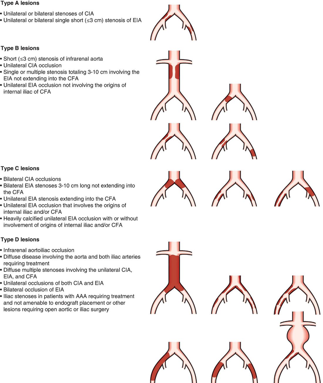
- Rutherford grades 0 (asymptomatic), I categories 1–3 (mild, moderate, severe claudication: category 1 completes a 5-minute treadmill at 2 mph on a 12% incline with post-exercise ankle pressure over 50 but at least 20 mmHg below rest, category 3 cannot complete it with ankle pressure under 50), II category 4 (rest pain: ankle under 40, toe under 30 mmHg, flat pulse volume) and III categories 5–6 (minor and major tissue loss beyond the transmetatarsal level: ankle under 60, toe under 40 mmHg), grades II–III constituting chronic critical ischaemia; chronic limb ischaemia requires objectively proven occlusive disease with symptoms over 2 weeks, rest pain usually below an ankle pressure of 50 or toe pressure of 30 mmHg, and ulcer healing needs more (critical ischaemia is suggested by ankle under 70 or toe under 50 mmHg with tissue loss) though no consensus haemodynamic definition exists [6].
- TASC femoropopliteal type A is a single stenosis ≤10 cm or occlusion ≤5 cm; B multiple lesions each ≤5 cm, a single lesion ≤15 cm sparing the infrageniculate popliteal, lesions without continuous tibial vessels to improve inflow for a distal bypass, a heavily calcified occlusion ≤5 cm or a single popliteal stenosis; C multiple lesions totalling over 15 cm or recurrence after two endovascular treatments; D chronic total occlusion of common femoral or SFA over 20 cm involving the popliteal, or of the popliteal and proximal trifurcation; infrapopliteal A is a single lesion under 1 cm sparing the trifurcation, B multiple sub-centimetre lesions or one at the trifurcation, C extensive trifurcation disease or 1–4 cm stenoses or 1–2 cm occlusions, D occlusions over 2 cm or diffuse disease; endovascular therapy is recommended for A and surgery for D with no ruling on B and C [6].
- Aortoiliac TASC A is common iliac stenosis or a single short (≤3 cm) external iliac stenosis; B a short infrarenal aortic stenosis, unilateral common iliac occlusion, 3–10 cm of external iliac stenosis sparing the common femoral, or external iliac occlusion sparing internal iliac and common femoral origins; C bilateral common iliac occlusions, bilateral 3–10 cm external iliac stenoses, external iliac disease extending into the common femoral or involving the internal iliac origin, or heavily calcified external iliac occlusion; D infrarenal aortoiliac occlusion, diffuse aorto-bi-iliac disease, diffuse unilateral common–external–common femoral disease, unilateral occlusion of both iliacs, bilateral external iliac occlusion or iliac stenoses in a patient needing open aneurysm surgery; endovascular treatment is the choice for A, preferred for B, surgery preferred for good-risk C and the choice for D, tempered by comorbidity, patient preference and the operator's own results [6].
- Aortoiliac disease is type I (5–10%: distal aorta and common iliacs, younger patients in their mid-50s with type IV hyperlipoproteinaemia rather than hypertension or diabetes, bilateral thigh and buttock claudication and impotence (Leriche's syndrome when femoral pulses are absent) rarely limb-threatening, sometimes presenting as acute aortic occlusion or "trash foot"), type II (25%: diffuse aorta and common iliac) or type III (65%: multilevel above and below the ligament, older, male 6:1, diabetic, hypertensive, with coronary, cerebral and visceral disease, presenting with advanced ischaemia for limb salvage and with shorter 10-year life expectancy) [6].
Treatment and Management
- The two principal aims of treating claudication are prevention of major cardiovascular morbidity through risk-factor modification and symptom relief [2].
- A supervised exercise programme of at least 2 hours per week for 3 months, combined with smoking cessation, produces sustained improvement in claudication distance and cardiovascular risk [2].
- Medical therapy includes an antiplatelet agent (aspirin or clopidogrel 75 mg/day) and a statin regardless of baseline lipid level, as it stabilises atherosclerotic plaque and reduces cardiac death; beta-blockers may exacerbate claudication [2].
- First-line therapy for claudication is medical: smoking cessation (the top priority), aspirin, statin, and exercise to the point of pain to promote collateral formation [5].
- Only about a quarter of claudicants deteriorate symptomatically over their lifetime, and the risk of progression to CLTI/amputation is <5% over 5 years, so most patients do not require invasive treatment [2][4].
- Surgical indications for PAD include rest pain, ulceration or gangrene, severe lifestyle-limiting claudication despite medical therapy, and atheromatous embolisation [5].
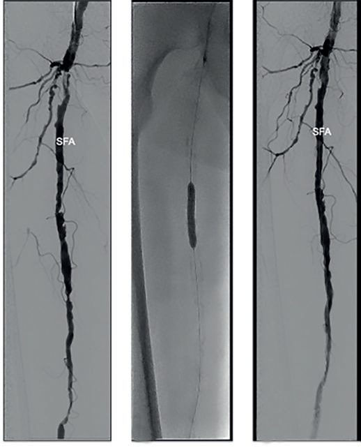
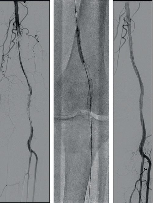
- Percutaneous transluminal angioplasty (PTA) with or without stenting is highly successful in the aorto-iliac segment (>90%) and good in the superficial femoral segment, but is less durable in popliteal/tibial segments; complications occur in about 5% (failure, haematoma, thrombosis, distal embolisation) [2][4].
- Isolated iliac lesions are treated first with PTA/stent; if this fails, femoral-to-femoral crossover is considered [5].
- Endovascular stenting is not favoured at joint-crossing sites (common femoral, popliteal) owing to kinking risk [5].
- In CLTI, all efforts should be made to revascularise if the patient's general condition allows; a single level of disease may need treatment to relieve rest pain, whereas tissue loss requires restoration of in-line flow [4].
- There is little evidence of benefit from prostacyclin or sympathectomy (surgical or chemical) in CLTI [4].
- Acute limb ischaemia requires immediate anticoagulation (heparin) to prevent clot propagation, urgent assessment of viability (irreversible vs immediately threatened vs marginally threatened), and either amputation (non-salvageable), emergency revascularisation with fasciotomy, or continued heparinisation with imaging and thrombolysis/angioplasty/surgery depending on urgency [4].
- There is no clear superiority of thrombolysis over surgery for 30-day limb salvage or mortality in acute limb ischaemia, though surgery is used three- to five-fold more often than thrombolysis in the United States [8].
Supervised exercise comes before any intervention, and CG147 specifies the dose. Offer a supervised exercise programme to all people with intermittent claudication, and consider one involving 2 hours of supervised exercise a week for a 3-month period, encouraging people to exercise to the point of maximal pain [3].
Angioplasty for claudication has three preconditions, all of which must be met [3]:
| Precondition |
|---|
| --- |
| Advice on the benefits of modifying risk factors has been reinforced |
| A supervised exercise programme has not led to a satisfactory improvement in symptoms |
| Imaging has confirmed that angioplasty is suitable for the person |
Table reformats the CG147 preconditions for angioplasty in claudication [3]. Bypass surgery is offered for severe lifestyle-limiting claudication only when angioplasty has been unsuccessful or is unsuitable and imaging has confirmed bypass is appropriate [3].
On stents, CG147 is restrictive and the exception is precise. Do not offer primary stent placement for intermittent claudication caused by aorto-iliac disease (except complete occlusion) or by femoro-popliteal disease; consider primary stent placement for claudication caused by complete aorto-iliac occlusion rather than stenosis; and use bare metal stents where stenting is used [3]. The identical three rules are repeated for critical limb ischaemia [3]. Use an autologous vein whenever possible for infra-inguinal bypass in both claudication and critical limb ischaemia [3].
- On drugs for claudication, NICE recommends one agent and rejects three.
- Consider naftidrofuryl oxalate, starting with the least costly preparation, only when supervised exercise has not led to satisfactory improvement and the person prefers not to be referred for angioplasty or bypass
- Review progress after 3 to 6 months and discontinue if there has been no symptomatic benefit [3]. Cilostazol, pentoxifylline and inositol nicotinate are not recommended for treating intermittent claudication [3].
- For critical limb ischaemia, the gateway is a team rather than a test. Ensure that all people with critical limb ischaemia are assessed by a vascular multidisciplinary team before treatment decisions are made [3].
- Offer angioplasty or bypass surgery to those requiring revascularisation, taking into account comorbidities, pattern of disease, availability of a vein, and patient preference [3].
- For ischaemic pain, offer paracetamol and either weak or strong opioids depending on severity [3].
Managing acute ischaemia and choosing between lysis and surgery in Schwartz's account
- Heparin is given as soon as acute ischaemia is suspected to stop propagation, with fluids, a urinary catheter, baseline creatinine and a thrombophilia screen before heparin if suspected; imaging is ideal, especially after previous reconstruction, but must not delay treatment, and the table is prepared for fluoroscopy of the whole inflow and outflow; randomised trials show no clear superiority of thrombolysis over surgery for 30-day limb salvage or mortality and US registries show surgery used three to five times more often, but lysis is preferred first-line for Rutherford I and IIa ischaemia because it spares endothelium, clears branch vessels too small for balloons and may reduce reperfusion injury, and is the only option for small-vessel occlusion without bypass targets; it is less effective for thrombosis older than 2 weeks, absolutely contraindicated by cerebrovascular events within 2 months, active bleeding, GI bleeding within 10 days, neurosurgery or intracranial trauma within 3 months and intracranial malignancy, relatively by CPR, major surgery or trauma within 10 days, blood pressure over 180/110, non-compressible puncture, intracranial tumour and recent eye surgery, and minor contraindications are hepatic failure, endocarditis, pregnancy and diabetic haemorrhagic retinopathy; percutaneous mechanical or pharmacomechanical thrombectomy extends treatment to IIb ischaemia and to those who cannot receive lytics [6].
- When thrombosis complicates atheroma or a failed graft, embolectomy catheters will not pass, so prompt angiography defines the occlusion, inflow and outflow: good distal targets with a usable saphenous vein favour bypass (fast, durable, reliable), absent targets or vein or high surgical risk favour lysis; prosthetic graft thrombectomy is likelier to succeed than vein graft thrombectomy, which fails unless a retained valve or side branch is corrected, so acute vein graft failure is revised by valve lysis, interposition or extension [6].
- For chronic disease there is no effective drug therapy beyond risk-factor control, antiplatelets and graduated exercise, which helps little in aortoiliac disease, so buttock claudication with weak femoral pulses that fails exercise and drugs is revascularised; indications are disabling claudication, rest pain, non-healing wounds and microembolisation; endovascular treatment is first-line for lower extremity disease and bypass is reserved for failed endovascular therapy or long femoropopliteal occlusion, with long occlusions, heavy calcification, ostial lesions and uncrossable lesions faring poorly; clinical success is judged by walking distance, symptoms, quality of life and patency; Nolan's Markov analysis found angioplasty and stenting surpass bypass for TASC C lesions when their 5-year primary patency exceeds 32%, the patient is over 80 or vein bypass mortality exceeds 6% [6].
- Primary amputation without attempted revascularisation is rarely needed except for neglected class III ischaemia or critical ischaemia in non-ambulatory patients with contractures, disabling stroke or dementia [6].
Surgeries
- Aortobifemoral bypass with a Dacron graft is the standard operation for aortoiliac occlusion, with good long-term patency (90% at 5 years) but perioperative mortality of about 5% and systemic morbidity (stroke, cardiorespiratory failure, renal injury) of about 15%; an axillobifemoral bypass is an alternative in unfit patients, though with lower patency [2].
- When performing aorto-bifemoral repair, flow to at least one internal iliac (hypogastric) artery must be preserved to prevent vasculogenic impotence and pelvic ischaemia [5].
- Isolated unilateral iliac disease can be treated with a femorofemoral crossover graft [2].
- Superficial femoral artery disease is treated with femoropopliteal bypass, ideally using autologous great saphenous vein (reversed or in situ after valve disruption); if no vein is available, a PTFE prosthetic graft is used, often with a Miller cuff/St Mary's boot to improve patency [2].
- Isolated common femoral or profunda disease can be treated with endarterectomy and patch angioplasty [2].
- Femorodistal (tibial) bypass is reserved for limb salvage in CLTI, particularly with diabetic tibial disease, requiring a vein conduit of minimum 3 mm diameter and adequate run-off; early graft failure with limb loss occurs in about 30% at 30 days [2].
- Extra-anatomic grafts (axillofemoral, femorofemoral crossover) avoid a hostile abdomen in frail, multiply-operated patients [5].
- Obturator bypass is used to reconstruct arterial anatomy through the anteromedial portion of the obturator membrane in patients with groin sepsis from infected prior grafts, intra-arterial drug abuse or irradiation, with reported 57% 5-year patency and 77% 5-year limb salvage [8].
- For prosthetic infrageniculate bypass, vein cuffs, precuffed grafts or heparin-bonded grafts (e.g., Gore Propaten using Carmeda bioactive surface technology) improve patency compared with plain PTFE [8].
- Below-knee bypasses require saphenous vein, as PTFE has reduced patency below the knee [5].
- Fasciotomy is performed for compartment syndrome following reperfusion, releasing all four compartments of the lower leg via medial and lateral incisions, left open for 5–10 days [5].
- Amputation (below-knee, BKA, or above-knee, AKA) is indicated for gangrene, large non-healing ulcers or unrelenting rest pain not amenable to revascularisation; BKA has 80% healing and 70% ambulation with 5% mortality, while AKA has 90% healing and 30% ambulation with 10% mortality [5].
Endovascular technique and results in Schwartz's detail
- Access is through the common femoral over the medial third of the femoral head under fluoroscopy with an 18-gauge Seldinger needle for 0.035-inch wires or 21-gauge micropuncture for 0.018-inch, single-wall puncture preferred (double-wall risks posterior bleeding), retrograde femoral commonest (puncture above the ligament risks retroperitoneal haemorrhage, below the bifurcation SFA or profunda thrombosis and pseudoaneurysm), antegrade for lesions unreachable across the bifurcation, and left brachial when aortoiliac access fails (avoiding the carotid origin, but with more thrombosis and nerve injury); wires (0.011–0.038 inch, 180–260 cm) include stiff Amplatz for large devices and tortuosity, hydrophilic Glidewires primed in saline for tight lesions, angled tips for steering and the soft-curled Rosen for renal stenting; sheaths of 5–9F (up to 26F with open exposure) carry a haemostatic valve and side-port; low-compliance balloons are the mainstay, oversized 10–20% and inflated until the waist disappears, severe pain warning of rupture or dissection, with the wire kept across the lesion for completion angiography; self-expanding Elgiloy or nitinol stents foreshorten, come long, keep expanding to fit tapering vessels (ideal for the internal carotid) and are oversized 1–2 mm, while balloon-expandable stainless steel stents are rigid, precise, crush-resistant and endothelialise faster, suiting short ostial common iliac and renal lesions; heparin 80–100 units/kg to an ACT above 250 seconds with 1000 units hourly, aspirin afterwards and clopidogrel for at least 6 weeks after stenting [6].
- Iliac lesions are crossed retrograde from the ipsilateral femoral (over 90% of occlusions with simple wires; subintimal wires that will not re-enter at the bifurcation are handled by re-entry catheters, intravascular ultrasound or contralateral antegrade crossing with a 4F Berenstein catheter and snaring, 5–20% failure), dilated with 8–10 mm balloons in the common and 6–8 mm in the external iliac, stented for over 30% residual stenosis, flow-limiting dissection or a gradient of 5 mmHg or more, and primarily stented for lesions over 5 cm (1-, 2- and 3-year patency 96%, 90%, 72% versus 46%, 46%, 28% selective), all TASC C and D lesions, chronic occlusions, restenosis and eccentric, calcified, ulcerated or dissected plaques, since direct stenting traps embolic debris (embolisation up to 24% with angioplasty of ulcerated, bifurcation or occlusive lesions, up to 10% with stents), though the Dutch Iliac Stent Trial found angioplasty with selective stenting (43% needing stents) equal to primary stenting at 2 years (77% vs 78%) and 5 years (82% vs 80% free of reintervention); the aortic bifurcation is treated with kissing balloons or kissing stents (95–100% technical success, 79% 3-year patency), focal aortic stenoses with a single stent sized by CT to 12–18 mm at low pressure (hypoplastic aortas ≤12 mm recanalised to 8–9 mm) with an occlusion balloon and covered stent ready for rupture, and stent grafts have treated complex iliac lesions with 91.1% 1-year patency after gentle 5 mm predilatation [6].
- Iliac angioplasty of stenoses under 4 cm gives 86% 2-year patency with about 2% complications; technical success exceeds 90% for stenoses (5-year patency 54–92%) and 78–98% for occlusions (3-year 48–85%); Becker's 2697 cases gave 72% 5-year patency (79% in claudicants); external iliac lesions and women do worse (common iliac 81% at 1 and 52% at 6 years versus external 74% and 48% at 1 and 4); stenting gives 41–92% 3-year patency for stenoses and 64–85% for occlusions, improves long-term patency 39% over angioplasty in Bosch and Hunink's meta-analysis of 2116 patients, and Park reported 87%, 83%, 61% and 49% primary patency at 3, 5, 7 and 10 years across all TASC types while Leville found no TASC-dependent difference (76% primary, 90% secondary at 3 years) and urged exhausting endovascular options first; iliac angioplasty carries 2–4% major and 4–15% minor complications, embolisation in 2–10% (catheter aspiration for calf vessels, surgical embolectomy for profunda or common femoral plaque), 0.5% pseudoaneurysm (thrombin injection if over 2 cm) and 0.3% rupture (balloon tamponade and covered stent) [6].
- Femoropopliteal angioplasty succeeds technically in 80–90% (failing to cross 7% of stenoses and 18% of occlusions) with 38–58% 5-year primary patency, poor beyond 7–10 cm and good under 3 cm, 12% at 5 years for occlusions over 5 cm versus 32% for shorter, 53% for stenoses and 36% for occlusions with good runoff versus 31% and 16% with poor, 12–68% overall, and infrapopliteal angioplasty is reserved for limb salvage with straight-line flow to the foot (77% 1-year salvage, over 80% at 2 years in favourable anatomy); subintimal angioplasty (described 1987) deliberately dissects from proximal to the occlusion and re-enters distally with a looped hydrophilic wire, suits long, calcified and diffuse occlusions, succeeds in over 80% (Bolia: 71% 1-year femoropopliteal and 53% tibial patency, over 80% limb salvage; Treiman 92% patency at 13 months; Ingle 94% 3-year salvage), stents residual stenosis over 30% and damages collaterals in 1–1.5% [6].
- Femoropopliteal stents give 18–72% 3-year patency, poor after suboptimal long-lesion angioplasty (22% at 1 year) but better with nitinol, SMART 76% at 12 and 60% at 24 months for 12.2 cm lesions, Zilver 90%, 78%, 74%, 69% at 1–4 years for 19 cm recanalisations, 82% versus 37% 6-month patency against Wallstents, BLASTER 83% clinical patency at 1 year; Schillinger's trial found primary stenting cut 2-year restenosis to 45.7% from 69.2%; sirolimus-eluting SMART stents in SIROCCO gave 0% versus 23.5% 6-month restenosis, and PaRADISE below-knee drug-eluting stents in 106 patients gave 6% 3-year amputation and 68% amputation-free survival; below-knee Xpert nitinol stents gave 76.3% 12-month patency, 95.9% limb salvage and 20.5% binary restenosis; stent fracture from torsion, compression, flexion and extension in the adductor canal occurred in 18.2% in SIROCCO without symptoms; Viabahn ePTFE–nitinol stent grafts equal prosthetic above-knee bypass (74% 1-year patency each, stay 0.9 vs 3.1 days; at 48 months 59% vs 58% primary and 74% vs 71% secondary patency for 25.6 cm lesions) but cover collaterals and occluded in 28.6% at 7 months in an early Hemobahn series; directional, orbital, rotational and laser atherectomy (SilverHawk, Jetstream, Diamondback, Rotablator, Excimer) achieve high technical success (TALON registry 90% and 80% freedom from target-lesion revascularisation at 6 and 12 months with stenting in only 6.3%; PELA 83.2% success but 33.6% 1-year patency; CELLO 59% and 54% at 6 and 12 months) but restenose often, with 15–43% complication rates including spasm, thrombosis, dissection, perforation (2.2–4.3% with laser), embolisation (3–4%) and no-reflow [6].
- Angioplasty complications total 7.3% in the STAR registry (75% minor: emboli and haematoma 41.7% each, contained rupture and vagal reactions 8.3%) or 4.2% significant (retroperitoneal bleed 0.2%, false aneurysm 0.2%, acute ischaemia 1.5%, perforation 1.7%), subintimal angioplasty 15% (stenosis, rupture 6%, embolisation 3%), cryoplasty 13% and stent grafts 23% (embolisation 7.7%, haematoma 13.5%, AV fistula 1.9%) [6].
Open reconstruction in Schwartz's detail
- Aortobifemoral bypass relieves symptoms in 70–80% with 2–3% mortality, 85–90% 5-year and 70–80% 10-year patency; both femorals are exposed first, the aorta approached transperitoneally (retroperitoneal left flank as an alternative, contraindicated with acute right renal occlusion, harder for right-limb tunnelling, perhaps better for the reoperated abdomen and severe lung disease with less ileus and third-spacing), a collagen-impregnated knitted Dacron graft sewn with 3-0 polypropylene as close to the renals as possible, end-to-end for aneurysm or occlusion to the renals (less turbulence and competitive flow but no proven patency gain) or end-to-side when large aberrant renals, a big IMA with poor back-bleeding or bilateral external iliac occlusion would otherwise devascularise the pelvis and cause impotence, colonic and buttock ischaemia or paraplegia, at the cost of embolisation from a partial clamp, later aortic occlusion and more aortoenteric fistula from difficult coverage; limbs are tunnelled retroperitoneally to end-to-side 5-0 anastomoses on the common femoral bifurcation with profunda endarterectomy or patch as needed, declamping hypotension is anticipated, and 10–15% need simultaneous outflow reconstruction unless the profunda accepts a 4 mm probe and a No. 3 Fogarty passes 20 cm [6].
- Aortoiliac endarterectomy, now rarely done because of blood loss, sexual dysfunction and difficulty (though prosthesis-free with equal patency, and contraindicated in aneurysm or with external iliac involvement), has ceded localised disease to angioplasty; axillofemoral bypass with 6–8 mm externally supported PTFE from the infraclavicular axillary artery to the ipsilateral common femoral, plus femorofemoral crossover, suits limb salvage in the unfit under local anaesthesia after checking both arm pressures, with 30–80% 5-year patency and paradoxically 10% mortality; iliofemoral bypass for unilateral distal common or external iliac occlusion matches aortounifemoral patency with lower mortality via a retroperitoneal approach without aortic clamping; femorofemoral bypass gives 60–70% assisted 5-year patency with no proven difference between PTFE and Dacron and no steal unless the donor iliac is diseased, and is often replaced or supplemented by iliac stenting; obturator bypass through the anteromedial obturator membrane (sparing the posterolateral obturator vessels and nerve) to the distal SFA or popliteal treats groin sepsis from infected grafts, drug injection, tumour or radiation with 57% 5-year patency and 77% limb salvage in some series; thoracofemoral bypass from the descending thoracic aorta behind the left kidney through the diaphragm and inguinal ligament, right limb through the space of Retzius, salvages failed or infected infrarenal reconstructions with patency equal to aortofemoral grafting [6].
- Embolectomy through a vertical groin incision exposes the common femoral bifurcation, a transverse arteriotomy admits Fogarty catheters until back- and antegrade bleeding are good, the cast-like embolus goes for culture and histology, and completion angiography confirms clearance; popliteal emboli are cleared from the femoral (wider closure, but fluoroscopy and over-the-wire catheters help enter each tibial vessel) or a separate popliteal exposure, with fasciotomy considered [6].
- Common femoral and profunda disease is endarterectomised with a medial cleavage plane and a tacked distal endpoint, but long SFA endarterectomy has essentially no role because of restenosis; above-knee femoropopliteal bypass needs at least 4 cm (ideally 10 cm) of normal popliteal above the knee and one continuous vessel to the foot, and although PTFE and vein patencies are comparable there, vein remains ideal (5-year primary patency 74% vein vs 39% prosthetic) because a thrombosed prosthetic damages outflow more and saving vein for later is a "flawed argument"; outflow targets in order are above-knee popliteal, below-knee popliteal, posterior tibial, anterior tibial and peroneal (often spared in diabetics and acceptable via collaterals; dorsalis pedis often spared), with 60% 5-year assisted patency for infrapopliteal vein bypass, 84% 3-year limb salvage and 74% secondary patency for plantar bypasses, but only 30.5% 5-year primary patency for infrapopliteal prosthetic; reversed and in situ vein give equal results, a distal AV fistula does not improve patency but vein cuffs or patches on below-knee prosthetics do (52% vs 29% 2-year patency, 84% vs 62% limb salvage), precuffed Distaflo equals vein-cuffed PTFE, and heparin-bonded Propaten gave 55% versus 42% 3-year above-knee patency and 80% 1-year below-knee patency; vein is unavailable in up to 30%, conduits ranking ipsilateral then contralateral great saphenous, small saphenous, arm vein and spliced vein (or an in-continuity basilic–median cubital–cephalic conduit), with cryopreserved allografts (endothelium lost, thrombosis and aneurysmal degeneration, reserved for infected fields) and glutaraldehyde-stabilised Dacron-wrapped human umbilical vein (better than PTFE, worse than vein above the knee; better than cryopreserved vein at 1 year) as alternatives [6].
- Wolf's randomised trial found 1-year patency of 43% after angioplasty and 82% after bypass for long SFA lesions, while BASIL's 452 critical-ischaemia patients showed equal 6-month amputation-free survival, recommending endovascular treatment first in the unfit but surgery for fit patients surviving beyond 2 years [6].
Complications
- Complications of angioplasty/stenting include failure, haematoma, bleeding, thrombosis and distal embolisation [2].
- Complications of DSA include bleeding, haematoma, false aneurysm, thrombosis, arterial dissection, distal embolisation, renal dysfunction and allergic reaction, in up to 5% of procedures [2].
- Reperfusion after revascularisation of an ischaemic limb can cause compartment syndrome, lactic acidosis, hyperkalaemia and myoglobinuria/rhabdomyolysis, which can progress to renal failure if untreated [5].
- Early swelling after lower extremity bypass reflects reperfusion injury/compartment syndrome (treated with fasciotomy); late swelling suggests DVT [5].
- Technical error is the leading cause of early reversed saphenous vein graft failure, while vein atherosclerosis (intimal hyperplasia) is the leading cause of late graft failure [5].
- Acute limb ischaemia itself carries a mortality of around 20% and a limb-loss rate of around 40%, with irreversible tissue damage from severe ischaemia within 6 hours [4].
- Untreated CLTI carries a 1-year all-cause mortality of 22%, with limb loss rates of 20–40% [1].
Complications of reconstruction and revascularisation in Schwartz's account
- After aortoiliac surgery haemorrhage occurs in 1–2%, early limb thrombosis in 1–3% (cleared through the femoral hood of the graft), intestinal ischaemia in about 2% (more on colonoscopy; IMA reimplantation for a large IMA or prior colectomy, mesenteric revascularisation when the angiogram shows coeliac or SMA disease), plus declamping shock, retroperitoneal bleeding, groin haematoma, embolisation, erectile dysfunction, lymph leak, chylous ascites and paraplegia early, and 21% need secondary operations over 22 years: limb occlusion in 5–10% within 5 years and 15–30% beyond 10, anastomotic pseudoaneurysm in 1–5% of femoral anastomoses (degeneration, tension, infection; repaired promptly), graft infection in 1% (femoral and axillofemoral anastomoses most prone; local measures for an isolated groin, otherwise excision and extra-anatomic or in situ revascularisation) and aortoenteric or aortourinary fistula with 50% death or limb loss, commoner after end-to-side anastomoses and treated by removing all prosthetic material, closing the aorta, repairing the gut and extra-anatomic bypass [6].
- Thrombolysis causes haemorrhagic stroke in 1–2.3% (half during the infusion, so many avoid it over 80), puncture haematoma in 12–17%, GI bleeding in 5–10%, transfusion in about 25% and occult retroperitoneal bleeding, and haematuria prompts a search for urinary tumours; revascularisation risks rethrombosis, re-embolisation, balloon injury, reperfusion syndrome (ARDS, shock, DIC, renal failure, hypotension, hyperkalaemia, myoglobinuria, rhabdomyolysis in 20%, treated by urinary alkalinisation, forced saline diuresis and excision of dead muscle) and compartment syndrome, in which capillary leak within a non-distensible fascial envelope exceeds capillary perfusion pressure despite palpable pulses, presenting with pain, pain on passive stretch and sensory loss, first-web numbness from deep peroneal compression is diagnostic of the commonest anterior compartment, with pressures over 20 mmHg (measured by an arterial line) the disputed threshold for medial (superficial and deep posterior) and lateral (anterior and peroneal) fasciotomies of adequate length [6].
- Fifteen percent of vein grafts develop intrinsic stenoses within 18 months, so 3-monthly duplex seeks velocities over 300 or under 45 cm/s and stenoses over 50% (especially with a falling ABI) are patched, interposed or angioplastied, giving assisted-primary patency equal to primary whereas a thrombosed vein graft has poor secondary patency, though a randomised trial found no cost or quality-of-life benefit from surveillance of femoropopliteal vein grafts at 18 months; an occluded graft within 30 days has only 25% limb salvage, salvage being better when the original indication was claudication; limb swelling from lymphatic, interstitial and venous disruption settles over 2–3 months but worsens with reoperation; groin infection occurs in 7%, managed locally with vein grafts but requiring excision and vein revascularisation with prosthetics because of thrombosis and anastomotic disruption [6].
Prognosis
- At 32%, 5-year mortality in PAD is higher than that for many cancers, reflecting the systemic burden of atherosclerosis and concomitant coronary and cerebrovascular disease [1].
- Fifty per cent of claudicants die within 10 years, chiefly from myocardial infarction or stroke, and the annual risk of a major cardiovascular event in claudicants exceeds 5% [2].
- One-third of claudication patients improve, one-third remain stable, and one-third deteriorate; 4% require intervention and 2% progress to amputation [4].
- Smoking cessation improves 5-year mortality substantially (14% vs 31% in claudicants; 18% vs 43% in CLTI) and amputation-free survival (hazard ratio 0.43) [1].
- Overall mortality after major leg amputation is 50% within 3 years [5].
Survival and durability in Schwartz's account
Patients with aortoiliac reconstruction live 10–15 years less than matched controls, 25–30% of those with coronary, carotid or visceral atherosclerosis dying within 5 years and 50–60% by 10; a meta-analysis of 5358 patients found open aortoiliac bypass more durable than endovascular treatment (primary patency 94.8% vs 86.0%, 86.0% vs 80.0% and 82.7% vs 71.4% at 1, 3 and 5 years; secondary 95.7% vs 90.0%, 91.5% vs 86.5%, 91.0% vs 82.5%) at the cost of 13 versus 4 days in hospital, 18.0% versus 13.4% complications and 2.6% versus 0.7% 30-day mortality, with poorer runoff in the open group (50% vs 24.6%); femoropopliteal disease is stable in over 70% with risk-factor control, progressing chiefly in diabetics and continuing smokers [6].
References
- Sabiston Textbook of Surgery, 22nd ed., Ch. 103 Peripheral Occlusive Disease
- Bailey & Love's Short Practice of Surgery, 28th ed., Ch. 61 Arterial disorders
- NICE Clinical Guideline CG147: Peripheral arterial disease — diagnosis and management (2012, last updated December 2020), 1.1.1; 1.2.1; 1.2.2; 1.2.3; 1.2.4; 1.3.1; 1.3.2; 1.3.3; 1.3.4; 1.3.5; 1.4.1; 1.4.2; 1.4.3; 1.5.1; 1.5.2; 1.5.3; 1.5.4; 1.5.5; 1.5.6; 1.5.7; 1.5.8; 1.5.9; 1.5.10; 1.6.1; 1.6.2; 1.6.3; 1.6.4; 1.6.5; 1.6.6; 1.6.7; Recommendations www.nice.org.uk
- Oxford Handbook of Clinical Surgery, 5th ed., Ch. 19 Peripheral vascular disease, Critical limb ischaemia
- The ABSITE Review, 2022, Peripheral Arterial Disease
- Schwartz's Principles of Surgery, 11th ed., Ch. 23, Arterial Disease, Fig. 23-43
- Browse's Introduction to the Symptoms and Signs of Surgical Disease, 6th ed., Ch. 10 The arteries, veins and lymphatics
- Schwartz's Principles of Surgery: ABSITE and Board Review, Ch. 23 Arterial Disease