



Aortic Aneurysm
Summary
- An aneurysm is an abnormal localised dilatation of a blood vessel, most commonly affecting the infrarenal abdominal aorta, and is associated with structural abnormalities of collagen and elastin in the vessel wall [1].
- Abdominal aortic aneurysm (AAA) is the most common large-vessel aneurysm, found in 2% of the population at autopsy, and is usually asymptomatic until rupture, which carries a mortality of 75–95% [1][2].
- Screening, surveillance, and elective repair (open or endovascular) once a threshold diameter is reached form the basis of management, while rupture is a surgical emergency requiring immediate transfer to theatre [2].
- NICE NG156 is the most contested vascular guideline NICE has published, and its central recommendation runs against international practice.
- For unruptured aneurysms meeting the repair criteria, NG156 says offer open surgical repair, and reserves endovascular repair for people in whom open repair is contraindicated [3].
- The guideline itself carries an unusual institutional footnote recording how contested this was: "NICE amended recommendations 1.5.1 to 1.5.7, after the committee's proposed recommendations were reviewed by NICE's Board" [3].
- The published version is therefore already a softening of what the guideline committee originally proposed.
- The screening backdrop is the NHS Abdominal Aortic Aneurysm Screening Programme, which offers a single ultrasound scan to men during the screening year, 1 April to 31 March, in which they turn 65 [4].
- NG156 extends that reach in two directions the programme itself does not cover: inform all men aged 66 or over who have not already been screened that they can self-refer, and encourage self-referral where they have COPD, coronary, cerebrovascular or peripheral arterial disease, a family history of AAA, hyperlipidaemia, hypertension, or a current or past smoking history [3]. Consider an aortic ultrasound for women aged 70 and over with those same risk factors, if AAA has not already been excluded on abdominal imaging [3], women are not screened by the national programme at all.
- Two epidemiological facts are stated for awareness: people of European family origin are at higher risk of an AAA, and AAAs are more likely to rupture in women than men [3].
Definition
- An AAA is a pathologic focal dilatation of the aorta that is >30 mm, or 1.5 times the adjacent normal aortic diameter [5].
- True aneurysms contain all three layers of the artery wall and may be fusiform (symmetrical dilatation) or saccular; false aneurysms do not contain all three layers and are lined by surrounding connective tissue/adventitia, usually secondary to penetrating trauma including iatrogenic injury [1].
- Aortic dissection is a distinct entity from aneurysm, arising from disruption of the intima and media, presenting acutely with tearing pain, and is classified separately from aneurysmal disease [1][6].
Pathophysiology
- Ninety-five per cent of AAAs are associated with atheromatous degeneration and occur below the renal arteries [2].
- The most common cause of AAA is atherosclerosis, which results in degeneration of the medial layer of the aortic wall [7].
- Predisposing mechanisms for aortic dissection include hypertension, connective tissue disorders, vascular inflammation, and disruption of the intima and media (e.g., penetrating aortic ulcer and intramural haematoma); dissection occurs within the medial layer of the vessel wall [6][8].
- Genetic syndromes associated with aortic dissection and aneurysm include Marfan syndrome (FBN1 mutation) and Loeys-Dietz syndrome (TGFB1, TGFBR2, TGFB mutations), as well as less common genes MYH11, ACTA2 and SMAD3 [6].
- Inflammatory aneurysms (5–10% of AAAs) show gross connective tissue changes around the aortic wall in the retroperitoneum and are not secondary to infection but a distinct inflammatory process that resolves after aortic graft placement [1][7].
- Mycotic aneurysms arise when bacteria (Staphylococcus most common, then Salmonella) infect an atherosclerotic plaque, causing aneurysmal degeneration and a high rupture rate [1][7].
Causes, growth and rupture risk in Schwartz's account
- Over 50,000 Americans have elective repair yet about 15,000 die of rupture each year, the tenth leading cause of male death, incidence rising with imaging and an ageing population; degeneration of the wall with abundant matrix metalloproteinases is the usual cause, associated with atherosclerosis, age, male sex, smoking, family history, hypertension, coronary disease and COPD and negatively with diabetes and black race; inflammatory aneurysms (5–10%) show marked wall thickening, retroperitoneal fibrosis and rigid adherence of adjacent structures to the anterior wall, with male sex and smoking even stronger risk factors and smoking cessation the first step before repair; mycotic aneurysms are rare but lethal; Marfan's and Ehlers–Danlos syndromes produce larger, more extensive aneurysms at younger ages [9].
- Growth is "staccato", quiescence alternating with expansion at an aggregate 3–4 mm a year, larger aneurysms tending to grow faster; rupture risk follows Laplace's law and maximal transverse diameter remains the standard despite finite-element wall-stress research, annual risk is about 1% at 4–5 cm (5–10% over 5 years), 2–5% at 5–6 cm (30–40%), 3–10% at 6–7 cm (over 50%) and over 10% above 7 cm (approaching 100%), and over 25% of patients judged unfit for repair die of rupture within 5 years [9].
- Ninety percent of aneurysms are infrarenal and fusiform, women have more juxtarenal and suprarenal aneurysms, common iliac or hypogastric aneurysms coexist in 20–25% and significant peripheral occlusive disease in under 10%; the infrarenal neck ranges 18–30 mm, common iliacs 8–16 mm and external iliacs 6–10 mm, the neck is often angulated and conical, reverse-conical or barrel-shaped rather than tubular, and calcified tortuous iliacs challenge endovascular access [9].
Clinical features
- Most AAAs are asymptomatic and detected incidentally on clinical examination, ultrasound, abdominal X-ray or CT [1].
- Symptomatic aneurysms may cause minor back and abdominal discomfort before sudden, severe pain develops with expansion or rupture; rarely, symptoms arise from erosion or compression of surrounding structures, e.g. aortoenteric fistula or ureteric obstruction [2].
- About 3% of aneurysms are painful because of inflammation of the aneurysm itself, and pain may signal imminent rupture, warranting urgent surgery [2].
- Ruptured AAA classically presents with the triad of severe abdominal and/or back pain, hypotension, and a pulsatile abdominal mass; pain typically begins centrally and radiates to the back, or along the genitofemoral nerve to the groin [2][10].
- Cullen's sign and Grey Turner's sign (periumbilical and flank bruising respectively) are late indicators, appearing 3–4 days after a long-standing rupture [10].
- A clear separation between the upper end of a palpable aneurysm and the costal margin suggests an infrarenal origin, and additional iliac fossa pulsatile masses suggest associated iliac aneurysms; dilated popliteal arteries strengthen the suspicion of AAA [10].
- Large amounts of free intraperitoneal blood, obesity, guarding and hypotension may render a leaking aneurysm impalpable [10].
- Ascending thoracic aortic aneurysms are often asymptomatic and picked up on routine chest X-ray, but can cause vertebral compression (back pain), recurrent laryngeal nerve compression (voice changes), bronchial compression (dyspnoea/pneumonia), or oesophageal compression (dysphagia) [6][8].
- Aortic dissection presents with tearing chest pain, may mimic myocardial infarction, and can show unequal pulses or blood pressures in the upper extremities; 95% of patients have severe hypertension at presentation [8].
- Absence of the brachial or femoral pulse suggests a dissecting aneurysm, especially when pain began in the chest [10].
Presentation and the symptomatic intact aneurysm in Schwartz's account
Most aneurysms are asymptomatic and found during work-up of back pain or renal stones; examination misses large aneurysms in the obese and mistakes normal pulsation for aneurysm in the thin; back or abdominal pain with a tender pulsatile mass is treated as rupture until proven otherwise, a stable patient with an intact aneurysm on CT is admitted for intravenous blood pressure control and repaired within 12–24 hours or at least the same admission, while an unstable patient with acute back pain or syncope and a known aneurysm or pulsatile mass goes straight to theatre [9].
Etiology
- Risk factors for AAA development include tobacco use, hypercholesterolaemia, hypertension, male sex, family history (male predominance), age, concurrent aneurysms and height, while female sex, Black race and diabetes appear protective against development [6].
- Risk factors for AAA expansion include advanced age, severe cardiac disease, previous stroke, tobacco use, and cardiac or renal transplantation; risk factors for rupture include female sex, reduced FEV1, larger initial diameter, higher mean blood pressure, and current tobacco use [6].
- Prevalence of AAA is 4% of men aged 65, increasing with age, with a male-to-female ratio of roughly 5:1 (4:1 by screening data), and 90% are infrarenal [1][5].
- Fifteen per cent of AAAs extend to involve the common iliac origins ('aortoiliac') [1].
- Concomitant common iliac and/or hypogastric artery aneurysms occur in 20–25% of patients, and there is a higher predilection for juxtarenal and suprarenal AAAs in women than men [5].
- Thoracoabdominal aneurysm risk factors and associations mirror generalised atherosclerosis; patients with abdominal aortic aneurysms are invariably current or former smokers and may have a family history of atherosclerotic aneurysms [10].
Diagnosis
- Ultrasonography is the standard screening and surveillance tool for AAA, the UK national screening programme offers a single scan to men in their 65th year [2].
- CT scanning (with three-dimensional reconstruction) is the best modality to assess aneurysm morphology for planning intervention; the aneurysm sac often contains circumferential thrombus, which produces a falsely narrowed appearance on digital subtraction angiography, so DSA should not be used to assess aneurysm size [2].
- Preoperative work-up includes full blood count, electrolytes, liver function tests, coagulation and lipid studies, cross-matched blood, ECG, chest radiograph, and, where indicated, echocardiography, cardiopulmonary exercise testing and spirometry [2].
Suspected rupture and dissection
- For ruptured AAA, diagnosis is clinical (pain, hypotension, pulsatile mass); if the patient is stable and the diagnosis uncertain, contrast CT confirms rupture and assesses suitability for EVAR, CT shows retroperitoneal fluid and extraluminal contrast [1][7].
- Rupture is most likely from the left posterolateral wall, 2–4 cm below the renal arteries [7].
- For aortic dissection, CT with contrast is the diagnostic test of choice; chest X-ray is often normal but may show a widened mediastinum [8].
- CT and MRI both achieve sensitivities and specificities up to 100% for dissection; transoesophageal echocardiography is more sensitive (98%) and specific (63–96%) than transthoracic echocardiography for thoracic aortic dissection [6].
- Before widespread CT availability, aortic dissection was diagnosed at autopsy in >25% of affected patients [6].
- Absent or differential pulses, considered a pathognomonic sign of dissection, are present in only a minority of patients (139/526 in one series), and over 25% of dissection patients show ECG changes of myocardial ischaemia/infarction, risking misdiagnosis as acute coronary syndrome [6].
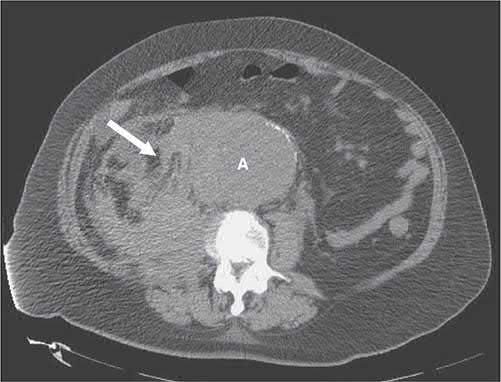
UK measurement and referral standards
- The single detail most often lost when UK aneurysm sizes are quoted is how the diameter is measured.
- NG156 requires that, when measuring aortic size with ultrasound, you report the inner-to-inner maximum anterior-posterior aortic diameter, in accordance with the NHS AAA screening programme, documenting clearly any additional measurements taken [3].
- Every threshold below, 3.0 cm, 5.5 cm, the 1 cm growth rule, is an inner-to-inner AP measurement, which reads systematically smaller than the outer-to-outer convention used in much of the literature.
Referral timings are set by size at diagnosis [3]:
| Ultrasound finding | Action |
|---|---|
| AAA 5.5 cm or larger | Refer to a regional vascular service, to be seen within 2 weeks of diagnosis |
| AAA 3.0 cm to 5.4 cm | Refer to a regional vascular service, to be seen within 12 weeks of diagnosis |
Table reformats the NG156 referral thresholds [3]. Surveillance itself uses the same frequency as the NHS AAA screening programme [3], whose care pathway sets three bands [4]:
| Aortic diameter | Screening programme action |
|---|---|
| Under 3.0 cm | No further scans required, end of pathway |
| 3.0 cm to 4.4 cm (small AAA) | Repeat ultrasound in surveillance in 12 months |
| 4.5 cm to 5.4 cm (medium AAA) | Repeat ultrasound in surveillance in 3 months |
| 5.5 cm or larger (large AAA), or grown more than 1 cm in 12 months | Refer to a vascular surgeon |
Table reformats the NHS AAA screening programme surveillance pathway [4]. Note that these two-band intervals (12 months and 3 months) are considerably simpler than, and differ from, the three- and six-monthly schemes quoted in the textbook accounts above.
- On imaging technique, offer thin-slice contrast-enhanced arterial-phase CT angiography to people being evaluated for elective AAA repair, and consider it for suspected ruptured AAA in people being evaluated for repair [3].
- For a person in whom symptomatic or ruptured AAA is being considered, offer an immediate bedside aortic ultrasound, and discuss immediately with a regional vascular service if the ultrasound shows an AAA, or if it is not immediately available or non-diagnostic and AAA is still suspected [3].
- Ruptured AAA should be considered in anyone with new abdominal or back pain, cardiovascular collapse or loss of consciousness, and is more likely with an existing AAA diagnosis, age over 60, current or past smoking, or a history of hypertension [3].
Imaging for planning in Schwartz's account
History must cover symptoms, pelvic surgery or radiation (relevant to retroperitoneal exposure or hypogastric interruption), claudication, prior femoral reconstruction, renal insufficiency and contrast allergy; ultrasound is the screening tool but CT is the gold standard for endovascular eligibility, sizes may differ by up to 1 cm between the two so follow-up compares like with like, multidetector timed-bolus 2.5–3 mm CT of chest, abdomen and pelvis takes under 30 seconds in one breath-hold with sub-millimetre resolution, window settings separate wall, calcium, thrombus and lumen, contrast nephropathy is the only major drawback, three-dimensional reconstruction gives centre-line diameters and lengths, and angiography is reserved for concomitant iliac occlusive disease (under 10%) or unusual renal anatomy [9].
Scoring and Severity
- The Crawford classification (extents I–V) grades thoracoabdominal aortic aneurysms by the extent of thoracic/abdominal aortic involvement and guides management in specialist centres [1][6].
- Aortic dissection is classified by the DeBakey system, type I (entire ascending aorta, arch, and descending aorta), type II (ascending aorta only), and type III (descending aorta only, with IIIa confined above and IIIb extending below the diaphragm) (and by the Stanford system) type A (any ascending aortic involvement, generally surgical) and type B (distal to the left subclavian artery, generally managed medically/endovascularly) [6][8].
- Dissections are further staged by time from onset: hyperacute (<24 hours), acute (2–7 days), subacute (8–30 days) and chronic (>30 days), with the classical acute/chronic cut-off at 2 weeks [6].
- Endoleaks after EVAR are classified types I–V by failure site: type I (proximal/distal attachment site), type II (retrograde collateral flow, e.g. lumbar or IMA vessels), type III (junction between graft components or fabric tear), type IV (graft wall porosity), and type V/endotension (aneurysm expansion without a demonstrable leak) [7].
- Risk of AAA rupture rises with diameter: <0.5%/year at <4.0 cm, 1%/year at 4–5.5 cm, and >3%/year at >5.5 cm [1]; similarly, annual rupture risk is quoted as ≤1% below 55 mm, 5–10% at 55–60 mm, and ~25% at ≥70 mm [2].
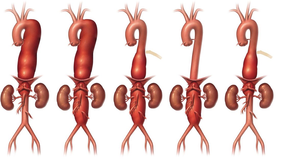
UK risk tools and repair thresholds
- NG156 bans ten named risk-assessment tools outright.
- For an asymptomatic unruptured AAA, do not use the British Aneurysm Repair score, Carlisle Calculator, Comorbidity Severity Score, Glasgow Aneurysm Scale, Medicare risk prediction tool, Modified Leiden score, POSSUM, Vascular-POSSUM, the Vascular Biochemical and Haematological Outcome Model, or the Vascular Governance North West risk model to determine whether repair is suitable [3].
- For a ruptured AAA the prohibition is broader still: do not use any single symptom, sign or patient-related risk factor, and do not use patient risk assessment tools of any kind, to determine whether repair is suitable [3].
- What NICE does permit is cardiopulmonary exercise testing, considered when assessing people for elective repair if it will assist in shared decision making [3].
The repair thresholds are three, and any one of them suffices [3]. Consider aneurysm repair for an unruptured AAA that is:
| Criterion |
|---|
| --- |
| Symptomatic |
| Asymptomatic, larger than 4.0 cm, and grown by more than 1 cm in 1 year (inner-to-inner maximum AP diameter on ultrasound) |
| Asymptomatic and 5.5 cm or larger (inner-to-inner maximum AP diameter on ultrasound) |
Table reformats the NG156 repair criteria [3]. Note that NICE sets a single 5.5 cm threshold irrespective of sex, it does not adopt the lower 5.0 cm threshold for women that the textbook accounts describe, despite stating elsewhere that AAAs are more likely to rupture in women [3].
- NG156 also names the uncertainty a clinician must convey.
- When discussing repair, explain the uncertainties around estimates of risk for AAAs larger than 5.5 cm, alongside aneurysm size and morphology, the person's age, life expectancy and fitness, rupture risk without repair, and the disadvantages of repair including hospital stay, recovery, potential further procedures and lifelong surveillance appointments [3].
- Where repair is not currently suitable, the reason must be explained, including the blunt statement that on average, people with poor overall health do not benefit from AAA repair, there is no reliable way to assess whether a particular person will benefit or be harmed, and repair for people with poor overall health is an unnecessary risk even if their AAA meets the size criteria [3].
Repair thresholds and endovascular eligibility in Schwartz's account
- Rupture risk is low below 5.5 cm and rises exponentially thereafter, so 5.5 cm is the threshold for elective repair provided surgical mortality is under 5%; women rupture at smaller sizes so 4.5–5.0 cm is reasonable in good-risk women, expansion over 0.5 cm in 6 months is a relative indication, smaller aneurysms are safely followed by CT or ultrasound every 6 months with equivalent long-term outcome, and in the ADAM study 80% of surveilled aneurysms came to repair within 5 years [9].
- Endovascular eligibility rests on the neck (18–28 mm diameter (up to 32 in the ideal table) at least 15 mm long, angulation under 45–60°, calcification and thrombus each under 50% of circumference, measured mid-wall to mid-wall at several levels), the common iliac landing zone (8–20 mm diameter, at least 20 mm of uniform patent artery, or the external iliac when the common iliac is aneurysmal) and access (at least one femoral–external iliac axis of 7 mm or more for the main sheath); about 60% of aneurysms qualify, the main body is oversized 10–20% and the limbs 1–4 mm, and length from renal to hypogastric arteries is the hardest measurement, plumb-line axial measurements underestimate, centre-line overestimates and marker-catheter angiography ignores graft stiffness, with too long a device covering the hypogastric and too short needing extra components [9].
- Short or absent necks are addressed by fenestrated or branched grafts (precise alignment with visceral vessels, customised and confined to high-volume centres, with promising short- and mid-term results for juxtarenal aneurysms and low mortality with less renal and cardiac morbidity than open repair, which remains safe for good-risk patients), surgeon-modified fenestrations, and chimney, snorkel and periscope techniques [9].
Treatment and Management
- Asymptomatic AAA in a fit patient should be considered for repair once the diameter exceeds 55 mm (measured anteroposteriorly by ultrasound), balancing the roughly 5% mortality of elective open surgery against rupture risk; regular ultrasound surveillance is used below this threshold [2].
- Surveillance intervals by size are: 3.0–3.9 cm every 3 years, 4.0–4.9 cm yearly, and >5.0 cm every 6 months [7].
- Repair is also indicated for growth >0.5 cm in 6 months (or >1.0 cm/year), symptomatic aneurysms, and infected (mycotic) aneurysms, with a lower threshold (≥5.0 cm) in women or high-risk patients (severe COPD, strong family history of rupture, poorly controlled hypertension, eccentric shape) [1][7].
Open repair versus EVAR
- Bailey & Love notes that the UK and European guideline positions differ, the European Society for Vascular Surgery 2019 guidelines favouring endovascular repair as first line with open repair reserved for patients with a long life expectancy; the UK position is set out in the note below [2].
- EVAR is associated with reduced early mortality compared with open repair but has a higher reintervention rate, requires lifelong imaging surveillance, and has shown no long-term survival advantage [1][2].
- About 75% (or ~60% by other estimates) of infrarenal aneurysms are anatomically suitable for EVAR; unsuitability is caused by a short, flared or angulated neck or difficult iliac access [2][5].
- Ideal AAA morphology for EVAR includes neck length >10–20 mm, neck diameter <32 mm, neck angulation <60 degrees, common iliac length >10 mm and diameter 7–18 mm, and non-tortuous, non-calcified iliac arteries [5][7].
Schwartz summarises the trials: DREAM (345 patients, 28 European centres) found 30-day mortality of 4.6% open versus 1.2% endovascular and combined death or severe complication 9.8% versus 4.7%, largely from pulmonary complications, with more graft-related complications after endovascular repair; EVAR-1 (1082 patients, 34 UK centres) found 30-day mortality 4.7% versus 1.7%, in-hospital mortality 6.2% versus 2.1% and reintervention 9.8% versus 5.8% favouring surgery; both enrolled only patients fit for either repair so cannot be generalised to the unfit or anatomically unsuitable; the Veterans Affairs OVER trial (881 patients followed up to 9 years) found the perioperative survival advantage sustained to 3 years but not beyond, equal long-term all-cause mortality, six ruptures after endovascular and none after open repair, and better long-term survival with endovascular repair in younger but not older patients, while being cost-effective for at least the first 2 years [9]. Device series report 30-day mortality of about 1% (Excluder, with 17% and 20% endoleak at 1 and 2 years, 7% annual reintervention and 14% sac growth at 2 years), 3.5% (Zenith, equal to open, 7.4% and 5.4% endoleak, 5.3% migration at 1 year, 100% and 98.9% freedom from rupture in low- and high-risk patients), 2.8% (AneuRx, 13.9% endoleak, 11.5% enlargement, 9.5% migration and 98.4% freedom from rupture at 4 years) and 0.8% (Talent, 10% endoleak, three deployment failures); early cost analyses found no saving because devices cost $5000–6000 and endovascular repair cost 1.74 times open repair before counting surveillance [9].
Ruptured aneurysm
For ruptured AAA, management is emergency: permissive hypotension (systolic <100 mmHg, or SBP 80–100), two large-bore IV cannulae, urinary catheter, cross-matched blood, high-flow oxygen, analgesia, and immediate transfer to theatre, "the treatment of ruptured aneurysm is an operation, not monitoring and resuscitation" [1][2]. EVAR should be considered first-line for anatomically suitable ruptured AAA (REVAR) [2].
Schwartz gives overall rupture mortality of 71–77% against 2–6% for elective open repair; nearly half die before hospital and operative mortality for the rest is 45–50%, unchanged in 30 years [9].
Thoracic aneurysm and dissection
Indications for ascending aortic aneurysm repair include acute symptoms, diameter ≥5.5 cm (≥5.0 cm in Marfan syndrome), or rapid growth >0.5 cm/year; descending/thoracoabdominal aneurysms are repaired at >5.5 cm if endovascular repair is feasible or >6.5 cm if open repair is required [8]. All ascending aortic dissections (Stanford A) require open surgical repair; descending dissections (Stanford B) are managed operatively only if there is visceral/extremity malperfusion or contained rupture, otherwise medically with IV beta-blockade (e.g. esmolol) and nitroprusside to control blood pressure [8].
NICE recommendations on repair
The open-versus-EVAR recommendation reverses between unruptured and ruptured aneurysms, and getting the two the right way round is the examinable point.
For unruptured AAA meeting the repair criteria [3]:
| Patient | Recommendation |
|---|---|
| Meets repair criteria, no contraindication | Offer open surgical repair, unless contraindicated by abdominal copathology, anaesthetic risks or medical comorbidities |
| Abdominal copathology, a hostile abdomen, horseshoe kidney, a stoma, or other person-specific considerations | Consider EVAR |
| Anaesthetic risks or comorbidities contraindicating open repair | Consider EVAR or conservative management |
For ruptured infrarenal AAA the position inverts [3]:
| Group | Recommendation |
|---|---|
| Most people, especially men over 70 and women of any age | EVAR provides more benefit than open surgical repair |
| Men under 70 | Open surgical repair is likely to provide a better balance of benefits and harms |
Tables reformat the NG156 repair recommendations [3]. Where standard EVAR is unsuitable for a ruptured AAA, consider open surgical repair [3].
- Complex EVAR is restricted to a research footing.
- If open repair and complex EVAR are both suitable, only consider complex EVAR if the risks and the uncertainties around whether complex EVAR improves perioperative survival or long-term outcomes have been discussed, and if it is performed with special arrangements for consent and for audit and research, with all patients entered onto the National Vascular Registry [3].
- The same conditions apply where open repair is contraindicated [3].
- For a ruptured AAA, do not offer complex EVAR if open surgical repair is suitable, except as part of a randomised controlled trial comparing the two [3].
Three perioperative recommendations are negatives. Do not routinely offer preoperative beta blockers to people having AAA repair; do not offer remote ischaemic preconditioning; and for open repair of an unruptured AAA, consider epidural analgesia in addition to general anaesthesia [3]. For EVAR of a ruptured AAA, consider using local infiltrative anaesthesia alone [3].
- On emergency transfer, NICE sets a clock and states a futility threshold.
- When a person with a suspected ruptured or symptomatic unruptured AAA has been accepted by a regional vascular service, ensure they leave the referring unit within 30 minutes of the decision to transfer [3], under a protocol agreed between emergency departments, ambulance services and regional vascular services and reviewed at least every 3 years [3]. Consider a restrictive approach to volume resuscitation (permissive hypotension) during that transfer [3].
- But there is no evidence that any single symptom, sign or prognostic risk assessment tool can determine whether a person with suspected or confirmed ruptured AAA should be transferred [3], and the one clear futility statement is that people with a confirmed ruptured AAA who have a cardiac arrest and/or persistent loss of consciousness have a negligible chance of surviving AAA repair [3].
Surgeries
- Open AAA repair uses a full-length midline or supraumbilical transverse incision; the aorta is exposed just below the renal arteries, clamps are applied proximally and distally, and an inlay synthetic (Dacron/PTFE) tube or bifurcated ('trouser') graft is sutured within the opened aneurysm sac, which is then closed over the graft to separate it from the bowel [1][2].
- A straight tube Dacron graft is typically used; if performing aorto-bifemoral repair, flow to at least one internal iliac (hypogastric) artery must be preserved (with visible back-bleeding) to avoid vasculogenic impotence and buttock claudication, reimplanting the internal iliac if necessary [7].
- The inferior mesenteric artery is reimplanted if back-pressure is <40 mmHg, if there has been previous colonic surgery (disrupting collaterals such as the Arc of Riolan or Marginal Artery of Drummond), if there is SMA stenosis, or if left colon perfusion looks inadequate [7].
- Emergency proximal aortic control for rupture is obtained by compressing the aorta against the spine through the gastrohepatic ligament (supraceliac aorta beneath the diaphragmatic crus), dividing the crus further if needed [7].
- EVAR uses a modular stent-graft (main body plus limbs, made of Dacron/PTFE with metallic stents) deployed via bilateral femoral access under fluoroscopic guidance, anchored by hooks/barbs with infrarenal or suprarenal fixation, requiring lifelong duplex/CT surveillance for endoleak, disconnection or migration [2].
- For mycotic aneurysms and infected aortic grafts, an extra-anatomic bypass (axillo-bifemoral with femoral-to-femoral crossover) is performed first, followed by resection of the infected infrarenal aorta/graft and oversewing of the aortic stump [7].
- Aortoenteric fistula, where a graft erodes into the third or fourth part of the duodenum near the proximal suture line, is treated the same way: bypass through a non-contaminated field, graft resection, and closure of the duodenal defect [7].
- Repair of descending thoracic/thoracoabdominal aneurysms carries a risk of paraplegia from spinal cord ischaemia (occlusion of intercostal arteries and the artery of Adamkiewicz), reduced by lumbar CSF drainage, maintaining spinal perfusion pressure (MAP minus spinal pressure) with vasopressors, and reimplanting intercostal arteries below T8; paraplegia risk is <5% with endovascular repair versus about 20% with open repair [8].
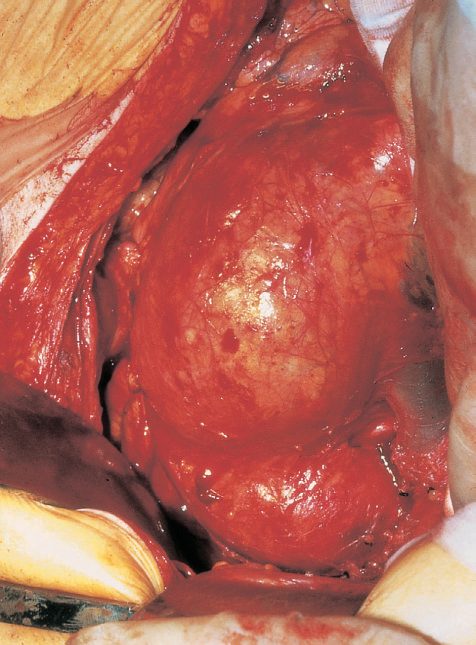
Open and endovascular technique in Schwartz's detail
Open repair needs general anaesthesia, an epidural for analgesia and usually a midline transabdominal incision (retroperitoneal is accepted); small bowel and transverse colon are retracted, the retroperitoneum opened, proximal and distal segments isolated, heparin 100 IU/kg given, the aneurysm clamped and opened, and a tube graft or, for iliac involvement, a bifurcated aorto-bi-iliac or aorto-bifemoral graft sewn in, the sac and retroperitoneum closed over the graft to keep bowel off it; open repair permanently removes the aneurysm and the need for surveillance, allows direct assessment of colonic perfusion with concomitant mesenteric bypass if needed and exploration for other pathology, and carries hospital stays of 5–10 days [9]. Endovascular repair, first proposed conceptually after Dotter's 1964 transluminal angioplasty and first performed by Parodi in 1991, fixes a modular stent graft (main body plus one or two iliac limbs, over fifteen devices approved worldwide) to non-aneurysmal aorta and iliac to exclude the sac, leaving lumbar and inferior mesenteric branches that can pressurise it; it is done in a surgical suite for sterility, with N-acetylcysteine and bicarbonate for renal impairment, under general, regional or even local anaesthesia, through femoral cutdown or percutaneous "preclose" suture closure (95% success for 12–16F sheaths, 87% for 18–24F); stiff wires to the arch, heparin 80 IU/kg to an ACT of 200–250 seconds, the main device (usually from the right) advanced to L1–L2 where the renals lie, a road-map aortogram, deployment just below the lowest renal, cannulation of the contralateral gate with a directional catheter and angled wire, confirmation of intrastent passage, docking of the contralateral limb, and completion angiography for renal and hypogastric patency, fixation and endoleak, with extra cuffs, balloons or bare stents as needed; patients recover 2–4 hours, go to a general ward, eat that evening and usually leave the next morning after 1–3 days total, benefiting particularly those with severe pulmonary disease and avoiding ileus, hernia and adhesions [9].
Complications
Postoperative complications after open AAA repair are most commonly cardiac (ischaemia, infarction, the leading cause of acute death after surgery) and respiratory (atelectasis, lower lobe consolidation); colonic ischaemia occurs in about 10% of patients (usually self-limiting) due to loss of IMA collateral supply, and acute kidney injury (the leading cause of late death) is more likely with preoperative renal impairment or ruptured presentations [2][7]. Ischaemic colitis after AAA repair presents with (often bloody) diarrhoea; the middle and distal rectum are spared because of internal iliac collateral supply, and diagnosis is by lower endoscopy (best test) or CT, with colectomy and colostomy required if there is diffuse peritonitis, sepsis, or black/necrotic mucosa [7].
Mortality, graft and late complications
- Elective AAA repair carries about 3–7% (Oxford Handbook) or 5% (ABSITE Review) operative mortality; a third of patients experience impotence secondary to disruption of autonomic nerves and pelvic blood flow [1][7].
- Major vein injury (retro-aortic left renal vein) can occur with proximal cross-clamping [7].
- Graft infection occurs in about 1% of cases (Staphylococcus epidermidis most common), and pseudoaneurysm formation after graft placement occurs in about 1%; atherosclerotic graft limb occlusion is the most common late complication [7].
- Aortoenteric fistula typically presents more than 6 months after surgery with a herald bleed (haematemesis, then melaena/haematochezia, then exsanguination) [7].
- Risk factors for perioperative mortality include creatinine >1.8 (the leading factor), congestive heart failure, ECG evidence of ischaemia, pulmonary dysfunction, older age, and female sex [7].
Schwartz quantifies open-repair complications: cardiac events (infarction, arrhythmia) in 2–6%, renal failure under 2% electively but over 20% after rupture (hypotension, atheroembolism, ureteric injury, contrast, suprarenal clamping), partial-thickness ischaemic colitis in 5% without sequelae but 90% mortality if it becomes full-thickness gangrene with peritonitis (highest risk after prior colectomy and ruptured repair), graft infection in 1–4% (commoner after rupture), and aortoenteric fistula years later with massive haematemesis when the graft is not covered [9]. After endovascular repair a meta-analysis of 1118 patients found 24% endoleak, rupture under 0.8%, limb thrombosis from kinking as the sac remodels and shortens or from progressive iliac disease (treated by lysis or thrombectomy and relining), renal occlusion from malposition or migration, component separation, pelvic ischaemia (buttock claudication, impotence, gluteal sloughing, colonic ischaemia) in 20–45% after hypogastric coil embolisation, groin haematoma and infection, and deployment failure needing open conversion [9].
Dissection and rupture outcomes
Death from ascending aortic dissection is usually due to cardiac failure from aortic insufficiency, cardiac tamponade, or aortic rupture; aortic insufficiency occurs in 70% of ascending dissections, from annular dilatation or cusp shearing [8]. Rupture into the peritoneal cavity (20% of ruptures) causes free bleeding with very few patients reaching hospital alive, whereas posterolateral retroperitoneal rupture (80%) may be temporarily contained, allowing transfer to hospital [2].
Post-EVAR surveillance and endoleaks
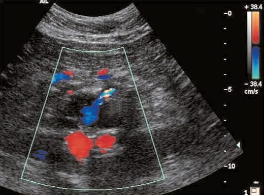
- Schwartz calls lifelong follow-up essential (its absence "tantamount to not having had a repair") with triple-phase CT and four-view abdominal radiographs within a month, 6-monthly for 1–2 years then yearly, the CT detecting endoleak, migration and size change and the plain film showing limb conformation, impending separation and stent fractures or suture breaks invisible on CT [9].
- Endoleak occurs in 20–30% early, over half resolving within 6 months to leave about 10% chronic; angiography is least sensitive but most specific, CT most sensitive but least specific and the de facto standard, duplex and MRA limited by equipment and expertise; type I attachment-site leaks are under 5% of endoleaks, seen as an early blush at completion, over 80% seal within 6 months but persistence demands prompt treatment; type II retrograde branch leaks (lumbar, inferior mesenteric, accessory renal, hypogastric) are the commonest at 20–30%, half resolve, and they are treated only with sac growth; type III junctional or fabric failures (early from inadequate overlap, late from fabric tear or separation with remodelling) are repaired promptly; type IV porosity blush disappears within 48 hours of heparin reversal and becomes type III by definition if it persists [9].
- Endotension, sac growth without demonstrable leak in about 5%, possibly transudation through certain ePTFE fabrics, with sacs found bloodless at conversion, has no proven rupture risk but merits elective open conversion in fit patients; secondary interventions run 10–15% a year (extender cuffs for migration, selective or translumbar embolisation of type II leaks, surgical or laparoscopic branch ligation, bridging cuffs for separation, late conversion); late rupture occurs in about 1–1.5% a year, 2.3% over 15.4 months with an endoleak versus 0.3% without in EUROSTAR, 63% of delayed ruptures following type I leak with migration, 11% type I without migration and 19% type II, open conversion for rupture carries 43% mortality so emergency endovascular relining is preferred [9].
- Post-EVAR surveillance is mandatory and its failure mode is named. Enrol people who have had EVAR into a surveillance imaging programme, with the frequency based on the individual's risk of EVAR-related complications [3].
- Consider contrast-enhanced CT angiography or colour duplex ultrasound for assessing aneurysm diameter and device limb kinking; use contrast-enhanced CT angiography if an endoleak is suspected, and contrast-enhanced ultrasound if CT angiography is contraindicated [3].
- The trap is stated explicitly: do not exclude endoleaks based on a negative colour duplex ultrasound alone [3].
Endoleak management is graded by type, and type 2 is treated only on evidence of sac expansion [3]:
| Endoleak type | NICE recommendation |
|---|---|
| Type 1 and type 3 | Consider open, endovascular or percutaneous intervention |
| Type 2 | Consider intervention only in people who have AAA expansion following EVAR |
| Type 5 | Consider further investigation |
Table reformats the NG156 endoleak recommendations [3].
Abdominal compartment syndrome is flagged after either operation, not just open repair. Be aware that people can develop abdominal compartment syndrome after EVAR or open surgical repair of a ruptured AAA, and assess for it if their condition does not improve after either [3].
Secondary cardiovascular prevention is treated as part of aneurysm care rather than a separate matter: offer people with an AAA information, support and interventions for secondary prevention of cardiovascular disease, covering smoking cessation, diet and exercise, lipid modification and statin therapy, diabetes management and hypertension [3]. Offer referral to a stop smoking service to anyone with an AAA who smokes [3].
Prognosis
- Less than 50% of patients with ruptured AAA reach hospital alive, and overall combined community-and-hospital mortality is 75–95% [1][2].
- Operative mortality for ruptured AAA is around 50% [2][7].
- Without operation, rupture is virtually always fatal [2]; 80% of patients with ruptured AAA will die if not operated on [1].
- Outcome is best with an experienced dedicated vascular team and rapid transfer from emergency department to theatre [1].
- Elective thoracoabdominal aneurysm surgery carries up to 20% mortality and a risk of paraplegia, with about 10% requiring dialysis afterward [1].
- About 30% of aortic dissection survivors eventually develop aneurysmal degeneration requiring surgery, warranting lifetime serial imaging surveillance (preferring MRI to reduce radiation exposure) [8].
References
- Oxford Handbook of Clinical Surgery, 5th ed., Ch. 19, Treatment of abdominal aortic aneurysm
- Bailey & Love's Short Practice of Surgery, 28th ed., Ch. 61 Arterial disorders
- NICE Guideline NG156: Abdominal aortic aneurysm — diagnosis and management (2020), 1.1.1; 1.1.2; 1.1.3; 1.1.4; 1.1.5; 1.1.7; 1.1.8; 1.1.9; 1.1.10; 1.1.11; 1.1.12; 1.1.14; 1.2.1; 1.2.3; 1.3.1; 1.3.2; 1.3.4; 1.3.5; 1.3.6; 1.4.1; 1.4.3; 1.4.4; 1.4.5; 1.4.6; 1.4.7; 1.4.8; 1.5; 1.5.1; 1.5.2; 1.5.3; 1.5.4; 1.5.5; 1.5.6; 1.5.7; 1.5.8; 1.6.1; 1.6.2; 1.6.3; 1.6.4; 1.6.5; 1.6.6; 1.7.1; 1.7.2; 1.7.3; 1.7.4; 1.7.5; 1.8.1; 1.8.2; 1.8.3 www.nice.org.uk
- NHS Abdominal Aortic Aneurysm Screening Programme (NAAASP): programme overview and care pathway, NHS England, Care pathway; Programme overview www.gov.uk
- Schwartz's Principles of Surgery: ABSITE and Board Review, Ch. 23 Arterial Disease
- Sabiston Textbook of Surgery, 22nd ed., Ch. 102 Aortic Disease
- The ABSITE Review, 2022, Abdominal Aortic Disease
- The ABSITE Review, 2022, Thoracic Aortic Disease
- Schwartz's Principles of Surgery, 11th ed., Ch. 23, Figs. 23-30 and 23-31
- Browse's Introduction to the Symptoms and Signs of Surgical Disease, 6th ed., Ch. 15 The abdomen, Ruptured abdominal aortic aneurysm
- Maingot's Abdominal Operations, 13th ed., Ch. 20