



Zenker's Diverticulum
Summary
- Zenker's (pharyngeal, pharyngoesophageal) diverticulum is a false diverticulum that protrudes through Killian's triangle, a weak area of the posterior pharyngeal wall between the oblique fibres of thyropharyngeus and the transverse fibres of cricopharyngeus [1][2].
- It is the most common oesophageal diverticulum, typically affecting patients over age 60, more common in men than women [2][3].
- Treatment centres on division or myotomy of the cricopharyngeus, with or without excision or suspension of the pouch itself [3].
There is no NICE clinical guideline on pharyngeal pouch, but two NICE HealthTech guidances cover the endoscopic procedures (HTG8 for rigid endoscopic stapling and HTG367 for flexible endoscopic treatment) and the BSG oesophageal dilatation guideline covers dilatation of the upper oesophageal sphincter in this setting [4][5][6]. Both NICE guidances were migrated from earlier interventional procedures guidance (HTG8 from IPG22 and HTG367 from IPG513) with recommendations unchanged [4][5].
Definition
- Zenker's diverticulum is a false pulsion diverticulum, only mucosa and submucosa herniate through Killian's triangle, which is bounded inferiorly by the cricopharyngeal muscle and laterally by the oblique fibres of the inferior pharyngeal constrictor [3].
- It is classified among the pharyngeal (as opposed to midoesophageal or epiphrenic) oesophageal diverticula [1].
- It is a cervical pulsion diverticulum located posteriorly, occurring specifically between the superior pharyngeal constrictors and the inferior cricopharyngeus (Killian's triangle) [7].
History and epidemiology
The pouch was originally described by Ludlow in 1769; the eponym comes from Zenker's classic clinicopathological description of 34 cases published in 1878 [8]. Pharyngo-oesophageal diverticula are reported in about 1 of every 1000 routine barium examinations and classically occur in elderly white men [8].
Pathophysiology
- The diverticulum is usually secondary to a lack of coordination between pharyngeal contraction and opening of the upper oesophageal sphincter (UES), a pulsion diverticulum resulting from increased intraluminal pressure during swallowing against a cricopharyngeus that fails to relax appropriately [1][3][7].
- The mechanisms are muscular weakness together with UES dysfunction, the latter characterised by incomplete UES relaxation, increased intrapharyngeal pressure, and discoordinated pharyngeal contractions [9].
- The condition is also associated with lower cranial nerve dysfunction, for example motor neurone disease or a previous cerebrovascular accident [10].
- Videofluoroscopic and manometric studies have not fully elucidated the cause: many patients show normal UES relaxation with swallowing, while others show incomplete pharyngeal relaxation, early cricopharyngeal contraction, and abnormal pharyngeal contraction waves [2].
- As the diverticulum enlarges it almost invariably deviates toward the left side of the neck, and the fundus can descend into the mediastinum [1][2].
- The left-sided predominance is attributed to the slight convexity of the oesophagus to the left [9].
- The physiological upper oesophageal sphincter mechanism is formed by the oblique thyropharyngeus fibres, the transverse cricopharyngeus fibres, and the circular fibres of the upper oesophagus [2].
Association with reflux
Gastro-oesophageal reflux is present in up to 95% of patients and may relate to reflex contraction of the oesophageal longitudinal muscle, which widens the gap between the pharyngeal constrictors and cricopharyngeus, or to spasm of the UES [9]. About 30% of patients have associated GERD in other series [3].
Restrictive cricopharyngeal myopathy
- Zenker's diverticulum is one of the anatomical manifestations of transit dysphagia, disorders of the pharyngeal phase of swallowing that arise from inadequate oropharyngeal bolus transport, inability to pressurise the pharynx, inability to elevate the larynx, discoordination of pharyngeal contraction with cricopharyngeal relaxation, or decreased compliance of the pharyngo-oesophageal segment secondary to neuromuscular disease [8].
- Standard manometry has often failed to show a consistent motility abnormality in these patients; the abnormality most apt to be present is a loss of compliance in the pharyngo-oesophageal segment, manifested by an increased bolus pressure [8].
- Using simultaneous manometry and videofluoroscopy, Cook and colleagues showed that the cricopharyngeus is only partially relaxed, enough for its pressure to drop to oesophageal baseline on manometry, but not enough to allow unimpaired passage of the bolus [8].
- This decreased compliance is recognised on manometry as a "shoulder" on the pharyngeal pressure wave, whose amplitude correlates directly with the degree of outflow obstruction [8].
- Muscle biopsies from patients with Zenker's diverticulum show histological evidence of a restrictive myopathy of the cricopharyngeus, supporting the view that the diverticulum develops as a consequence of the outflow resistance to bolus transport through the non-compliant muscle [8].
- The pouches tend to enlarge progressively with time because the compliance of the skeletal-muscle portion of the cervical oesophagus falls with ageing [8].
Clinical features
- Dysphagia is the most common symptom, present in about 80–90% of patients [3].
- Cervical dysphagia is often associated with regurgitation, halitosis, choking, chronic cough, hoarseness, gurgling, or aspiration pneumonia [9].
- As the diverticulum enlarges, patients may experience regurgitation of undigested food, sometimes hours after eating, particularly when bending or lying down, and may wake at night with throat tightness and coughing fits [2].
- Recurrent, unexplained chest infections from aspiration of pouch contents are a recognised presentation [2].
- Patients can also experience halitosis and gurgling sounds in the neck on swallowing, and as the pouch grows it may form a visible neck swelling [2][3].
- Symptoms per the ABSITE Review include upper esophageal dysphagia, choking, halitosis, and regurgitation of non-digested food [7].
Physical signs and red flags
- Physical examination may show the Boyce sign (a neck mass that gurgles on palpation) and weight loss [9].
- However, most patients have no palpable swelling at all, since a pouch is often smaller than the 5–10 cm needed to become detectable; when present, it lies behind sternomastoid, below the thyroid cartilage, dull to percussion, and neither fluctuant nor transilluminant.
- It is fixed deeply and cannot be moved in the neck, its neck and oesophageal attachment cannot be felt, and although compression can empty it with an accompanying gurgle, this does not meet the usual definition of a swelling being "reduced" [11].
- A long antecedent history of halitosis and recurrent sore throats often precedes the onset of regurgitation [11].
- Progressive dysphagia, odynophagia, haemoptysis and haematemesis are more suspicious of malignancy, which may be a squamous cell carcinoma arising within the diverticulum, with an incidence of up to 1.1% [9].
Etiology
The underlying mechanism is failure of the cricopharyngeus to relax normally during swallowing, generating a pulsion force that herniates mucosa through the natural weak point of Killian's triangle [1][7]. Another area of potential weakness where a similar diverticulum can form is the "Killian-Jamieson area," located just below the cricopharyngeal muscle [3].
Diagnosis
A barium swallow (thin barium emulsion), ideally as part of a videofluoroscopic swallowing study, shows the position (at the level of C5–C6) and size of the diverticulum, or a prominent cricopharyngeal bar without an associated diverticulum; it also provides information on pharyngeal contraction waves and UES performance [2][3][7]. The barium study also assesses the size of the diverticular neck and the distance from the diaphragm [9].
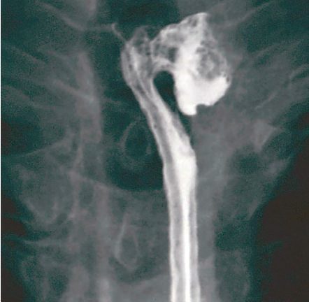
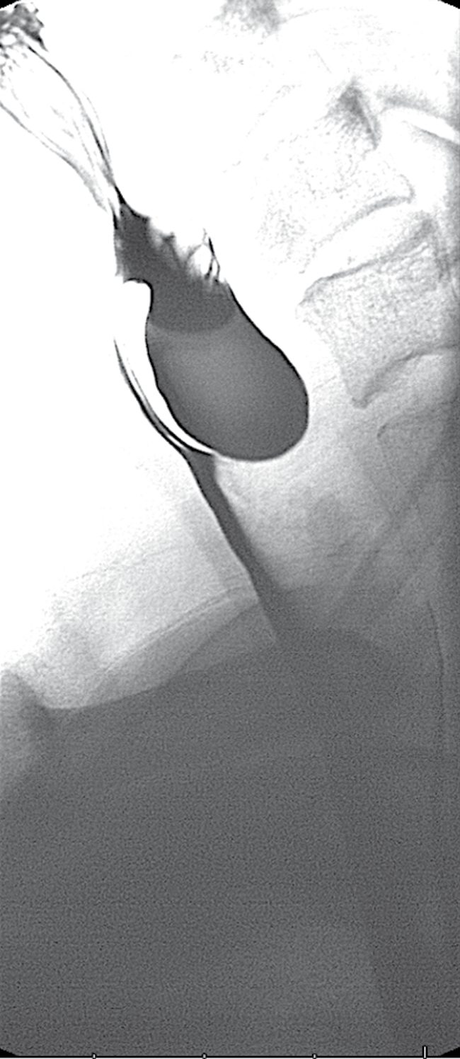
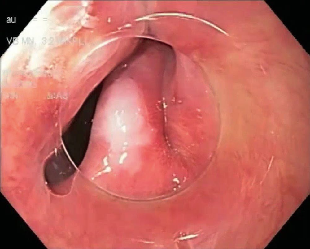
Endoscopy to exclude other pathology (including the rare occurrence of carcinoma within the pouch) must be performed with great caution given the risk of iatrogenic perforation [2][3][7]. Upper endoscopy is nonetheless regarded as mandatory to rule out cancer or other oesophageal disease and to assess for signs of reflux [9].
Oesophageal manometry can show lack of coordination between the pharynx and UES and a hypertensive UES, and also localises the LES for ambulatory pH monitoring in patients suspected of concurrent GERD [3]. Manometry defines any underlying oesophageal motility disorder (most commonly achalasia, followed by diffuse oesophageal spasm and nutcracker oesophagus) though some surgeons do not regard it as mandatory, believing a primary motility disorder is always present [9].
Videoradiography and the criteria for myotomy
- Video- or cineradiography is the most objective test of oropharyngeal bolus transport, pharyngeal compression, relaxation of the pharyngo-oesophageal segment and airway protection during swallowing; it readily identifies a diverticulum, stasis of contrast in the valleculae, a cricopharyngeal bar, or narrowing of the pharyngo-oesophageal segment [8].
- Combined with manometry using specially designed catheters, it identifies the cause of pharyngo-oesophageal dysfunction in most patients: inadequate pharyngeal pressurisation, insufficient or absent cricopharyngeal relaxation, discoordination between pharyngeal pressurisation, cricopharyngeal relaxation and cervical oesophageal contraction, or a raised hypopharyngeal bolus pressure suggesting loss of compliance [8].
- The requirements for a successful pharyngo-oesophageal myotomy are (a) adequate oropharyngeal bolus transport, (b) an intact swallowing reflex, (c) reasonable coordination of pharyngeal pressurisation with cricopharyngeal relaxation, and (d) a cricopharyngeal bar, Zenker's diverticulum or narrowed pharyngo-oesophageal segment on video-oesophagogram and/or excessive pharyngo-oesophageal "shoulder" pressure on motility study [8].
Scoring and Severity
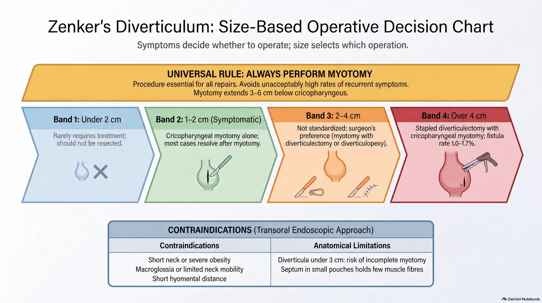
There is no formal severity score, but diverticulum size is the practical classification because it determines which operation is appropriate [3][9].
| Size | Approach |
|---|---|
| Under 2 cm | Rarely requires treatment; should not be resected |
| 1–2 cm, symptomatic | Cricopharyngeal myotomy alone, most resolve after myotomy |
| 2–4 cm | Not standardised; surgeon's preference, most commonly myotomy with diverticulectomy or diverticulopexy |
| Over 4 cm | Stapled diverticulectomy with cricopharyngeal myotomy is the approach of choice, with fistula rates of 1–1.7% |
| Under 3 cm | Poorer outcomes with endoscopic stapling; endoscopic diverticulotomy may give an incomplete myotomy because the septum contains few muscle fibres |
Table compiled from the size-based recommendations above [3][9]. Notably, the decision to intervene at all is made regardless of size, since it is mostly the underlying motility disorder that determines symptom severity [9].
Treatment and Management
- Surgery is indicated for progressive symptoms, particularly with a prominent cricopharyngeal bar and abnormal UES mechanism causing significant dysphagia; in most patients the pouch itself is contributing to underlying debilitation, so surgery is generally recommended in all but the most unwell patients, mindful of the risk of recurrent aspiration pneumonia [2].
- Diverticula smaller than 2 cm rarely require treatment [3].
- The main indication is to address the patient's symptoms, though some advise operating even in the absence of symptoms to prevent aspiration [9].
- Preoperative optimisation includes chest physiotherapy and attention to respiratory, cardiovascular, and nutritional status [2].
- Postoperatively, if the preoperative evaluation shows pathologic gastroesophageal reflux, medical or surgical antireflux therapy should be given, as aspiration risk increases after UES resection [3].
Consider upper oesophageal sphincter dilatation in the treatment of dysphagia with disordered upper oesophageal sphincter opening, post-cricoid web, or cricopharyngeal bar (with or without the presence of a Zenker's diverticulum) or to permit passage of radiofrequency ablation catheters [6].
Patient selection and expected success
The low morbidity and mortality of cricopharyngeal and upper oesophageal myotomy have encouraged a liberal approach to its use for almost any problem in the oropharyngeal phase of swallowing, and that attitude has produced an overall symptom-relief rate of only 64%; when patients are selected using radiographic or motility markers of disease, a much higher proportion benefit [8]. Patients with low pharyngeal pressure (poor "piston" function of the pharynx) or increased resistance of the pharyngo-cervical oesophageal segment from loss of skeletal-muscle compliance are the ones improved by myotomy, which enlarges the segment and reduces outflow resistance [8].
Surgeries
The essential surgical step in all approaches is myotomy of the cricopharyngeus (and the upper 3 cm of the posterior oesophageal muscle wall), which eliminates the functional obstruction of the UES; this is considered the key therapeutic manoeuvre, and the diverticulum itself may either be resected or simply suspended [3][7]. Any procedure performed without a cricopharyngeal myotomy carries an unacceptably high incidence of recurrent symptoms; the myotomy relieves the functional obstruction distal to the diverticulum, reducing both the risk of leak after diverticulectomy and the risk of recurrence, and its extent usually ranges between 3 and 6 cm below the cricopharyngeus [9].
Open transcervical approach
- Via a left-sided neck incision along the sternocleidomastoid, the cricopharyngeus muscle is divided completely with the myotomy extended onto the oesophageal muscle wall for about 3 cm below the diverticulum; circumferential dissection is avoided to reduce risk to the left recurrent laryngeal nerve [3].
- Visualisation of the muscular layers and mucosa may be enhanced by placing a bougie dilator in the oesophagus before starting the myotomy [9].
- Small diverticula (<2 cm) should not be resected; larger ones can be resected (typically stapled, with a bougie in place to avoid narrowing the lumen) or suspended/fixed upward to the prevertebral fascia with the apex pointing cranially, which avoids a suture line and lowers leak risk [1][3].
- Diverticulopexy (suspension), rather than diverticulectomy, is associated with a lower risk of fistula formation, though not of hematoma, recurrent nerve paralysis, phonation difficulty, or Horner syndrome [13].
- A left cervical incision is standard, with drains left in place and an esophagogram obtained on postoperative day 1 [7].
- The three open options handle the pouch differently.
- Diverticulectomy was historically hand-sewn and technically challenging with a leak risk; staplers have significantly reduced the risk of oesophageal leak and mediastinitis to 1.7% [9].
- Diverticulopexy fixes the sac to the prevertebral fascia or the pharyngeal muscles above its neck, leaving the hypopharyngeal mucosa intact so that leakage risk is eliminated and hospital stay is shorter [9].
- Diverticular inversion invaginates the pouch into the oesophageal lumen and closes the neck with a purse-string suture, again without breaching the mucosa, with shorter stay and lower complication rate than diverticulectomy [9].
- There are no randomised controlled trials comparing the open approaches, so evidence for one over another is limited [9].
Endoscopic approaches
- Rigid endoscopy uses a bivalve diverticuloscope with one blade in the diverticulum and one in the oesophagus, exposing the cricopharyngeal "bar," which is then divided with an endoscopic linear stapler fired repeatedly until the pouch base is reached, opening the pouch into the oesophageal lumen and dividing the cricopharyngeus simultaneously [2][3].
- The goal is to divide the common wall or septum between the oesophageal lumen and the diverticulum, which performs a cricopharyngeal myotomy automatically because that septum contains the cricopharyngeal fibres [9].
- Endoscopic stapling requires a diverticulum of at least 2–3 cm to accommodate the stapler; both rigid and flexible endoscopic techniques report about a 90% success rate [3][7].
- Flexible endoscopic division uses a needle knife, hook knife, or argon plasma coagulation to divide the septum, with endoclips placed afterward to close the mucosa [3].
- CO2 laser division of the bar is an emerging alternative to stapling in some centres [2].
- Peroral endoscopic myotomy (Z-POEM) has also been applied to Zenker's diverticulum with good results (roughly 90% success), with a complication rate around 6%, mostly perforation or bleeding [3].
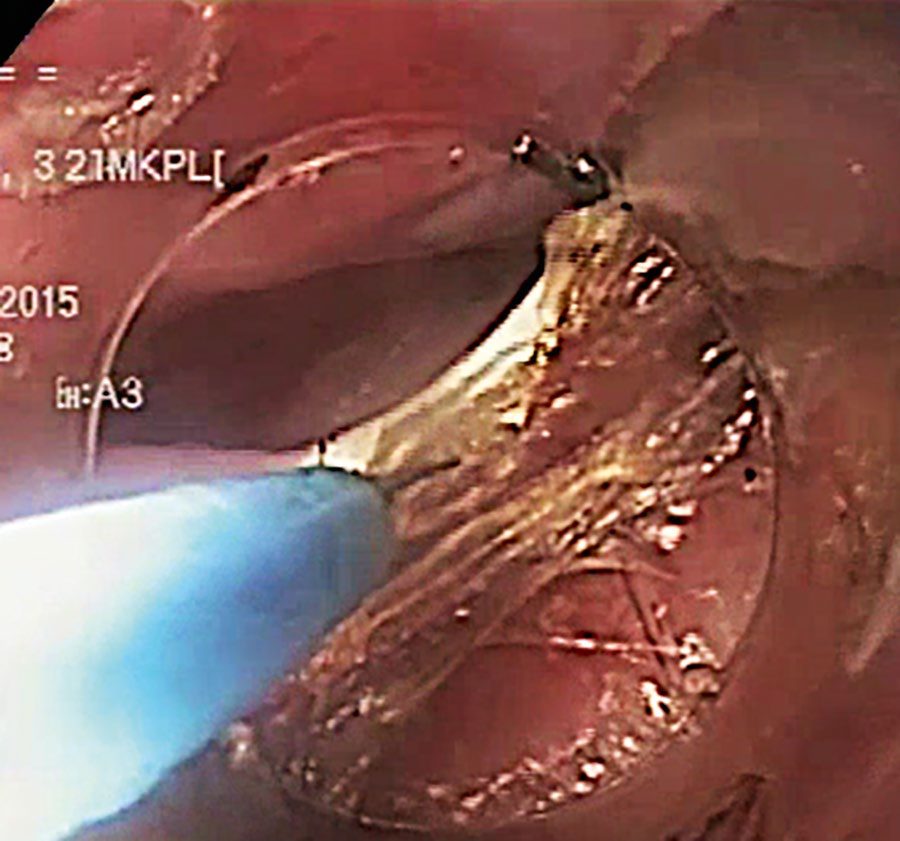
| Technique | Overall morbidity | Mortality | Commonest complications |
|---|---|---|---|
| Rigid, electrocautery | ~8% | 0.2% | Subcutaneous emphysema, mediastinitis, largely replaced by laser and stapling |
| Rigid, CO2 laser | 9.3% | 0.2% | Subcutaneous emphysema, mediastinitis, fistula, bleeding |
| Rigid, linear stapler | 7.1% | 0.3% | Dental injury, oesophageal mucosal injury, oesophageal perforation |
| Flexible endoscopic | 15% | None reported | Subcutaneous cervical emphysema, oesophageal perforation, bleeding |
Table reproduces the reported outcomes by technique [9]. Flexible diverticulotomy can be done under conscious sedation without general anaesthesia or neck extension, which suits patients with comorbidities or anatomy that prevents good exposure, and repeat sessions are common to reduce perforation risk [9].
Selecting between approaches
- Endoscopic diverticulotomy requires adequate oral access and the absence of both neck-mobility limitation and macroglossia; exposure is very limited in patients with a short neck, a short hyomental distance, or severe obesity, leading to a high rate of conversion to open surgery [9].
- It may also produce an incomplete myotomy in diverticula under 3 cm, and some surgeons regard a very large diverticulum as a contraindication, since laser, argon plasma coagulation and diathermy carry higher bleeding risk and multiple staple cartridges raise leak risk [9].
- Comparative studies, none randomised, show endoscopic surgery has shorter operative time, lower morbidity and shorter hospital stay than open surgery with similar symptom relief, but their heterogeneity prevents definitive conclusions, so a tailored approach based on patient selection is advocated [9].
Current evidence on the safety and efficacy of endoscopic stapling of pharyngeal pouch appears adequate to support use of the procedure, provided that normal arrangements are in place for consent, audit and clinical governance [4].
Current evidence on the efficacy and safety of flexible endoscopic treatment of a pharyngeal pouch is adequate to support its use provided normal arrangements are in place for clinical governance, consent and audit, and it should only be done by experienced interventional endoscopists with training in the procedure [5].
The efficacy evidence NICE reviewed for flexible treatment includes a case series of 150 patients in which the mean dysphagia score fell from 1.9 at baseline to 0.3 at one month, maintained at a median 43 months, with no improvement at discharge in 5% (8 of 150); a series of 42 patients of whom 93% had no dysphagia at a mean 38 months; and a series of 31 patients in which 61% were successfully treated by a single procedure, with an intention-to-treat clinical success rate of 84% [5].
Reported safety outcomes include oesophageal perforation in 27% (6 of 22) in one small series, all managed conservatively; macroscopic perforation in 11% (3 of 28) treated by cap-assisted flexible technique, closed immediately with endoclips; suspected perforation in 2% (3 of 150); bleeding in 2% (2 of 125); and infection with fever lasting more than 24 hours in 10% (4 of 41). Specialist advisers considered septicaemia and death to be theoretical adverse events [5].
Technical details of the open and stapled procedures
- The open myotomy can be done under local or general anaesthesia through an incision along the anterior border of the left sternocleidomastoid; the sternocleidomastoid and carotid sheath are retracted laterally and the thyroid, trachea and larynx medially [8].
- A benefit of local anaesthesia is that the patient can swallow to demonstrate the area of persistent narrowing, and gelatin can be fed before closure to confirm that the previously narrowed segment now opens; under general anaesthesia, and when no diverticulum is present to mark the segment, a nasogastric tube placed to the level of the manometrically determined sphincter helps localise the cricopharyngeus [8].
- The myotomy is extended cephalad by dividing 1–2 cm of the inferior constrictor and caudad through the cricopharyngeus and cervical oesophagus for 4–5 cm [8].
- The wound is closed only once all oozing has stopped, because a haematoma is common after this operation and is often associated with temporary dysphagia while it absorbs; oral intake starts the day after surgery and the patient is usually discharged on the first or second postoperative day [8].
- A diverticulum large enough to persist after the myotomy may be sutured in the inverted position to the prevertebral fascia with a permanent suture (diverticulopexy); one so large that it would be redundant if suspended, or with thickened walls, is excised, under general anaesthesia, with a 48 Fr Maloney dilator in the oesophagus after the neck of the pouch has been controlled and the myotomy completed, a linear stapler is placed across the neck and the pouch excised distal to the staple line [8].
- The security of the staple line and the effectiveness of the myotomy can be tested with a water-soluble contrast oesophagogram before discharge [8].
- Endoscopic stapled cricopharyngotomy is most effective for larger pouches (>2 cm) and may be impossible for a small one; each firing of the endoscopic linear stapler divides the common septum over a length of 30 mm and places three rows of staples on each side, so more than one application may be needed depending on the size of the pouch, after which liquids are resumed immediately [8].
Complications
- Postoperative complications after open diverticulectomy include fistula formation, abscess, hematoma, recurrent laryngeal nerve paralysis, phonation difficulties, and Horner syndrome, occurring in about 2–3% of cases; the recurrence rate of the diverticulum is 5–10% [3][13].
- Perioperative complications of cricopharyngeal myotomy include recurrent laryngeal nerve injury, pharyngocutaneous fistula, mediastinitis, and haemorrhage; risks of stapled diverticulectomy specifically include recurrent laryngeal nerve injury, recurrence, and wound haematoma [9].
- Endoscopic stapling/division carries a small risk of perforation or bleeding [3].
Schwartz lists the postoperative complications of the open approach as fistula formation, abscess, haematoma, recurrent laryngeal nerve paralysis, difficulties with phonation and Horner's syndrome, and notes that the incidence of the first two is reduced by performing a diverticulopexy rather than a diverticulectomy [8].
Prognosis
- Recurrence after open diverticulectomy is 5–10% [3].
- Short-term studies of flexible endoscopic diverticulotomy report recurrence rates ranging between 0% and 35%, and long-term studies are needed to understand the role and outcomes of this approach [9].
- Symptom relief rates are similar after open and endoscopic approaches, and because each option carries different advantages and disadvantages, patient selection is the key determinant of satisfactory short- and long-term outcomes [9].
- The recurrence data NICE reviewed for flexible endoscopic treatment show symptom recurrence in 23% (31 of 134) after a median 7 months in the largest series, of whom 23 had a second treatment and 5 a third, leaving one patient symptomatic; 7% (3 of 42) in another series at 12, 22 and 60 months, all improved by re-treatment; and 15% (5 of 34) in a third.
- In a non-randomised comparison, recurrence occurred in 1 patient after flexible treatment and 2 after rigid endoscopic stapling, and re-treatment of the residual bridge relieved symptoms in all three [5].
- The specialist advisers described the key efficacy outcome as resolution or reduction of dysphagia [5].
References
- Bailey & Love's Short Practice of Surgery, 28th ed., Ch. 66, The oesophagus
- Bailey & Love's Short Practice of Surgery, 28th ed., Ch. 52, The pharynx, larynx and neck
- Sabiston Textbook of Surgery, 22nd ed., Ch. 83, Benign Esophageal Disorders
- NICE HealthTech Guidance HTG8: Endoscopic stapling of pharyngeal pouch (2003, migrated from IPG22), 1.1; Overview www.nice.org.uk
- NICE HealthTech Guidance HTG367: Flexible endoscopic treatment of a pharyngeal pouch (2015, migrated from IPG513), 1.1, 1.2; 4.1, 4.2; 4.3; 4.4; 5.1, 5.2, 5.4, 5.6; Overview www.nice.org.uk
- British Society of Gastroenterology: UK guidelines on oesophageal dilatation in clinical practice. Gut 2018, 3.9; 3.9; GRADE of evidence: moderate; strength of recommendation: strong www.bsg.org.uk
- The ABSITE Review, 2022, Ch. Esophagus
- Schwartz's Principles of Surgery, 11th ed., Ch. 25, Esophagus and Diaphragmatic Hernia
- Maingot's Abdominal Operations, 13th ed., Ch. 21, Esophageal Diverticula and Benign Tumors
- Oxford Handbook of Clinical Surgery, 5th ed., Ch. 8, Upper gastrointestinal surgery
- Browse's Introduction to the Symptoms and Signs of Surgical Disease, 6th ed., Ch. 12, The neck
- Sabiston Textbook of Surgery, 22nd ed., Ch. 12
- Schwartz's Principles of Surgery: ABSITE and Board Review, Ch. 25, The Esophagus and Diaphragmatic Hernia