



GERD and Hiatal Hernia
Summary
- Gastro-oesophageal reflux disease (GERD/GORD) is defined by the Montreal definition as a condition that develops when reflux of stomach contents causes troublesome symptoms and/or complications [1].
- It is the most common benign gastroesophageal disorder, closely linked to hiatal hernia, obesity, and esophageal adenocarcinoma, and most patients respond to medical therapy, though surgery has an established role in medically refractory disease [2].
- Hiatal hernias occur when the stomach herniates through the diaphragmatic hiatus into the mediastinum and are classified into four types, with type I (sliding) accounting for 70–95% of cases [1][2].
In the UK, this condition is managed along a defined NHS pathway: primary care screens for red-flag features and trials acid suppression, secondary care confirms pathological reflux objectively, and surgery is offered to a selected minority [3][4].
Definition
- GERD is defined by the Montreal definition, reached by a 44-expert international consensus in 2006, chosen so that patients can be diagnosed independent of the technology used for evaluation, as reflux of gastric contents causing troublesome symptoms and/or complications [1][6].
- Within this definition, nonerosive reflux disease (NERD) is classic GERD symptoms without mucosal complications, and accounts for 30–70% of patients presenting for endoscopy with reflux symptoms [6].
- A hiatal hernia is the presence of part or all of the stomach within the thoracic cavity, usually through protrusion via the oesophageal hiatus in the diaphragm [7].
- GERD may arise if gastroesophageal junction (GEJ) pressure is lower than the transdiaphragmatic pressure gradient; GEJ pressure is not determined by any single structure but by a complex of anatomic features including the intrinsic LES muscle, gastric sling fibers, diaphragmatic crura, and the angle of His [2].
Pathophysiology
- The antireflux mechanism in humans is composed of three components: a mechanically effective lower esophageal sphincter (LES), efficient esophageal clearance, and an adequately functioning gastric reservoir; a defect in any one can lead to increased esophageal acid exposure and mucosal injury [8].
- GERD develops from an imbalance between the transdiaphragmatic pressure gradient (positive abdominal pressure vs. negative thoracic pressure) and the valve mechanism at the EGJ, which comprises the LES, the diaphragmatic crus, the angle of His, mucosal (Gubaroff) valves, and the intra-abdominal esophageal segment [2].
- Hiatal hernias are the most common cause of a defective EGJ valve because they eliminate intra-abdominal esophageal length, obtuse the angle of His, enlarge the hiatus, and cause the diaphragm to pinch the stomach rather than the esophagus; transient LES relaxations (TLESRs) are also more frequent with hiatal hernias [2].
- Hormonally, gastrin and motilin increase LES pressure, while cholecystokinin, estrogen, glucagon, progesterone, somatostatin, and secretin decrease it; drugs such as anticholinergics, calcium channel blockers, caffeine, and theophylline also decrease LES pressure [8].
- Hiatal hernias themselves result from laxity of the diaphragmatic crura and phrenoesophageal ligaments, worsened by high intra-abdominal pressure (obesity, weightlifting) and low intrathoracic pressure (chronic pulmonary disease); they are more common in females and with increasing age [2].
The sphincter, gastric distension and the origin of reflux in the stomach
- The high-pressure zone at the oesophagogastric junction has no distinct anatomical landmarks; architecturally it is a specialised thickening made up of the collar-sling musculature on the greater-curvature side and the clasp fibres on the lesser-curvature side, which remain in tonic opposition until swallowing, and which also open when the fundus distends with gas or liquid, unfolding the valve to vent a belch [9].
- Resistance to reflux is a function of both resting LES pressure and the length over which it is exerted, so a shorter sphincter needs a higher pressure to stay competent; much like the neck of a balloon being inflated, sphincter length falls as the stomach fills, and a sphincter left permanently short by repeated fundal distension from large-volume meals becomes incompetent with minimal further distension [9].
- Normal manometric values (n = 50) are a median pressure of 13 mmHg (2.5th–97.5th percentile 5.8–27.7), overall length 3.6 cm (2.1–5.6) and abdominal length 2 cm (0.9–4.7); a permanently defective sphincter, mean resting pressure under 6 mmHg, overall length under 2 cm or intra-abdominal length under 1 cm, each below the 2.5th percentile, is irreversible, its commonest cause is inadequate abdominal length, and mucosal injury may heal with antisecretory drugs while reflux continues [9].
- Reflux with a normal resting LES pressure is usually a functional problem of gastric emptying or excessive air swallowing: gastric distension raises intragastric pressure and shortens or unfolds the LES, a mechanical explanation for "transient LES relaxation", and with repeated distension the collar-sling musculature attenuates and the physiological venting of gas is replaced by severe postprandial reflux [9].
- Patients then swallow more often to neutralise refluxed acid with saliva (pH 7.0), which increases air swallowing and gastric distension in a vicious cycle of aerophagia, bloating and belching; fatty meals compound the problem by delaying emptying [9].
- As the collar-sling and clasp fibres attenuate the junction assumes an "upside-down funnel" appearance with progressive opening of the angle of His, stretching the phreno-oesophageal ligament, enlarging the hiatus and producing axial herniation; the yield pressure of the sphincter falls as hernia size increases [9].
- Repeated exposure of the squamous epithelium at the distal LES produces inflammation and columnar epithelium at the cardia, carditis, which explains the epigastric pain of early reflux disease and why early oesophagitis is mild and confined to the very distal oesophagus, and the inflammation can form a fibrotic Schatzki ring at the squamocolumnar junction or extend into the muscularis propria with progressive loss of sphincter length and pressure, which is why severe oesophagitis is almost always associated with a defective LES [9].
Duodenal juice and the composition of the refluxate
- Experimental evidence shows that acid alone does minimal damage to the oesophageal mucosa, while acid combined with pepsin is highly deleterious; duodenal juice alone does little harm, but duodenal juice combined with gastric acid is particularly noxious, and maximal injury occurs with bile salts plus acid and pepsin [9].
- Oesophagitis, stricture and Barrett's metaplasia occur in the presence of two predisposing factors (a mechanically defective LES and increased exposure to fluid containing duodenal content) and measurement of oesophageal bilirubin exposure as a marker of duodenal juice shows that 58% of GERD patients have increased duodenal exposure, occurring most commonly when the oesophageal pH is between 4 and 7 [9].
- In a series of 150 consecutive patients with proven GERD, a structurally defective sphincter was found in 42% of those without complications, 77% of those with erosive oesophagitis, 89% with stricture and 100% with Barrett's oesophagus; the finding of a defective sphincter in 42% of uncomplicated patients suggests that a vigorously contracting oesophageal body can compensate until all three components fail [9].
Clinical features
- Typical oesophageal symptoms include heartburn (retrosternal burning) and regurgitation, which are the most common complaints, with regurgitation indicating more severe disease [1][2].
- Symptoms are often provoked by large meals, fatty/spicy foods, recumbency, and bending or heavy lifting [1][10].
- Regurgitation of gastric acid with central chest pain is not seen in cardiac ischaemia and is typical of GERD, whereas breathlessness favours cardiac disease [11].
- Extraoesophageal manifestations include chronic cough, laryngitis, asthma, hoarseness, dental erosions, aspiration pneumonia, and are also associated with sinusitis, pulmonary fibrosis, pharyngitis, and recurrent otitis media [1][7].
- Dysphagia may result from a large hiatal hernia, peptic stricture, or oesophageal carcinoma; bleeding may present as haematemesis from erosive oesophagitis, Cameron ulcers (ischaemic ulcers at the diaphragmatic constriction within large hiatal hernias), or tumour [1].
Hiatal hernia presentations
Sliding hiatal hernias predispose to GERD and may be asymptomatic or found incidentally on chest imaging; true paraesophageal hernias more often cause obstructive symptoms (dysphagia, early satiety, pain, regurgitation, bloating) and may cause dyspnea, cardiac compression, anemia (30–40%, from chronic gastric compression/Cameron ulcers), and rarely acute gastric volvulus [2]. Acute gastric volvulus classically presents with "Borchardt triad": sudden chest/epigastric pain, dry heaving, and inability to pass a nasogastric tube [2].
Chest pain of oesophageal origin
- Nearly 50% of patients with severe chest pain, normal cardiac function and normal coronary arteriograms have positive 24-hour pH studies, and exercise-induced reflux can produce exertional pain indistinguishable from angina [9].
- When Nevens and colleagues tested experienced cardiologists, 48 (26%) of 185 patients thought to have typical angina had normal angiograms and 16 of 63 with "atypical" pain had abnormal ones (the clinical impression was wrong 25% of the time) and among 10,689 patients presenting to an emergency department with acute chest pain, about 17% had acute ischaemia, 6% stable angina, 21% other cardiac causes and 55% non-cardiac causes [9].
- Pain precipitated by meals, occurring at night when supine, non-radiating, responsive to antacids, or accompanied by dysphagia or regurgitation should raise the thought of an oesophageal origin; heartburn occurs monthly in as many as 40–50% of the Western population, and a Gallup poll found 50 million Americans with night-time heartburn at least weekly, 80% of sufferers having nocturnal symptoms and 63% reporting that it affected sleep and next-day work [9].
- Regurgitation from an obstructed junction, as in achalasia, is bland "as if the food had been put into a blender", and most patients can distinguish it from acid regurgitation; bronchospasm can be precipitated by oesophageal acidification and cough by either acid stimulation or distension of the oesophagus [9].
Respiratory manifestations
- Up to 50% of patients with asthma have endoscopic oesophagitis or abnormal distal acid exposure, and GERD may present as laryngopharyngeal symptoms, adult-onset asthma or even idiopathic pulmonary fibrosis, with or without heartburn [9].
- Two mechanisms are proposed: the reflux theory, direct laryngopharyngeal exposure and aspiration, supported by the correlation between idiopathic pulmonary fibrosis and hiatal hernia, more frequent proximal acid exposure on pH testing in patients with respiratory symptoms, scintigraphic aspiration of ingested isotope, and impedance-detected proximal fluid movement correlating with cough, and the reflex theory, a vagally mediated afferent reflex producing bronchoconstriction when acid is infused into the distal oesophagus, reflecting the shared embryological origin of the tracheo-oesophageal tract and vagus; both probably operate together [9].
- Diagnosis traditionally uses dual-probe pH monitoring with a distal and a proximal (tracheal, pharyngeal or proximal oesophageal) probe, but sensitivity is poor, the temporal relation between reflux events and laryngeal or pulmonary symptoms is complex, and refluxate neutralised by saliva as it travels proximally goes undetected, so impedance testing may be added [9].
Etiology
- Risk factors include obesity (increases intra-abdominal pressure and augments the transdiaphragmatic pressure gradient), pregnancy, athletic abdominal straining, hiatal hernia, connective tissue disease, and elevated intra-abdominal pressure from tight garments or chronic straining [1][2].
- An "acid pocket" (an area of unbuffered gastric acid accumulating in the proximal stomach after meals) together with a hiatal hernia can exacerbate GERD severity [1].
- Global GERD incidence is rising, attributed to increasing obesity and declining rates of Helicobacter pylori infection (postulated to reduce oesophageal acid exposure via corpus gastritis/atrophy) [1].
- Male gender, older age, and obesity are established risk factors for GERD and Barrett's oesophagus [7].
- Predisposing factors for hiatal hernia include obesity and previous surgery, and hiatal hernias are more common in females [2][7].
Hiatal hernia epidemiology
- Sliding hernias are seven times commoner than paraoesophageal hernias on radiography for GI symptoms, pure type II hernias are quite rare, and in many cases a type I sliding hernia evolves into a type III mixed hernia as the abdominothoracic pressure gradient enlarges it over time [9].
- The median age is 61 for paraoesophageal against 48 for sliding hernias, and paraoesophageal hernias affect women by 4:1 [9].
- Structural deterioration of the phreno-oesophageal membrane with age, thinning of its upper fascial layer (the supradiaphragmatic continuation of the endothoracic fascia) and loss of elasticity in its lower layer (the continuation of the transversalis fascia), allows it to yield to cranial stretching from persistent intra-abdominal pressure and the tug of oesophageal shortening on swallowing; the stretching occurs more anteriorly and posteriorly, with the left crus fixed to the stomach at the 3 o'clock position, creating anterior and posterior sacs, the latter often filled with epiphrenic and retroperitoneal fat [9].
- Barrett's oesophagus, first described by Norman Barrett in 1950 (who wrongly believed it congenital), occurs in 10–15% of GERD patients and is the end stage of the disease; it was traditionally defined as columnar mucosa extending at least 3 cm into the oesophagus but is now diagnosed on any length of endoscopically visible columnar mucosa showing intestinal metaplasia with goblet cells on biopsy, the only tissue predisposed to malignant degeneration, and patches of intestinal metaplasia at the cardia appear to carry similar malignant potential as precursors of cardia carcinoma [9].
- Barrett's patients usually have markedly increased acid exposure, deficient LES characteristics, poor body function and a high prevalence of duodeno-gastro-oesophageal reflux, and gastric hypersecretion occurs in 44% [9].
Diagnosis
Endoscopy and contrast studies
- Barium swallow evaluates hiatal hernias and obstructing lesions and can identify reflux events but cannot alone establish the diagnosis of GERD [2].
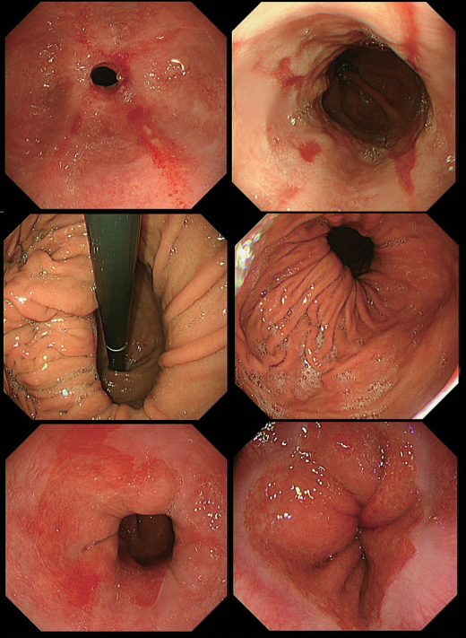
- Upper endoscopy is essential before antireflux surgery, establishing Barrett esophagus or reflux esophagitis and excluding malignancy; LA grade C/D esophagitis, peptic strictures, and Barrett esophagus are considered pathognomonic for GERD [2].
- The Los Angeles (LA) classification grades oesophagitis from A (mucosal breaks ≤5 mm not extending between mucosal folds) to D (mucosal breaks involving ≥75% of the circumference) [8].
- Hiatal hernia is typically identified on EGD, CT, or barium swallow; CT is the investigation of choice in acute presentations [2][7].
- Retroflexing the endoscope after passage through the GEJ allows the gastroesophageal flap valve to be graded by the Hill classification, from Grade I (a well-defined ridge of tissue closely wrapping the endoscope shaft) to Grade IV (no muscular ridge at all, the valve open at all times, with a hiatal hernia always present); an increasing Hill grade correlates with a more mechanically defective sphincter, abnormal acid exposure, erosive esophagitis, and Barrett's oesophagus [6].
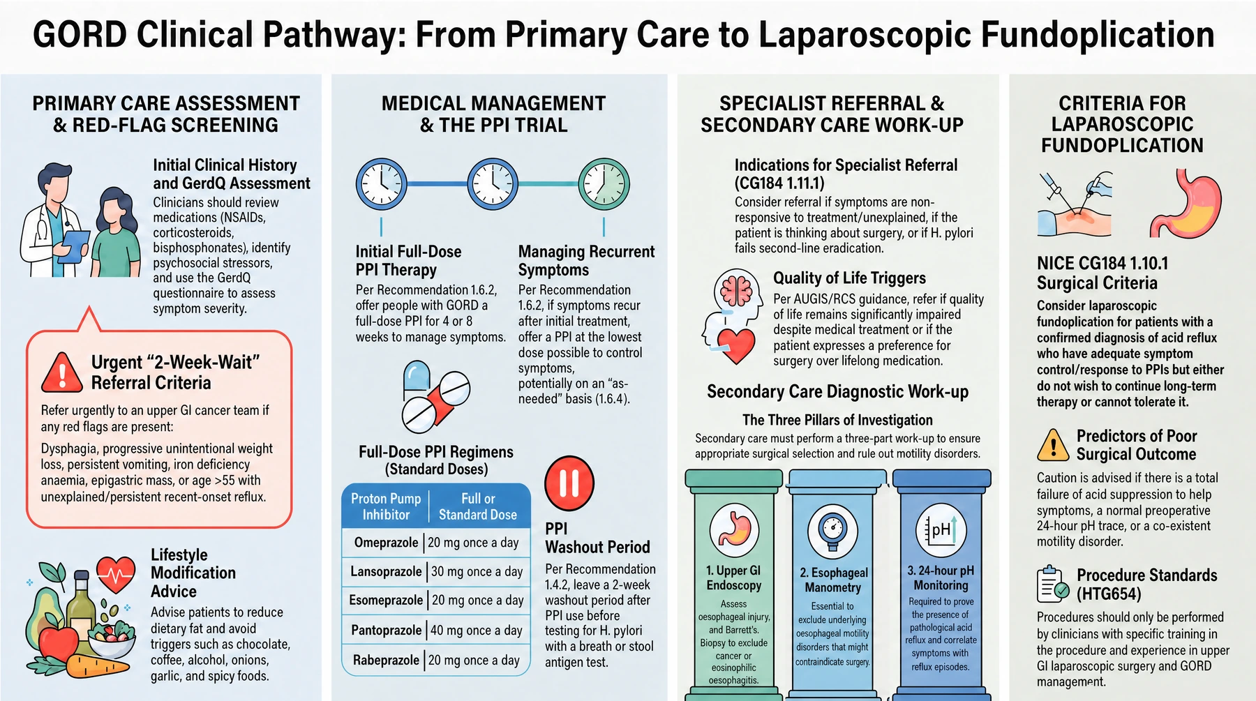
UK assessment before any endoscopy referral includes a review of all current medication for possible causes of dyspepsia (for example NSAIDs, corticosteroids, bisphosphonates, nitrates, theophyllines), identification of psychological and social stressors, and physical examination to rule out an upper abdominal mass [3][4]. Cardiac and biliary disease should be considered as part of the differential [3].
Refer immediately (same day) any person with dyspepsia and significant acute gastrointestinal bleeding [3]. Refer urgently to a team specialising in upper GI cancer if any of the following are present [4]:
- Dysphagia, progressive unintentional weight loss, or persistent vomiting
- Dyspepsia or reflux with iron-deficiency anaemia, or a chronic gastrointestinal bleed
- An epigastric mass, or a suspicious barium meal
- Age over 55 years with unexplained, persistent (>4–6 week) recent-onset reflux
- Worsening reflux with known Barrett's oesophagus, atrophic gastritis, intestinal metaplasia, dysplasia, previous peptic ulcer surgery, or a family history of upper GI cancer in more than two first-degree relatives
NICE does not recommend routinely offering endoscopy to diagnose Barrett's oesophagus, but it may be considered in a person with GORD, discussing their preferences and individual risk factors such as long symptom duration, increased symptom frequency, previous oesophagitis, previous hiatus hernia, oesophageal stricture or ulcers, or male gender [3].
Physiological testing
- Ambulatory pH monitoring is the gold-standard test for diagnosing GERD, typically performed off acid suppression, and calculates the DeMeester score from six parameters (number of reflux episodes, % total/upright/supine time pH<4, number of episodes ≥5 minutes, longest episode) [2].
- The 24-hour ambulatory pH monitoring is described as the most direct method of measuring esophageal gastric juice exposure [8].
- Ambulatory pH monitoring can be performed with a transnasal catheter, a wireless esophageal mucosal capsule, or intraluminal impedance measurements.
- Catheters carry multiple sensors and are cheaper and more dependable but less comfortable, while wireless capsules are more tolerable and allow monitoring beyond 24 hours at the cost of higher expense, possible chest pain, and data loss from radiofrequency interference or premature detachment [2].
- Clinical guidelines generally reserve pH monitoring for when GERD is not proven by endoscopic findings or after empirical pharmacologic treatment has failed [2].
- Esophageal manometry identifies achalasia and motility disorders before antireflux surgery but cannot itself diagnose GERD; it is unreliable in large paraesophageal hernias [2].
- A mechanically defective LES on manometry is defined by an average pressure of 6 mmHg or less, an average overall length of 2 cm or less, and an intra-abdominal length of 1 cm or less, though a hypotensive LES is not essential for a GERD diagnosis, and many patients with pathologic reflux have a normotensive sphincter [6].
- Pathologic acid exposure time (AET) is generally defined as esophageal pH <4 for greater than 6% of monitored time; the ABSITE Review states pH-probe positivity as >4.5% of total time with pH<4, and resting LES pressure <6 mmHg suggests GERD [12].
- Diagnosis is often assumed and treated empirically with PPIs, but a positive treatment response occurs in 69% of true GERD patients versus 51% of those without GERD by objective testing, risking overdiagnosis [1].
Diagnosing and classifying a hiatal hernia
- An upright chest radiograph diagnoses a hiatal hernia if it shows an air–fluid level behind the cardiac shadow, usually from a paraoesophageal hernia or intrathoracic stomach [9].
- Barium study is more accurate for a paraoesophageal than a sliding hernia because the latter often reduces spontaneously, whereas a paraoesophageal hernia is a permanent herniation and barium swallow provides the diagnosis in virtually every case, attention being paid to the position of the junction [9].
- On retroflexed endoscopy a sliding hernia is identified by a gastric pouch lined with rugal folds extending above the crural impression, or at least 2 cm between the crura (identified by asking the patient to sniff) and the squamocolumnar junction on withdrawal; a paraoesophageal hernia by a separate orifice adjacent to the junction into which rugal folds ascend; and a mixed hernia by a rugal-lined pouch above the diaphragm with the junction entering about midway up its side [9].
- A group of patients with sliding hernias, no reflux disease and dysphagia without endoscopic or manometric explanation have obstruction of the bolus by diaphragmatic impingement on the herniated stomach, seen on video barium studies and reflected manometrically by a double-humped high-pressure zone, the first rise from diaphragmatic impingement, the second from the true sphincter; impingement can propel supradiaphragmatic gastric contents into the pharynx, so the condition is often confused with GERD, and surgical reduction relieves the dysphagia in 91% [9].
Scoring and Severity
- The DeMeester score, calculated from ambulatory pH monitoring parameters, correlates clinically with GERD severity and pattern (supine, upright, or combined reflux) [2].
- The Los Angeles classification grades severity of reflux esophagitis from A (mildest) to D (most severe, ≥75% circumferential mucosal breaks) [7][8].
- The American Foregut Society Endoscopic Classification of Esophagogastric Junction Integrity (2023) grades hiatal integrity 1–4 based on hiatal axial length, hiatal aperture diameter, and presence/absence of the flap valve [2].
| Parameter | Threshold / range | Clinical significance |
|---|---|---|
| Acid exposure time (AET) | pH <4 for >6% of monitored time (>4.5% per ABSITE Review 2022) | Defines pathologic acid reflux on ambulatory pH monitoring |
| Resting LES pressure | ≤6 mmHg | One of three criteria for a mechanically defective LES |
| LES overall length | ≤2 cm | One of three criteria for a mechanically defective LES |
| LES intra-abdominal length | ≤1 cm | One of three criteria for a mechanically defective LES |
| LA Grade A oesophagitis | Mucosal breaks ≤5 mm, not extending between folds | Mildest endoscopic grade |
| LA Grade C/D oesophagitis | Breaks bridging folds (C) or involving ≥75% of the circumference (D) | Considered pathognomonic for GERD |
| DeMeester score | Composite of 6 pH-monitoring parameters | Correlates with GERD severity and reflux pattern (supine/upright/combined) |
| Hill grade I–II flap valve | Well-defined ridge, closes around the endoscope | Lower correlation with a defective sphincter, acid exposure, and erosive disease |
| Hill grade III–IV flap valve | Ridge barely present or absent; valve open at rest | Higher correlation with a defective sphincter, abnormal acid exposure, erosive esophagitis, and Barrett's oesophagus |
Table compiled from the diagnostic and severity criteria above [2][6][8][12].
UK primary care is advised to assess symptom severity with the GerdQ questionnaire, and to perform it before onward referral, since it is also useful in post-operative follow-up [4].
Treatment and Management
Lifestyle and medical therapy
- Lifestyle measures include weight loss, avoiding dietary triggers, elevating the head of the bed, and smoking/alcohol cessation [2][7].
- PPIs are the most potent antisecretory therapy, achieving symptom control and >90% mucosal healing of esophagitis after an 8-week course; AGA guidelines recommend a 4–8 week PPI trial, escalating to twice-daily dosing before further testing [1][2].
- H2-receptor antagonists are useful for breakthrough symptoms but are limited by tachyphylaxis [2].
- Long-term PPI use carries a modestly increased risk of chronic kidney disease progression, Clostridioides difficile infection, and community-acquired pneumonia [2].
- Most patients are treated empirically with PPIs (99% effective per the ABSITE review), with escalating doses over 3–4 weeks before pursuing further diagnostic studies if refractory [12].
NICE advises simple lifestyle advice on healthy eating, weight reduction, and smoking cessation, and advises avoiding known precipitants (smoking, alcohol, coffee, chocolate, fatty foods, and being overweight) noting that raising the head of the bed and taking the main meal well before bedtime may help some people [3]. The RCS/AUGIS guide adds decreasing dietary fat, avoiding recumbency for three hours after meals, and raising the head of the bed by 20 cm, while acknowledging that the evidence supporting this advice is weak [4].
Drug therapy in the UK escalates stepwise:
- Alginate–antacid combinations and H2RAs are useful for mild heartburn [4].
- Offer a full-dose PPI for 4 or 8 weeks; if symptoms recur, offer a PPI at the lowest dose that controls symptoms, and discuss "as-needed" self-management [3].
- If response to a PPI is poor, consider doubling the dose, with reassessment at two to three months with or without endoscopy [4].
- Offer H2RA therapy if there is an inadequate response to a PPI [3].
- People who have had dilatation of an oesophageal stricture should remain on long-term full-dose PPI therapy [3].
- Offer an annual review to people needing long-term management, encouraging them to step down or stop treatment unless an underlying condition or comedication requires it [3].
For severe oesophagitis, offer a full-dose PPI for 8 weeks to heal, and a full-dose PPI long-term as maintenance, taking into account the person's preference, tolerability, comorbidities, drug interactions, and acquisition cost; if initial treatment fails, consider a high dose of the initial PPI or switching to another PPI at full or high dose [3]. NICE specifies these doses for severe oesophagitis:
| PPI | Full / standard dose | Low (on-demand) dose | High / double dose |
|---|---|---|---|
| Esomeprazole | 40 mg once daily | 20 mg once daily | 40 mg twice daily |
| Lansoprazole | 30 mg once daily | 15 mg once daily | 30 mg twice daily † |
| Omeprazole | 40 mg once daily | 20 mg once daily | 40 mg twice daily |
| Pantoprazole | 40 mg once daily | 20 mg once daily | 40 mg twice daily † |
| Rabeprazole | 20 mg once daily | 10 mg once daily | 20 mg twice daily † |
Doses marked † are off-label for GORD [3].
Indications for intervention
Surgical indications include incomplete symptom control on medical therapy, intolerance of/unwillingness to continue long-term medication, regurgitation refractory to PPI, large hiatal hernia, GERD complications, and extraesophageal symptoms [1][12]. Endoscopic options include transoral incisionless fundoplication (TIF), the MUSE system, and Stretta radiofrequency ablation, though guidelines currently favor surgical fundoplication over these for most patients [1][2].
Consider referral to a specialist service for people of any age with gastro-oesophageal symptoms that are non-responsive to treatment or unexplained, or with suspected GORD who are thinking about surgery [3]. The RCS/AUGIS threshold is a significantly impaired quality of life with persistent symptoms despite medical treatment and lifestyle modification, or a patient preference for surgery over long-term medication [4].
NICE recommends considering laparoscopic fundoplication for people who have a confirmed diagnosis of acid reflux and adequate symptom control with acid suppression therapy but do not wish to continue that therapy long term, or a confirmed diagnosis of acid reflux with symptoms responding to a PPI but who cannot tolerate acid suppression therapy [3]. RCS/AUGIS indications for surgery additionally include volume reflux (especially affecting sleep or provoked by stooping), breakthrough heartburn despite optimal medical therapy, PPI intolerance, post-prandial chest pain or dysphagia from an incarcerated paraoesophageal hernia, and pH-confirmed atypical symptoms such as aspiration, cough, or hoarse voice [4].
Before deciding on intervention, secondary care should confirm pathological reflux objectively with upper GI endoscopy (assessing oesophageal injury, diagnosing Barrett's, and biopsying to exclude eosinophilic oesophagitis or malignancy) plus oesophageal manometry and 24-hour pH monitoring to exclude an underlying motility disorder [4].
Appropriate patient selection is the most important determinant of a good outcome. An adverse outcome is more likely where [4]:
- Acid suppression has failed to make any difference to symptom control, classical and volume reflux symptoms should be at least partially helped by acid suppression
- The pre-operative 24-hour pH tracing is normal
- There is a co-existent oesophageal motility disorder
- There is gastroparesis, or significant symptoms suggestive of irritable bowel syndrome
- Symptoms are atypical, this group has a lower success rate than classical or volume reflux
What medical therapy can and cannot achieve
- Patients first identified with mild uncomplicated symptoms can be given 12 weeks of simple antacids before diagnostic testing, together with head-of-bed elevation, loose clothing, small frequent meals, no meal immediately before bed, and avoidance of alcohol, coffee, chocolate and peppermint, which reduce resting LES pressure [9].
- Alginic acid reacts with sodium bicarbonate in saliva to form a viscous solution that floats like a raft on the gastric contents and is refluxed ahead of them as a protective barrier; prokinetics such as metoclopramide or domperidone help in early disease but are of little value in severe disease [9].
- High-dose PPI therapy (omeprazole up to 40 mg/day) reduces gastric acidity by 80–90% and usually heals mild oesophagitis, but severe oesophagitis heals in only about half; in patients who reflux mixed gastric and duodenal juice, acid suppression can relieve symptoms while mixed reflux continues to damage the mucosa of an asymptomatic patient [9].
- Within 6 months of stopping any medical therapy 80% of patients relapse, 40% of those with daily GERD eventually develop symptoms that break through adequately dosed PPIs, and most therefore need lifelong treatment [9].
- Castell, Triadafilopoulos and others showed that 40–80% of Barrett's patients continue to have abnormal acid exposure despite PPIs at up to 20 mg twice daily, and ablation trials needed mean omeprazole doses of 56 mg to normalise 24-hour pH; antireflux surgery eliminates acid and duodenal reflux more reliably, although as many as 25% of post-fundoplication patients have persistent pathological acid exposure on pH testing [9].
- Failure of medication suggests either relatively severe disease or a non-GERD cause; endoscopy at this stage assesses mucosal injury and Barrett's, and the choice then lies between long-term PPIs and antireflux surgery, which gives long-term symptom control in 85–90%, with 24-hour pH monitoring and manometry undertaken when surgery is considered [9].
For reflux-induced asthma, relief of respiratory symptoms can be anticipated in up to 50% of patients on antisecretory drugs, but objectively fewer than 15% improve their pulmonary function; in properly selected patients antireflux surgery improves respiratory symptoms in nearly 90% of children and 70% of adults, with measurable improvement in pulmonary function in around 30%, and the two randomised trials of medical against surgical therapy indicate that surgical valve reconstruction is the most effective treatment, its superiority most noticeable supine when PPI blood levels are at their nadir and asthma symptoms at their worst [9]. An antireflux operation will not prevent regurgitation and aspiration of swallowed food "upstream" of the valve in an asthmatic with an oesophageal motility disorder, so body function must be assessed before surgery [9].
Selection for surgery in Schwartz's terms
- Approximately 25–50% of GERD patients have persistent or progressive disease and it is they who are best suited to surgery; the key indications are objectively proven reflux, typical symptoms (heartburn and/or regurgitation) despite adequate medical management, or a younger patient unwilling to take lifelong medication [9].
- A good response to medical therapy predicts an excellent surgical outcome, whereas patients with atypical symptoms respond less predictably; patients with a structurally defective LES often relapse within 1–2 years of starting medical therapy and should be considered for surgery regardless of endoscopic oesophagitis, and a Veterans Administration cost analysis found surgery cheaper than medical therapy in patients under 49 [9].
- A stricture represents failure of medical therapy: malignancy and a drug-related cause are excluded, the stricture is progressively dilated to a 50–60 Fr bougie, and if dysphagia is relieved and manometry shows adequate distal contraction amplitude an antireflux procedure follows, with a partial fundoplication preferred when there is global loss of contractility [9].
- Barrett's oesophagus should be regarded as evidence of reflux, surgery is indicated for the treatment of symptoms, not cancer progression, since the evidence that fundoplication reduces progression is relatively weak, while high-grade dysplasia or intramucosal cancer redirects treatment to endoscopic ablation, endoscopic resection or oesophagectomy [9].
- Drug-induced strictures from lodgement of a capsule or tablet in the distal oesophagus should be distinguished from reflux strictures by documenting reflux; a long, string-like stricture from repeated pill injury on top of a reflux stricture is often resistant to dilatation, though this has become rarer since PPIs [9].
Surgeries
Principles and fundoplication technique
- Antireflux operations have three essential components: restoration of an intra-abdominal esophageal segment, crural repair, and reinforcement of the LES by fundoplication or a prosthesis [1].
- Laparoscopic Nissen (360°, complete) fundoplication mobilizes the fundus behind the esophagus over a large bougie (52–60 Fr) and secures it anteriorly, restoring the GE junction with at least 2–3 cm of intra-abdominal esophageal length and approximating the crura with permanent suture [2][12].
- Complete fundoplication is more durable for reflux control but carries a higher incidence of short-term dysphagia; partial fundoplications (posterior Toupet or anterior Dor) have fewer short-term side effects at the cost of a slightly higher long-term failure rate, and multisociety 2023 guidelines conditionally favor partial fundoplication due to less hiatal hernia recurrence, dysphagia, and inability to belch [1][2].
- The key dissection maneuver is identification of the right crus, and the key maneuver for the wrap is identification of the left crus [12].
- The Belsey Mark IV repair is performed through the chest [12].
- Collis gastroplasty (esophageal lengthening) uses linear staplers to create a neo-esophagus when insufficient intra-abdominal esophageal length can be achieved [2][12].
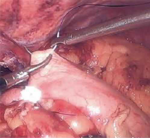
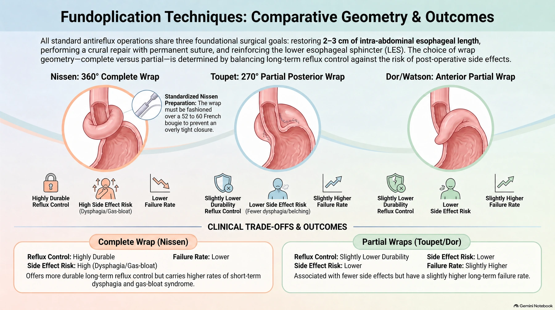
- Laparoscopic anti-reflux surgery can be performed as a day case or with a short inpatient admission, and has two components: repairing the hiatus (which fixes a hiatus hernia) and fundoplication, wrapping the gastric fundus around the lower oesophagus to create a sling barrier to reflux [4].
- The commissioned procedures are Nissen 360° fundoplication, Watson partial anterior fundoplication, and Toupet partial posterior fundoplication (each with hiatal repair) plus gastropexy with hiatus hernia repair [4].
- The guide explicitly declines to recommend any particular type of fundoplication over another, noting the approach depends on the training and personal experience of the operating surgeon [4].
- Where a large, possibly obstructing paraoesophageal hernia has prolapsed much of the stomach into the chest (which tends to occur in the elderly) it is sometimes only necessary to repair the diaphragmatic defect and fix the stomach in the abdomen by gastropexy, without a fundoplication [4].
- Quality specifications for providers are a median length of stay of two days and a demonstrated day-case rate for anti-reflux procedures [4].
Magnetic sphincter augmentation
Magnetic sphincter augmentation (Linx) is a titanium/magnetic bead ring placed around the esophagus, FDA-approved in 2012, indicated for grade A/B esophagitis, hiatal hernia <3 cm, BMI <35, and adequate motility; it is not indicated with paraesophageal hernia, grade C/D esophagitis, or Barrett esophagus [1][2].
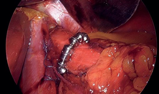
- NICE considers the evidence on the safety and efficacy of laparoscopic insertion of a magnetic ring for GORD adequate to support using the procedure, provided standard arrangements are in place for clinical governance, consent, and audit.
- Patient selection and the procedure should be done by clinicians with specific training in the procedure and experience in upper gastrointestinal laparoscopic surgery and managing GORD [5].
- This guidance replaced the earlier interventional procedures guidance IPG585 and was migrated from IPG749 without change [5].
Note that the 2013 RCS/AUGIS commissioning guide predates this: at that time Linx, along with Stretta endoscopic microwave ablation, EsophyX endoscopic plication, and EndoStim electrical LES stimulation, was placed under research regulation or restricted to long-term registry follow-up and was not recommended for commissioning outside registered research [4].
Hiatal hernia repair
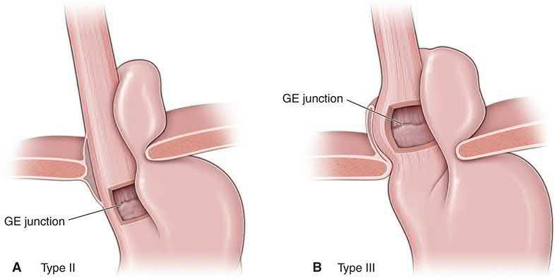
Hiatal hernia types are: Type I (sliding, 70–95% of cases, GEJ migrates into mediastinum); Type II (fundus migrates, GEJ intra-abdominal, uncommon "rolling"/paraesophageal); Type III (mixed, GEJ and stomach both herniated); Type IV (stomach plus another organ, e.g., colon, spleen, herniated) [1][2][12].
| Type | Frequency | Description |
|---|---|---|
| I, Sliding | 70–95% of cases | GEJ migrates into the mediastinum |
| II, Rolling / paraesophageal | Uncommon | Fundus migrates into the chest; GEJ remains intra-abdominal |
| III, Mixed | — | Both the GEJ and stomach are herniated |
| IV, Complex | — | Stomach plus another organ (e.g. colon, spleen) herniated |
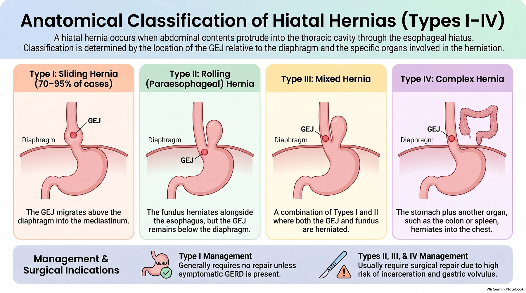
- [1][2][12] Type I hernias generally do not require repair unless GERD is present; Types II–IV generally require repair given risk of incarceration/volvulus, with mobilization and excision of the hernia sac to reduce recurrence, ± mesh for large defects [12].
- For paraesophageal hernia repair, evidence does not support routine biologic or permanent mesh use, an initial randomized trial showed reduced short-term recurrence with mesh, but 5-year follow-up and subsequent RCTs showed no benefit, and mesh carries risk of long-term complications including esophageal erosion [2].
- Obese patients (BMI >35) with GERD may be better served by Roux-en-Y gastric bypass rather than fundoplication as the antireflux procedure of choice, per a SAGES Foregut Task Force White Paper, given superior durability and treatment of metabolic comorbidity, though a subsequent consensus guideline recommends RYGB only (not a choice) for BMI >50 [2].
- Emergency management of acute gastric volvulus starts with nasogastric decompression (endoscopically if needed) to convert an emergent operation to a semi-elective one, followed by reduction and hernia repair; some surgeons defer fundoplication and instead perform gastropexy in the unstable or emergency setting [1][2].
Five principles of antireflux reconstruction
- Schwartz sets out five principles that any repair must satisfy [9].
- First, the operation should create a flap valve that raises distal sphincter pressure, after a Nissen to about twice resting gastric pressure (12 mmHg for a gastric pressure of 6 mmHg), less after a partial wrap, with a reconstructed valve at least 3 cm long in every type, which both augments a deficient sphincter and prevents unfolding of a normal one on gastric distension; the pressure rise is a function of the degree of wrap, but efficacy depends on the close apposition of fundus and oesophagus, not the tightness of the wrap [9].
- Second, it should place an adequate length of sphincter in the positive-pressure abdomen, permanent restoration of 2 cm or more of abdominal oesophagus, all popular procedures adding an average of at least 1 cm [9].
- Third, the reconstructed cardia must relax on deglutition, a vagally mediated relaxation lasting about 10 seconds: only the fundus should be used because it relaxes in concert with the sphincter, the wrap must not incorporate or encircle the body of the stomach, which does not relax, and the vagi must not be damaged [9].
- Fourth, the wrap must not raise the resistance of the relaxed sphincter above the peristaltic power of the oesophageal body, so a 360° wrap should be no longer than 2 cm and built over a large 50–60 Fr bougie, which is unnecessary for a partial wrap [9].
- Fifth, the fundoplication must sit in the abdomen without undue tension and be kept there by crural approximation above it, a wrap left in the thorax converts a sliding hernia into a paraoesophageal hernia, and a wrap under tension predisposes to recurrence [9].
- Rudolph Nissen described a 360° wrap over 4–5 cm of lower oesophagus without dividing the short gastric vessels; its side effects prompted the modifications now standard, fundus only, in a fashion analogous to a Witzel jejunostomy, sized over a 50–60 Fr bougie, 1–2 cm long, with or without short-gastric division [9].
- The essential elements are hiatal dissection with preservation of both vagi along their whole length, circumferential oesophageal mobilisation, hiatal closure (usually posterior) and a short floppy wrap over a dilator; five ports are used, dissection begins in the gastrohepatic omentum above and below the hepatic branch of the anterior vagus, which is usually preserved, the pillars are approximated with interrupted non-absorbable sutures from posterior to anterior, and the wrap is secured with either a single 2-0 polypropylene U-stitch buttressed with felt pledgets or 2–4 interrupted sutures, the first passed through the left fundic limb, the oesophagus 2.5 cm above the junction and the right limb [9].
- The Toupet wrap is a 270° posterior fundoplication around the distal 4 cm anchored posteriorly to the hiatal rim; the anterior 180° partial fundoplication rolls the anterior fundus over the front of the oesophagus and sutures fundus and oesophagus to the right hiatal rim, stabilising 3–4 cm of intra-abdominal oesophagus and never requiring short-gastric division, with excellent outcomes in about 90% at 10 years [9].
- When a short oesophagus is encountered a Collis gastroplasty staples the cardia and upper stomach parallel to the lesser curvature to create a gastric tube in continuity with the oesophagus, lengthening it by several centimetres, with the highest Nissen suture placed on native oesophagus, though its critics question the incidence of short oesophagus and the wisdom of leaving an amotile, acid-secreting gastric tube above the wrap [9].
What the randomised trials of technique show
- Six randomised trials totalling 438 patients found no difference in dysphagia or recurrent reflux with or without division of the short gastric vessels, but the three largest showed more flatulence, bloating and difficulty belching after division, and Engstrom's pooled analysis of 201 Australian and Swedish patients with 12-year follow-up in 170 confirmed equivalent reflux control with more bloating after division, the trials suggest division adds complexity and worsens outcome [9].
- Eleven trials comparing Nissen with posterior partial fundoplication consistently show equivalent reflux control with fewer wind-related side effects after the partial wrap, but less dysphagia in only 2 of 11: Lundell's 137 patients followed to 18 years had similar reflux control and dysphagia, more flatulence and more revision surgery (mainly for paraoesophageal herniation) after Nissen, and success above 80% for both with the mechanical side effects of Nissen improving progressively over time; Strate's 200 patients showed 85% satisfaction in each arm at 2 years but dysphagia in 19 versus 8 patients after Nissen, and subgroup analyses in three trials found no difference between patients with and without poor preoperative motility [9].
- Four trials of Nissen against anterior 180° fundoplication (Watson 107 patients, Baigrie 161, Cao 100, Raue 64) showed equivalent reflux control with less dysphagia and wind at up to 5 years, differences that disappeared at 10 years in Watson's trial as dysphagia declined; two trials of the anterior 90° variant found fewer side effects offset by slightly more recurrent reflux; and two trials of anterior against posterior partial wraps (Hagedorn 95 patients, Khan 103) found better reflux control but more side effects with the posterior wrap [9].
- Overall, laparoscopic fundoplication relieves typical symptoms in more than 90% at 2–3 years and 80–90% at 5 years or more, normalises pH tracings in over 90%, but relieves atypical symptoms (cough, asthma, laryngitis) in only two-thirds [9].
Barrett's oesophagus after antireflux surgery
- Symptomatic outcome in Barrett's patients is excellent to good in 72–95% at 5 years across the reported series [9].
- In Parrilla's randomised trial of 101 patients over 18 years with a median 6-year follow-up, omeprazole 20 mg twice daily and open Nissen fundoplication gave nearly identical symptomatic outcomes, but oesophagitis and/or stricture persisted in 20% of the medical group against 3–7% after surgery; about 15% had abnormal acid exposure after surgery, while of 12 medically treated patients monitored on treatment 3 (25%) had persistently high acid exposure and most (75%) persistently high bilirubin exposure [9].
- Bright and associates found about 50% regression in Barrett's length in 20 patients in the control arm of an ablation trial, so the belief that Barrett's cannot regress after surgery may be incorrect, but patients should remain under endoscopic surveillance with biopsies reviewed by an expert pathologist; confirmed low-grade dysplasia is re-biopsied after 12 weeks of high-dose acid suppression, high-grade dysplasia or intramucosal cancer on more than one specimen escalates to EMR, ablation or resection, and radiofrequency ablation halved short-term progression from high-grade dysplasia to invasive cancer in a randomised trial [9].
Paraoesophageal hernia repair in detail
- The classic report of Skinner and Belsey, in which 6 of 21 patients with a paraoesophageal hernia treated medically for minimal symptoms died of strangulation, perforation, exsanguinating haemorrhage or acute dilatation of the intrathoracic stomach, mostly without warning, underpinned the traditional advice to repair every one; more recent data are less alarming, Allen followed 23 patients for a median 78 months with only four worsening and a single death from aspiration during a barium study, and only three strangulations in 735 patient-years, though emergency repairs needed a median 48-day stay against 9 days for elective repair [9].
- Elective repair carries mortality below 1% whereas emergency repair is high-risk, so symptomatic patients are counselled to elective repair while watchful waiting of an asymptomatic hernia may be acceptable, the lifetime risk of incarceration or strangulation being under 5% [9].
- The transabdominal route facilitates reduction of the associated volvulus and laparoscopy has become the standard, but a pure type II or mixed type III repair is an order of magnitude harder than a Nissen and best avoided until considerable laparoscopic antireflux experience has accrued: vertical and horizontal volvulus obscures the anatomy, dissection of a large sac bleeds if the surgeon leaves the plane between peritoneal sac and endothoracic fascia, and the redundant epiphrenic fat pad and sac at the junction frustrate the wrap and must be excised [9].
- Primary crural closure with permanent suture recurs in 10–40%; randomised trials show mesh reinforcement reduces recurrence, but non-absorbable synthetic mesh must not be placed in keyhole fashion because of oesophageal or gastric erosion and infection, and the only randomised trial of biological mesh (acellular porcine or human dermis, porcine small-intestinal submucosa) failed to show superiority over suture at 5 years despite its far greater cost [9].
- Most surgeons add a fundoplication routinely because pH monitoring shows abnormal acid exposure in 60–70% of paraoesophageal hernias (nearly identical to the 71% in sliding hernias) symptoms bear no relation to the competence of the cardia, and dissection of the junction can itself cause reflux; surprisingly, peristalsis is normal in 88% despite the oesophagus being "accordioned" into the chest, and the incompetent cardia in these patients typically has normal pressure but shortened overall length displaced out of the abdomen [9].
- A short oesophagus, defined as failure to achieve 2.5 cm of intra-abdominal oesophagus with standard mediastinal dissection, accompanies 5–20% of giant hernias, accounts for 10–20% of repair failures, cannot be reliably predicted preoperatively and is diagnosed definitively only in the operating room; the laparoscopic stapled wedge Collis gastroplasty is the current lengthening technique, with the first fundoplication suture on the oesophagus immediately above the junction so that no acid-secreting mucosa lies above the wrap [9].
- Symptomatic improvement follows in 80–90% with symptomatic recurrence under 10–15%, but radiological recurrence is detected in 25–40% after giant hernia repair (against 5–10% after Nissen for GERD), and an asymptomatic recurrent hernia, like an asymptomatic primary one, does not need repair [9].
Complications
- Short-term surgical complications include mortality approaching 0.1–0.5%, and roughly 1% each of bleeding, infection, and gastric/esophageal perforation [1][2].
- Dysphagia occurs in up to 50% of patients in the first months after antireflux surgery, typically self-limited as edema resolves, but persistent dysphagia beyond 3 months warrants barium swallow evaluation for hiatal herniation, an overly tight closure, or malformed fundoplication [2][12]. "Gas-bloat syndrome" (abdominal distension with inability to belch or vomit) occurs more often after complete (Nissen) than partial fundoplication [1].
- The Horgan classification describes fundoplication failure types: Type IA (herniation of GEJ and wrap, causes heartburn ± dysphagia), Type IB (GEJ herniation alone), Type II (paraesophageal herniation of the wrap), and Type III (malpositioned wrap) [2].
- Long-term reoperation rates after fundoplication are 5–7%, chiefly for recurrent GERD or persistent dysphagia, with patient satisfaction falling from 91% at primary surgery to 76% at first reoperation and 49% at second reoperation [2].
- Radiographic recurrence after large paraesophageal hernia repair is approximately 30%, though most are small sliding recurrences not requiring reoperation [2].
- Magnetic sphincter augmentation carries risk of device erosion into the esophagus (0.3–1%, typically 1–4 years after placement) and device removal in 2.7–3.3% of patients [1][2].
- Complications from paraesophageal hernia include acute gastric volvulus with risk of ischemia and perforation, and chronic gastric compression can cause Cameron ulcers and anemia [2].
UK figures give laparoscopic anti-reflux surgery a low mortality of under 0.3%, with most deaths ascribed to postoperative cardiac events [4]. Complications are divided into immediate and delayed, the former being much rarer but tending to require operative intervention [4]:
- Immediate: bleeding; perforation of the oesophagus or proximal stomach; re-herniation of the stomach into the chest; slippage of the wrap
- Delayed: dysphagia, very common in the first few weeks and usually settling spontaneously; gas bloat, the sensation of trapped wind after eating from an inability to burp; diarrhoea, relatively rare and of unclear mechanism
Barrett's oesophagus surveillance
Consider surveillance to check progression to cancer for people with a diagnosis of Barrett's oesophagus confirmed by endoscopy and histopathology, taking into account the presence of dysplasia, the person's individual preference, and their risk factors such as male gender, older age, and the length of the Barrett's segment. Emphasise that the harms of endoscopic surveillance may outweigh the benefits in people at low risk of progression to cancer, for example those with stable non-dysplastic Barrett's oesophagus [3].
This general principle was superseded by dedicated, more specific guidance in 2023: NICE NG231 sets the surveillance interval at every 2 to 3 years for long-segment Barrett's (3 cm or longer) and every 3 to 5 years for short-segment Barrett's (under 3 cm) with intestinal metaplasia, and recommends against any surveillance for short-segment Barrett's without intestinal metaplasia once confirmed at two endoscopies [13]. See the Oesophageal Cancer page for the full NG231 surveillance and dysplasia-management pathway.
Side effects of fundoplication and failed repairs
- Temporary dysphagia is common after surgery and generally resolves within 3 months, though it can take up to 12 months, and dysphagia requiring ongoing dietary modification persists in up to 5% after Nissen fundoplication; most patients cannot vomit through an intact wrap, most cannot belch in the first 3–6 months but 80–90% regain the ability beyond 12 months, and hyperflatulence relates to the increased air swallowing present in most reflux patients [9].
- Failure means inability to swallow normally, upper abdominal discomfort with meals, or recurrent or persistent reflux; analysis of re-operations shows that a wrap placed around the stomach is the commonest cause after open surgery and herniation of the repair into the chest the commonest after laparoscopy, with partial or complete wrap breakdown, an over-tight wrap or an over-narrowed hiatus occurring after either [9].
- Recurrent heartburn and regurgitation without dysphagia in a patient with good motility is the most favourable situation for re-operation; immediate postoperative dysphagia is usually a technical failure (misplaced wrap or over-narrowed hiatus) that re-operation corrects, whereas dysphagia with poor motility after multiple repairs is unlikely to respond to further revision and oesophageal resection should be seriously considered, since each re-operation damages the oesophagus and its blood supply further and ischaemic necrosis can follow several mobilisations [9].
- Barrett's ulceration, unlike erosive reflux ulceration, resembles peptic ulceration of the stomach or duodenum with the same propensity to bleed, penetrate or perforate, and Barrett's strictures occur at the squamocolumnar junction, higher than peptic strictures without Barrett's; both were common before 1975 and have become rare with potent acid suppression, while adenocarcinoma, 0.2–0.5% per year, 40 times the general population, with about a third of Barrett's patients presenting with malignancy, has become commoner [9].
Paraoesophageal hernia complications and related lesions
- Approximately one-third of patients with a paraoesophageal hernia are anaemic from recurrent bleeding from ulcerated gastric mucosa in the herniated stomach even when ulcers are not seen at endoscopy, and repair corrects the anaemia in over 90%; new research suggests the dyspnoea of a giant hernia is more likely due to left atrial compression reducing cardiac output than to a restrictive pulmonary effect [9].
- Many patients lose their reflux symptoms as a sliding hernia evolves into the paraoesophageal type, because herniation of the stomach alongside the junction or twisting in the sac recreates the cardiophrenic angle, so repairing the hernia without an antireflux procedure can create extremely bothersome heartburn [9].
- A Schatzki ring is a thin submucosal circumferential ring at the squamocolumnar junction, often with a hiatal hernia, defined by Schatzki and Gary in 1953 with a prevalence of 0.2–14% depending on criteria; it produces brief episodes of dysphagia during hurried ingestion of solids, patients with a ring have less reflux and better LES function than hernia controls, most without proven reflux respond to a single dilatation while those with reflux need repeated dilatations, drug-induced pill injury is implicated in the former group, and a ring with proven reflux and a defective sphincter needs an antireflux procedure to avoid repeated dilatation [9]. Scleroderma involves the oesophagus in about 80% of patients, giving a poor pump and a poor valve, manometry shows normal peristalsis in the proximal striated portion with aperistalsis distally and progressive LES weakening, reflux leads to severe oesophagitis, stricture and shortening, and the majority opinion favours laparoscopic partial fundoplication (with Collis gastroplasty if shortened), because a total wrap risks severe dysphagia with aperistalsis; only 50% obtain a good-to-excellent result, and gastric resection with Roux-en-Y gastrojejunostomy has proved best after a failed repair with delayed gastric emptying [9]. Eosinophilic oesophagitis, first described in 1977 and immunologically akin to asthma, causes postprandial chest pain and solid-food dysphagia that do not respond to PPIs; barium shows a "ringed" or "feline" oesophagus, endoscopy stacked rings, biopsy at least 15 eosinophils per high-power field, and treatment is dietary elimination after allergy testing then inhaled or ingested corticosteroids, with dilatation (often rigid Maloney or Savary dilators given the length involved) reserved for persistent dysphagia and carried out with great care because the friable mucosa tears and full-thickness perforation has been reported [9].
Prognosis
- Patient satisfaction after both Nissen and Toupet fundoplication approaches 90% [2].
- Long-term outcomes from laparoscopic fundoplication are generally good with durable symptom control and objective reduction in acid exposure, although 20–40% of patients resume PPI use at 15–20 years post-surgery [2].
- When performed well in appropriately selected patients, 80–90% are satisfied with antireflux surgery, against a failure rate of 5–10% [1].
- The annual risk of acute volvulus in an untreated paraesophageal hernia is estimated at about 1% per year, with operative mortality after emergency repair around 5.4% based on Nationwide Inpatient Sample data, lower than previously assumed, which has shifted practice away from mandatory repair of all asymptomatic paraesophageal hernias [2].
References
- Bailey & Love's Short Practice of Surgery, 28th ed., Ch. 66, The oesophagus
- Sabiston Textbook of Surgery, 22nd ed., Ch. 83, Benign Esophageal Disorders
- NICE Clinical Guideline CG184: Gastro-oesophageal reflux disease and dyspepsia in adults: investigation and management (2014, updated 2019), 1.2.1–1.2.2; 1.3.1; 1.3.1, 1.4.2, 1.6.2, 1.6.4, 1.10.1, 1.11.1; 1.3.2; 1.3.3; 1.5.1; 1.6, 1.10, 1.11; 1.6.2–1.6.4; 1.6.5; 1.6.6; 1.6.7–1.6.10; 1.6.11; 1.10.1; 1.11.1; 1.12.1; Appendix A Table 2 www.nice.org.uk
- Royal College of Surgeons of England / Association of Upper Gastrointestinal Surgeons: Commissioning Guide — Gastro-oesophageal Reflux Disease (GORD) (2013), Primary Care; Primary Care; Secondary Care; Procedures Explorer; Quality Dashboard; Quality Specification/CQUIN; Secondary Care www.rcseng.ac.uk
- NICE HealthTech Guidance HTG654: Laparoscopic insertion of a magnetic ring for gastro-oesophageal reflux disease (2023), 1.1–1.2; Overview www.nice.org.uk
- Maingot's Abdominal Operations, 13th ed., Ch. 23
- Oxford Handbook of Clinical Surgery, 5th ed., Ch. 8, Upper gastrointestinal surgery
- Schwartz's Principles of Surgery: ABSITE and Board Review, Ch. 25, The Esophagus and Diaphragmatic Hernia
- Schwartz's Principles of Surgery, 11th ed., Ch. 25, Esophagus and Diaphragmatic Hernia
- Browse's Introduction to the Symptoms and Signs of Surgical Disease, 6th ed., Ch. 15, Causes of acute upper abdominal pain
- Browse's Introduction to the Symptoms and Signs of Surgical Disease, 6th ed., Ch. 2, The heart, lungs and pleura
- The ABSITE Review, 2022, Ch. Esophagus
- NICE Guideline NG231: Barrett's oesophagus and stage 1 oesophageal adenocarcinoma: monitoring and management (2023), 1.3.3, 1.3.5 www.nice.org.uk