



Achalasia
Summary
- Achalasia (from the Greek "khalasis," meaning failure to relax) is the most common oesophageal motility disorder, characterised by absent oesophageal peristalsis and failure of the lower oesophageal sphincter (LES) to relax with swallowing [1].
- It is a rare disease, with an incidence of roughly 1 per 100,000 individuals and a prevalence of 1.8–12.6 per 100,000 per year, affecting men and women equally, with a mean age at diagnosis around 50 years and peaks around 30 and 60 years [1][2].
- Because it is a chronic disease, prevalence accumulates to an estimated 9–10 per 100,000, and neither sex nor race appears to influence incidence [3].
- Treatment is palliative rather than curative, as no therapy reverses the underlying neuronal degeneration [1].
- There is no NICE clinical guideline on achalasia.
- The relevant UK guidance is the British Society of Gastroenterology's national guideline on oesophageal dilatation, which sets out how pneumatic dilatation should be delivered and by whom [4].
- NICE assessed peroral endoscopic myotomy for achalasia through its interventional procedures topic-prioritisation process and decided on 1 March 2024 not to develop guidance, on the grounds that it is an established procedure [5].
Definition
Achalasia is defined manometrically as absent esophageal peristalsis together with impaired relaxation of the LES (incomplete or absent relaxation), and is classified into three subtypes by the Chicago classification based on the pattern of esophageal pressurization: Type I (incomplete LES relaxation, aperistalsis, no esophageal pressurization), Type II (incomplete LES relaxation, aperistalsis, panesophageal pressurization in ≥20% of swallows), and Type III (incomplete LES relaxation, aperistalsis, spastic contractions in ≥20% of swallows) [2].
Pathophysiology
- Achalasia results from selective degeneration of inhibitory neurons of the esophageal myenteric (Auerbach's) plexus that contain nitric oxide and vasoactive intestinal polypeptide, which are required for peristalsis of the esophageal body and relaxation of the LES; excitatory (acetylcholine-containing) neurons are spared, so the LES fails to relax properly and is often hypertensive [2].
- Histology shows a reduced number of ganglion cells with variable chronic inflammation, postulated to result from a virus-induced autoimmune effect [1].
- Autoimmunity is the leading proposed mechanism, with infectious and genetic causes also implicated [6].
- Most researchers regard the process as autoimmune, supported by the finding that the ganglion cells which remain are often surrounded by lymphocytes and eosinophils [3].
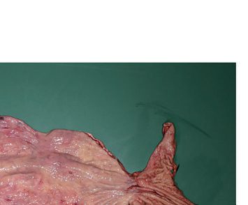
- The mismatch between excitatory and inhibitory neural activity causes failure of LES relaxation and absent peristalsis; over time the esophagus dilates, contractions disappear, and emptying occurs mainly by hydrostatic pressure, nearly always incompletely, leading to a progressively tortuous, dilated "megaoesophagus" [1].
Upper oesophageal sphincter dysfunction
Some patients also have dysfunction of the upper oesophageal sphincter and difficulty belching: normally, gas entering the oesophagus from the stomach triggers upper sphincter relaxation, but in achalasia this reflex may be lost, presumably through the same loss of inhibitory neurons, a likely contributor to the oesophageal distension seen in chronic disease [3].
Malignant transformation and secondary causes
- Persistent retention oesophagitis from fermenting food residue may predispose to oesophageal carcinoma [1], the late risk being squamous cell carcinoma [6].
- Secondary forms include Chagas disease, caused by Trypanosoma cruzi infection (destroys the myenteric plexus, with effects on the heart, brain, and GI tract as well), and pseudoachalasia caused by malignancy (especially cancer of the esophagus or gastric cardia), which should be excluded in older patients (>60 years) with short symptom duration and significant weight loss [1][2].
- Systemic sclerosis is a further motility mimic to exclude: it also causes poor oesophageal motility, but this leads to failure of acid clearance and reflux-related stricture, rather than achalasia's failure of LES relaxation [7].
- Achalasia is also part of Allgrove (triple A) syndrome, achalasia, alacrimia, and adrenal insufficiency [1][2].
Achalasia as a primary disorder of the LES
- Although complete absence of peristalsis in the oesophageal body was once proposed as the major abnormality, present evidence indicates that achalasia is a primary disorder of the LES: 24-hour outpatient oesophageal motility monitoring shows that, even in advanced disease, up to 5% of contractions can be peristaltic [8].
- The simultaneous waves develop as a result of the increased resistance to emptying caused by the non-relaxing sphincter, in experimental models a band placed loosely around the gastro-oesophageal junction left sphincter pressure unchanged but impaired LES relaxation, producing a marked increase in simultaneous waveforms, a fall in contraction amplitude and radiographic dilatation, all reversible on removing the band [8].
- Pseudoachalasia from tumour infiltration, a tight distal stricture or an over-tight antireflux repair, and the return of peristalsis in some patients after dilatation or myotomy, provide the same evidence [8].
- The presumed neurogenic degeneration is idiopathic or infective; in animals the disease has been reproduced by destroying the nucleus ambiguus and the dorsal motor nucleus of the vagus, and in patients degenerative change is found in the vagus nerve and in the myenteric ganglia of the oesophagus itself [8].
- The result is LES hypertension, failure of the sphincter to relax on swallowing, raised intraluminal pressure, oesophageal dilatation and a subsequent loss of progressive peristalsis; the dilatation comes from the combination of functional retention behind the non-relaxing sphincter and repetitive pharyngeal air-swallowing, and the height of the air–fluid level on radiographs reflects the degree of resistance the sphincter imposes [8].
- A subgroup with otherwise classic achalasia has simultaneous, sometimes high-amplitude, contractions of the oesophageal body (formerly "vigorous achalasia", now Chicago type 3) in whom chest-pain episodes are common and in whom videoradiography may show a corkscrew deformity and diverticulum formation that make the distinction from diffuse oesophageal spasm difficult [8].
Clinical features
- Achalasia has an insidious onset with gradual progression, most commonly dysphagia progressing from solids to liquids, and patients often go years before seeking medical attention, frequently being treated for other diseases such as gastro-oesophageal reflux first [3].
- Dysphagia for both solids and liquids is the most common symptom, present in approximately 95% of patients [2]; the Oxford Handbook notes initial dysphagia may be worse for liquids than solids as disease progresses [9].
- This oesophageal-body pattern, food sticking after the swallow has been initiated, is distinct from oropharyngeal dysphagia, in which conditions such as myasthenia gravis, multiple sclerosis, and cerebrovascular disease cause immediate difficulty initiating the swallow [7].
| Symptom | Frequency |
|---|---|
| Dysphagia to solids | 91% |
| Dysphagia to liquids | 85% |
| Regurgitation of food and saliva | 45–75% |
| Sore throat, hoarseness or postnasal drip | Up to 71% |
| Cough | 61% |
Table reproduces the reported symptom rates above [3]. Regurgitation of undigested food occurs in about 70% of patients and can cause aspiration with cough, hoarseness, and recurrent pneumonia [2]. Respiratory symptoms are attributed to chronic aspiration from failed oesophageal clearance, and most patients reporting them have had dysphagia for two or more years beforehand [3].
The reflux pitfall
- Heartburn occurs in 40–50% of patients but is due to stasis and fermentation of undigested food rather than true acid reflux; when misattributed to GERD, patients may be treated with acid-reducing medication or even referred for antireflux surgery, delaying correct diagnosis ("refractory GERD" pitfall) [1][2].
- Chest pain occurs in 40–50% of patients, thought to be secondary to esophageal distention [2].
- Weight loss may or may not occur, as patients often adjust their diet [1].
- Aspiration-related respiratory symptoms, halitosis from fermenting retained food, and regurgitation of previously ingested food are also reported [1].
Effect of patient demographics
Younger patients present with chest pain and heartburn more often than older patients, who tend to be less symptomatic overall; obese patients with a BMI of 30 kg/m² or more experience choking and vomiting more frequently before myotomy than non-obese patients [3].
Etiology
- The exact etiology of the degenerative process remains unknown; some viruses (varicella-zoster, human papilloma, herpes) have been implicated in triggering an inflammatory/autoimmune reaction [2].
- There is increasing evidence for a genetic contribution, drawn from twin and sibling studies and from the association of achalasia with Parkinson disease and Down syndrome, although genetic testing remains largely a research tool with limited diagnostic utility [3].
- Secondary causes include Chagas disease (Trypanosoma cruzi) and pseudoachalasia from malignancy; Allgrove syndrome is a rare genetic cause [1][2].
Diagnosis
Endoscopy
- Patients presenting with dysphagia are often trialled on a proton pump inhibitor first, which is appropriate, but failure to improve after 4 to 6 weeks warrants further evaluation, the next step being upper endoscopy with mucosal biopsy to exclude an inflammatory ring, erosive reflux, eosinophilic oesophagitis, and oesophageal cancer [3].
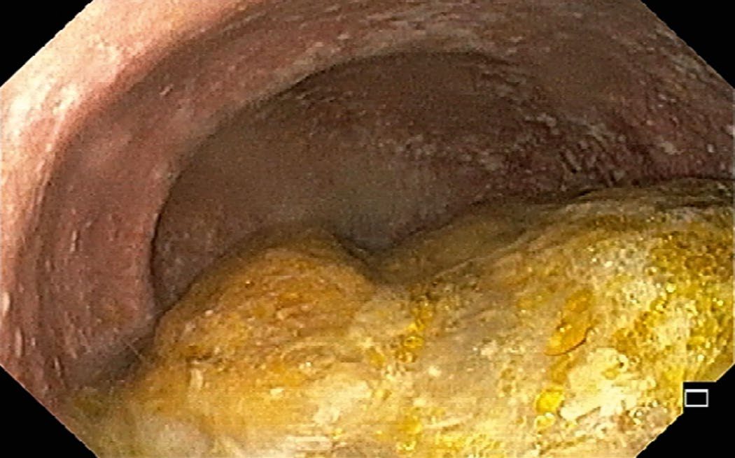
- Esophagogastroduodenoscopy is normal in about 40% of patients (30–40% per Bailey & Love); it may show retained food/saliva or stasis/Candida esophagitis, and is essential to exclude pseudoachalasia from malignancy [1][2][6].
- Other endoscopic findings include a dilated tortuous oesophagus, residual food and fluid, and difficulty passing the scope through the LES [3].
Contrast studies
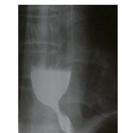
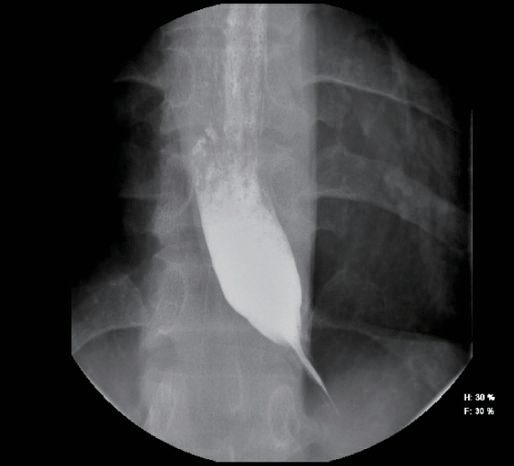
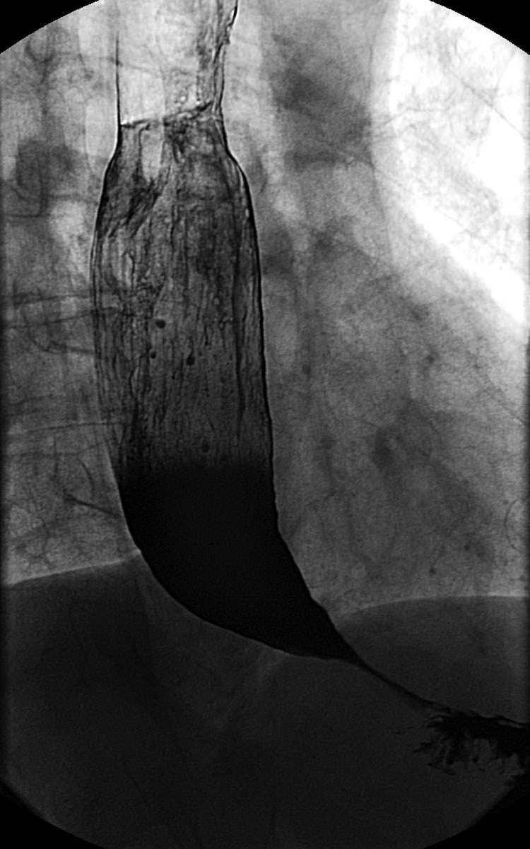
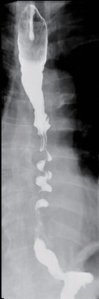
Barium swallow shows distal esophageal narrowing with a "bird's beak" or "rat's tail" tapering, an air-fluid level, slow emptying, tertiary contractions, and, with progression, a dilated tortuous "sigmoid" esophagus; a timed barium oesophagogram quantifies contrast retention height to assess severity [1][2]. These findings are suggestive but insufficient for definitive diagnosis, which requires manometry [3].
High-resolution manometry
- High-resolution esophageal manometry is the gold standard, establishing the diagnosis and classifying the achalasia subtype (I, II, or III), which has important treatment implications [1][2].
- High-resolution catheters carry sensors every 1 cm along their length, against every 3 to 5 cm in traditional water-perfused and strain-gauge systems, producing an oesophageal pressure topography that plots pressure on a colour scale against time and location [3].
- Manometry classically shows high/normal basal LES pressure with incomplete LES relaxation and poor or absent peristalsis [6][9].
- In normal individuals the LES relaxes completely during a swallow, to below 8 mmHg above gastric pressure; in achalasia relaxation is incomplete or absent, and additional findings are a resting LES pressure above 45 mmHg and aperistalsis in the distal two-thirds of the oesophagus [3].
- Incomplete LES relaxation is what distinguishes achalasia from other disorders associated with aperistalsis [3].
Adjunctive tests
- Ambulatory pH monitoring is recommended for patients with heartburn to distinguish achalasia from GERD; one study found 29% of patients eventually diagnosed with achalasia had been treated for an average of 29 months with PPIs beforehand [2].
- Functional lumen imaging probe (FLIP) is a newer complementary test assessing esophageal compliance and distensibility [2].
- Where the diagnosis remains unclear after endoscopy, barium oesophagram and manometry, endoscopic ultrasound of the LES or a timed barium oesophagram documenting contrast retention may help [3].
Conventional manometric criteria for the primary motor disorders
The classic (pre-Chicago) manometric definitions separate achalasia from the other primary oesophageal motility disorders, though Schwartz stresses that the boundaries are vague, intermediate types exist, and the disorders are best regarded as a spectrum reflecting progressive destruction of motor function [8].
| Disorder | Manometric characteristics |
|---|---|
| Achalasia | Incomplete LES relaxation (<75%); aperistalsis of the oesophageal body; elevated LES pressure; intra-oesophageal baseline pressure raised relative to gastric baseline |
| Diffuse oesophageal spasm | Simultaneous (non-peristaltic) contractions in >20% of wet swallows; repetitive, multi-peaked and spontaneous contractions; intermittent normal peristalsis; contractions may be of increased amplitude and duration |
| Nutcracker oesophagus | Mean distal peristaltic amplitude (10 wet swallows) ≥180 mmHg; mean contraction duration >7.0 s; normal peristaltic sequence |
| Hypertensive LES | LES pressure ≥26 mmHg with normal relaxation and normal body peristalsis |
| Ineffective oesophageal motility | Decreased or absent peristaltic amplitude (<30 mmHg); increased number of non-transmitted contractions |
- Table reproduces the manometric characteristics tabulated by Schwartz [8].
- A criterion of 30% or more peristaltic waveforms out of 10 wet swallows has been used to separate diffuse spasm from vigorous achalasia, although the figure is arbitrary and often debated [8].
- Botulinum toxin injection is also useful diagnostically when it is unclear whether a hypertensive LES is the primary cause of dysphagia: responsiveness to the injection may predict a good response to Heller myotomy [8].
Scoring and Severity
- The Eckardt score is a clinical scoring system used to assess achalasia severity and monitor treatment response, assigning 0–3 points each to four symptoms (dysphagia, regurgitation, weight loss, and chest/retrosternal pain) for a total range of 0–12; a score of ≤3 indicates successful treatment [1][2].
- The Chicago classification subdivides achalasia into Types I, II, and III based on high-resolution manometry pressurization patterns, which guides treatment choice and predicts outcome [1][2].
- The measurement that places a patient in that group is the integrated relaxation pressure (IRP), the relaxation pressure across the oesophagogastric junction in response to a swallow.
- An elevated IRP defines the disorders of OGJ outflow, which comprise the three types of achalasia and OGJ outflow obstruction (OGJOO), and separates them from the disorders of peristalsis, absent contractility, distal oesophageal spasm, and hypercontractile oesophagus [1].
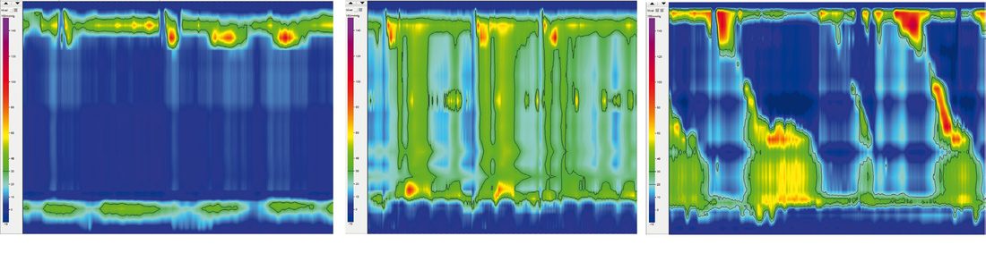
| Chicago type | Manometric pattern | Response to botulinum toxin | Response to pneumatic dilation | Response to Heller myotomy | Overall response |
|---|---|---|---|---|---|
| I (classic) | Impaired LES relaxation, absent peristalsis, normal oesophageal pressure | — | — | — | 56% |
| II | Impaired LES relaxation, absent peristalsis, increased panoesophageal pressure | 71% | 91% | 100% | Best of the three |
| III (spastic) | Impaired LES relaxation, absent peristalsis, distal oesophageal spastic contractions | — | — | — | 29% |
Type II patients are significantly more likely to respond to any therapy than type I or type III patients, which has improved the ability to discuss expected outcomes with patients [3].
Treatment and Management
Treatment is palliative, aiming to reduce the functional obstruction from the nonrelaxing, often hypertensive LES; the goal is early diagnosis and therapy to prevent late complications while preserving oesophageal function [2][3].
Medical therapy
- Pharmacologic therapy (calcium channel blockers such as nifedipine, phosphodiesterase-5 inhibitors such as sildenafil, nitrates) has limited efficacy and notable side effects (headache, oedema, hypotension), and is reserved for patients who are poor candidates for endoscopic or surgical treatment [1][2].
- It is the least invasive but least effective option: initial response is approximately 50%, and long-term success is limited by side effects including headache, orthostatic hypotension and oedema [3].
- Oral calcium channel blockers or nitrates can relax the LES in 47% to 64% of patients, the two most widely used agents being nifedipine 10–30 mg given 30–45 minutes before meals and isosorbide dinitrate 5–10 mg given 10–15 minutes before meals, with some evidence that isosorbide dinitrate acts more rapidly [3].
Botulinum toxin
Endoscopic botulinum toxin injection into the LES gives symptom relief in about 70–80% of patients at 1–3 months, falling to roughly 40% at 12 months, and is generally reserved for elderly/comorbid patients unsuitable for myotomy or dilatation, since repeated injections can cause scarring that complicates subsequent surgery [1][2]. It works by blocking the excitatory acetylcholine-releasing neurons that raise LES smooth muscle tone, so resting LES pressure falls and the oesophagus empties; initial improvement approaches 70%, comparable with pneumatic dilation, but symptoms recur in as little as 6 months [3].
Pneumatic dilatation
- Pneumatic dilatation uses a graded approach starting with a 30 mm balloon, reserving 35 mm and 40 mm balloons for persistent/recurrent symptoms; it is effective in roughly 80% of patients but with higher retreatment rates than surgery, and predictors of poor response include age <40, male sex, large esophageal diameter, and Chicago type I/III [2][6].
- A non-compliant cylindrical balloon tears the muscle fibres of the LES; commercially available balloons come in three sizes (3.0, 3.5 and 4.0 cm), against a largest through-the-scope balloon of 2.0 cm, and the balloon is insufflated with a hand-held pressure gauge for 15 to 60 seconds [3].
- Short-term success ranges from 60% to 90% and is generally sustained for up to 2 years, but nearly one-third of patients go on to require additional therapy; despite this, pneumatic dilatation is considered the most cost-effective treatment for achalasia [3].
- Success is influenced by age (better if over 45), sex (better in females), oesophageal diameter and achalasia subtype [3].
- Perforation risk with a 30 mm balloon is under 0.5%, rising with larger balloons; overall reported perforation incidence averages about 1.9% [1].
- Because of the risk of oesophageal rupture, patients should be considered appropriate surgical candidates before dilatation and surgical backup should be readily available [3].
The BSG national guideline sets the UK technique for pneumatic dilatation, and notes that bougie dilators are not used for achalasia [4].
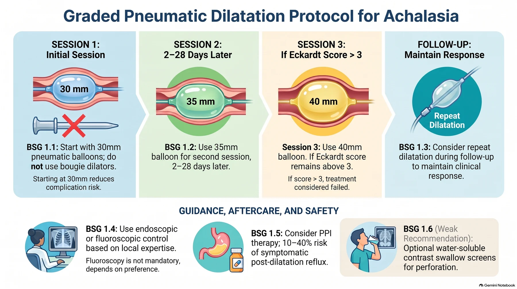
| Recommendation | GRADE evidence | Strength |
|---|---|---|
| 1.1 Perform dilatation with pneumatic balloons 30–40 mm in diameter, starting at 30 mm in the first session to reduce the risk of complications | High | Strong |
| 1.2 Perform a second dilatation session 2–28 days later with a larger balloon of 35 mm | High | Strong |
| 1.3 Consider repeat dilatation after the initial series during follow-up to maintain symptom response | High | Strong |
| 1.4 Perform dilatation under endoscopic or fluoroscopic control based on clinician preference and local expertise | Moderate | Strong |
| 1.5 Consider PPI therapy after dilatation, as the technique carries a 10–40% rate of symptomatic GORD or ulcerative oesophagitis | High | Strong |
| 1.6 Consider a water-soluble contrast swallow after dilatation to screen for perforation, but it is not essential | Moderate | Weak |
- Table reproduces recommendations 1.1 to 1.6 [4].
- Most authors advocate a third session, either routinely or where symptoms persist with an Eckardt score above 3, using a 40 mm balloon cautiously; if the Eckardt score remains above 3 after the third session the treatment is usually considered to have failed [4].
- The procedure is effective in 90% of patients in the first year, falling to 86% in the second, and up to a third relapse over 4 to 6 years, but the vast majority can be treated successfully by repeat dilatation, achieving remission in up to 97% at 5 years and 93% at 10 years [4].
Patients should fast at least 6 hours before dilatation, and those with achalasia are likely to have oesophageal stasis and so require a longer fast based on clinical judgement [4]. Dilatation should not be performed in active or incompletely healed oesophageal perforation, and requires careful weighing of benefit and risk after a recent healed perforation, recent upper gastrointestinal surgery, pharyngeal or cervical deformity, or bleeding disorders [4].
Comparative efficacy
A European RCT (PD vs. laparoscopic Heller myotomy with Dor fundoplication) found similar therapeutic success at 2 years (86% vs. 90%) and 5 years (82% vs. 84%), though 25% of PD patients needed additional dilatations [2]. Early studies suggested surgical myotomy gave better long-term symptom improvement than dilatation (68.2% versus 56.3%), but those patients underwent only a single dilatation, which is not current practice; in one of the most referenced randomised trials comparing surgical myotomy with graded pneumatic dilatation there was no significant difference at 2 years (92% for dilatation versus 87% for myotomy) [3].
The four therapeutic questions
- Schwartz frames the treatment decisions around four issues [8]. First, dilatation or myotomy for the newly diagnosed patient: long-term follow-up shows pneumatic dilatation achieves adequate relief of dysphagia and pharyngeal regurgitation in 50–60% of patients, so close follow-up is required and myotomy is indicated if dilatation fails; in a dilated, tortuous oesophagus or with an associated hiatal hernia balloon dilatation is dangerous and surgery is the better option, and the one controlled randomised study (38 patients) plus several trials of laparoscopic cardiomyotomy against balloon or botulinum toxin all favour surgery as primary treatment [8].
- The three methods differ in how reliably they reduce sphincter pressure to below 10 mmHg, patients whose pressure falls below 10 mmHg after balloon dilatation do as well as after myotomy, whereas botulinum toxin's effect lasts weeks or months rather than years [8].
- In Eckardt's series, post-dilatation LES pressure was the most valuable predictor of long-term response: <10 mmHg predicted a good result, the roughly 50% of patients left at 10–20 mmHg had a 2-year remission rate of 71%, 16 of 46 left above 20 mmHg had an unacceptable outcome, and overall only 30% of dilated patients remained in remission at 5 years [8].
- Myotomy after balloon dilatation is not usually more difficult unless the cardia has been ruptured in a saw-tooth manner; after botulinum toxin the submucosal inflammatory response is most intense in the first 6–12 weeks, so cardiomyotomy should wait at least 3 months after injection [8].
Second, abdomen or chest: without previous upper abdominal surgery most surgeons prefer the abdominal route, since laparoscopy gives less pain and a shorter stay than thoracoscopy and makes a long gastric myotomy easier to ensure [8]. Third, whether to add an antireflux procedure: in large retrospective cohorts more than 50% of patients had reflux symptoms 10 years after myotomy without fundoplication, and in a randomised trial 7% of patients with a Dor fundoplication had abnormal 24-hour pH studies against 42% after myotomy alone; if a wrap is added it must be partial (a 270° Belsey, a posterior Toupet or an anterior Dor) never a complete 360° fundoplication, whose own outflow obstruction produces long-term oesophageal dysfunction [8]. Fourth, whether cure is achievable: late deterioration occurs after myotomy, with or without an antireflux procedure, and after balloon dilatation even when pressure is reduced below 10 mmHg, probably because the underlying motor disorder of the body persists and progresses; the earlier an effective reduction in outflow resistance is achieved, the better the outcome and the more likely some body function is restored [8].
Surgeries
Laparoscopic Heller myotomy
- Laparoscopic Heller myotomy (LHM) with partial fundoplication is now the standard surgical treatment, dividing the circular muscle of the lower esophagus and gastric cardia, typically extending about 6 cm proximally on the esophagus and 2–3 cm distally onto the stomach [1][6].
- The original approach divided the circular and longitudinal muscle fibres through a thoracotomy with good results, but has been replaced by the laparoscopic transabdominal approach because of lower perioperative morbidity and faster recovery [3].
- The procedure uses a standard 4- to 5-port laparoscopic approach: after dividing the gastro-oesophageal ligament, both the outer longitudinal and inner circular fibres are divided by blunt traction or electrocautery for 4 to 6 cm proximally from the gastro-oesophageal junction and 2 cm distally onto the stomach, and the fundoplication then follows [3].
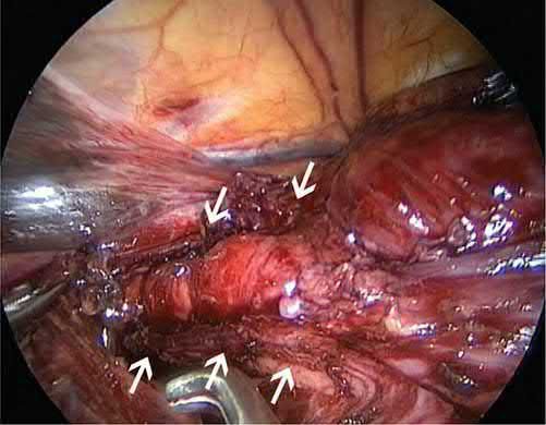
A partial fundoplication (anterior Dor or posterior Toupet) is added to reduce postoperative GERD, which can occur in up to 40–50% of patients after myotomy alone; a complete 360° (Nissen) fundoplication is contraindicated as it causes outflow resistance against an aperistaltic esophageal body, risking dysphagia [1][2][10]. A blinded randomised controlled trial found that adding a fundoplication reduced abnormal oesophageal acid exposure from 47% to 9%, and SAGES accordingly recommends including one after surgical myotomy, though whether an anterior Dor or posterior Toupet is preferable remains uncertain [3].
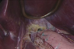
- The Padua group reported a 90% success rate among 407 LHM patients at 2.5-year median follow-up, with 87% 5-year actuarial probability of remaining asymptomatic; a Swedish RCT versus pneumatic dilatation showed 92% excellent results at 5 years and 80% at 10 years after LHM [2].
- Heller myotomy achieves efficacy of 88% to 95% against single pneumatic dilatation, though this superiority is less evident against serial graded dilatation, and efficacy falls over time from nearly 96% at 6 months to 57–92% at 6 years [3].
- Surgical outcomes are better for type I and II achalasia than type III, which may need a longer, more proximally extended myotomy [1][2].
Peroral endoscopic myotomy
- Peroral endoscopic myotomy (POEM) is a purely endoscopic myotomy via a submucosal tunnel, extending a minimum of 6 cm proximally and 2 cm onto the gastric cardia, closed with endoclips.
- It achieves symptom relief in >90% of patients, similar to LHM, and is particularly advantageous for type III achalasia because the myotomy length can be tailored [1][2].
- First described in 2010 by Inoue and colleagues, it uses a standard flexible gastroscope with a transparent cap to make a small mucosal cut roughly 14 cm proximal to the gastro-oesophageal junction, enters the submucosal plane, tunnels along the oesophagus and onto the stomach, divides the muscle fibres with electrocautery, and closes the mucosotomy with clips or sutures [3].
- Many series report success rates over 90% at one year, and a meta-analysis against surgical myotomy showed equivalent short-term outcomes with comparable complication rates [3].
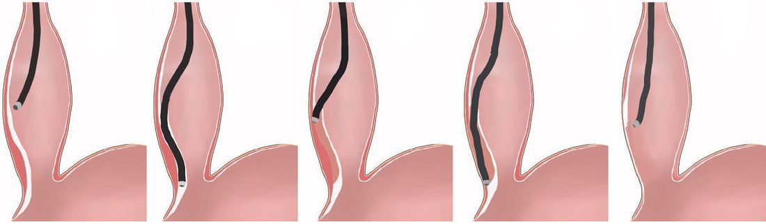
Because POEM has no antireflux component, postprocedure GERD is markedly more common than after LHM with fundoplication, pathologic reflux around 47–57% after POEM versus roughly 11–20% after LHM in comparative studies, including a multicenter RCT of 221 patients [1][2]. Reported reflux rates after POEM range from 10% to 46%, and most patients are placed on PPI therapy afterwards until pH testing can be completed [3].
Oesophagectomy
- Esophagectomy is reserved as a last resort for end-stage achalasia with a massively dilated, sigmoid ("megaoesophagus") that has failed other treatments, given the risk of aspiration and regurgitation from a poorly emptying esophagus.
- A 2001 series of 93 esophagectomies reported anastomotic leak in 10%, recurrent laryngeal nerve injury in 5%, and 2 deaths, with 50% developing anastomotic stricture [1][2].
- Pneumatic dilatation is often unsuccessful in these patients, who require surgical myotomy or POEM; where oesophagectomy with gastric pull-up is ultimately needed, uncontrolled studies report symptom improvement in 80% of patients with mortality ranging from 0% to 5.4% [3].
- UK standards for who may perform dilatation and where are set out alongside the technique itself.
- Oesophageal dilatation should be undertaken only by, or under the direct supervision of, an experienced operator performing sufficient numbers to maintain their skills, supported by at least two assistants in the room, one of whom must be a trained nurse [4].
- Trainees must have adequate knowledge of the indications for, contraindications to, and complications of the procedure, understand the steps for recognising and managing complications, and be familiar with different dilatation techniques as well as alternative and complementary treatment options [4].
The procedure should be performed in a dedicated, fully equipped endoscopy room with access to X-ray screening and surgical support, or a similarly equipped radiological suite [4]. Units must have an agreed protocol to follow in the event of a perforation, with clear identification of a qualified surgeon (on site or off) to manage it where luminal treatment such as a covered stent is not feasible or appropriate [4].
Operative principles and technical detail
- Four principles govern a surgical myotomy of the LES: complete division of all circular and collar-sling muscle fibres, an adequate distal myotomy to reduce outflow resistance, "undermining" of the muscularis to allow wide separation of the oesophageal muscle, and prevention of postoperative reflux [8].
- Ernst Heller, a German surgeon, described a "double myotomy" in 1913; the laparoscopic operation that bears his name uses the same trocar placement and hiatal exposure as a Nissen fundoplication [8].
- The short gastric vessels are divided in preparation for the wrap, the gastro-oesophageal fat pad is removed with the anterior vagus swept to the right, and the junction and distal 4–5 cm of oesophagus are bared; the myotomy is most easily begun 1–2 cm above the junction, above the zone of any previous botulinum toxin injection or balloon dilatation, using scissors or a hook electrocautery, and is carried across the junction onto the proximal stomach for about 2–3 cm, after which the muscle edges are separated bluntly from the mucosa over roughly 50% of the circumference [8].
- The Dor wrap is slightly easier and does not disturb the normal posterior gastro-oesophageal attachments, a theoretical advantage in preventing reflux [8].
- In vigorous (type 3) achalasia the myotomy must extend over the whole distance of abnormal motility mapped preoperatively, or dysphagia will persist [8].
- Open techniques are now rarely used outside re-operations.
- A modified Heller myotomy can be done through a left thoracotomy in the sixth intercostal space: the myotomy runs 1–2 cm onto the stomach and 4–5 cm up the oesophagus, a tongue of gastric fundus is sutured to the myotomy margins to prevent re-healing and give reflux protection (or a formal Belsey repair if the cardia has been extensively dissected), nasogastric drainage is traditionally kept for 6 days and oral diet resumed on day 7 after a barium swallow shows unobstructed passage without extravasation [8].
- Minimally invasive oesophagectomy for the end-stage, massively dilated oesophagus should combine thoracoscopic with abdominal dissection, because a transhiatal minimally invasive attempt can cause large-volume bleeding from mediastinal vessels enlarged by the dilatation [8].
Long-term outcome series
- In Csendes' randomised comparison of 81 patients, myotomy produced a greater reduction in sphincter pressure and improvement in contraction amplitude than forceful dilatation; 28% regained some peristalsis after surgery against 13% after dilatation, and at 5 years 95% of surgical patients were doing well against 54% of dilated patients, of whom 16% needed redilatation and 22% eventually required myotomy [8].
- Bonavina reported good-to-excellent results in 94% after transabdominal myotomy with Dor fundoplication at a mean 5.4 years, with no operative mortality [8].
- Malthaner and Pearson's 35 patients with at least 10 years' follow-up (22 after primary myotomy with Belsey hemifundoplication) had excellent-to-good results in 95% at 1 year, falling to 68%, 69% and 67% at 10, 15 and 20 years, the deterioration attributed to late complications of reflux; Ellis's 179 transthoracic short myotomies without an antireflux procedure showed 89% improved at a mean 9 years with symptom-free results falling from 54% at 10 years to 32% at 20, which he attributed to progression of the underlying disease rather than reflux [8].
| Reason for failure | Ellis, myotomy only (n = 81) | Goulbourne, myotomy only (n = 65) | Malthaner, myotomy + antireflux (n = 22) |
|---|---|---|---|
| Reflux | 4% | 5% | 18% |
| Inadequate myotomy | 2% | — | 9% |
| Megaoesophagus | 2% | — | — |
| Poor emptying | 4% | 3% | — |
| Persistent chest pain | 1% | — | — |
- Table reproduces the reasons for failure of oesophageal myotomy tabulated by Schwartz [8].
- For laparoscopic myotomy with hemifundoplication, two series of over 100 patients documented relief of dysphagia in 93%, and Richter's review of 254 published patients gave an average success rate of 93% at 2.5 years; conversion to open surgery occurs in 0–5%, complications in under 5% (largely mucosal perforation, more likely after botulinum toxin), and objective reflux on acid-exposure testing in under 10% [8].
- Symptoms alone are an unreliable endpoint because patients unconsciously modify their diet, and insufficient reduction in outflow resistance can allow slow progressive dilatation that masquerades as improvement; the best objective measures are LES pressure, oesophageal baseline pressure and scintigraphic oesophageal emptying time, though the latter two are rarely reported [8].
Complications
- Complications of pneumatic dilatation include esophageal perforation (roughly 1.9% average incidence, lower with smaller balloons) [1].
- Reported perforation rates range from 0% to 15% in the literature, though recent high-volume centre studies place it at approximately 1% to 5%; no pre-dilatation factors predict perforation, which is most likely due to an inappropriately positioned balloon [3].
- Perforation may be small and clinically insignificant, managed with antibiotics, parenteral nutrition or an oesophageal stent, or a major disruption requiring emergency surgical exploration and repair [3].
- The commonest complication after dilatation is gastro-oesophageal reflux disease, at rates approaching 20%, usually treatable with PPI therapy [3].
- Complications of Heller myotomy include mucosal perforation (increased risk in patients with prior botulinum toxin injection due to loss of normal tissue planes) and postoperative GERD, esophagitis, Barrett esophagus, and even adenocarcinoma with long-term reflux exposure, particularly relevant in younger patients [2].
- Reflux after surgical myotomy without fundoplication approaches 30% [3].
- POEM carries a high incidence of postprocedure reflux/esophagitis (up to 57% at 3 months in some series), potentially requiring lifelong acid suppression or a later antireflux operation [1].
- Recurrent dysphagia after treatment is common, particularly in younger patients, most often due to scarring at the distal myotomy or development of a peptic stricture from reflux; full re-evaluation (barium swallow, endoscopy, manometry, pH monitoring) is needed before further intervention [2].
Malignancy risk
- Long-standing achalasia with chronic retention oesophagitis is associated with an increased risk of oesophageal carcinoma, typically squamous cell carcinoma, occurring late in the disease course [1][6].
- The hazard ratio for developing squamous cell carcinoma is 28 compared with the general population, attributed to stasis and consequent inflammation; the risk of adenocarcinoma is also raised but substantially lower.
- The absolute risk nevertheless remains low, and there are insufficient data to support routine endoscopic surveillance [3].
- Perforation should be suspected when a patient develops pain, breathlessness, fever or tachycardia after dilatation; transient chest pain is not uncommon, but persistent pain should prompt a CT scan with oral contrast [4].
- Patients should be monitored for at least 2 hours in recovery and given clear written instructions on fluids, diet and medications, and should be well and tolerating water on leaving hospital [4].
- Imaging and contrast studies should not be performed routinely after the procedure unless the patient develops persistent chest pain, fever, breathlessness or tachycardia during recovery [4].
- If the patient becomes symptomatic while still in the procedure room, perform endoscopic re-inspection to assess for perforation and to undertake treatment, which may include immediate endoscopic stent placement [4].
- Provide patients with contact information for the on-call team should they experience chest pain, breathlessness or become unwell [4].
Prognosis
- Achalasia cannot be cured, only palliated, since neuronal degeneration is irreversible [1].
- With laparoscopic Heller myotomy, long-term symptom control is excellent, with several series reporting 84–92% success at 5 years and around 80% at 10 years [2].
- POEM and LHM show comparable clinical success rates (roughly 81–93% at 2 years across studies) though POEM carries a substantially higher rate of pathologic reflux [2].
- Considering the rarity and complexity of achalasia, best results are achieved through multidisciplinary evaluation with treatment tailored to the individual patient [2].
Diffuse oesophageal spasm
- Diffuse oesophageal spasm is characterised by uncoordinated, simultaneous contractions of normal amplitude in the distal oesophagus that fail to propel food, presenting with dysphagia, regurgitation and chest pain [3].
- On high-resolution manometry it shows 20% or more premature contractions; unlike achalasia the symptoms are intermittent, findings may be absent on some series of test swallows, and LES relaxation is usually normal [3].
- Contrast oesophagram may show a corkscrew or rosary bead appearance, and among patients undergoing manometry for non-cardiac chest pain the prevalence of diffuse oesophageal spasm was 4% [3].
References
- Bailey & Love's Short Practice of Surgery, 28th ed., Ch. 66, The oesophagus
- Sabiston Textbook of Surgery, 22nd ed., Ch. 83, Benign Esophageal Disorders
- Maingot's Abdominal Operations, 13th ed., Ch. 22, Achalasia and Other Motility Disorders
- British Society of Gastroenterology: UK guidelines on oesophageal dilatation in clinical practice. Gut 2018, 1.1; 1.1, 1.2, 1.3, 1.4, 1.5, 1.6; Achalasia dilatation; 1.1–1.6; 1.2; 1.3; 1.4; 4.1, 4.3; 4.2; 4.4; 4.5; 4.6; Achalasia dilatation; Contraindications; Disease-specific considerations 1.1–1.6; Fasting www.bsg.org.uk
- NICE Interventional Procedures topic prioritisation IP1229: Peroral endoscopic myotomy (POEM) for achalasia — decision not to develop guidance (2024), Decision www.nice.org.uk
- The ABSITE Review, 2022, Ch. Esophagus
- Browse's Introduction to the Symptoms and Signs of Surgical Disease, 6th ed., Ch. 15
- Schwartz's Principles of Surgery, 11th ed., Ch. 25, Esophagus and Diaphragmatic Hernia
- Oxford Handbook of Clinical Surgery, 5th ed., Ch. 8, Upper gastrointestinal surgery
- Schwartz's Principles of Surgery: ABSITE and Board Review, Ch. 25, The Esophagus and Diaphragmatic Hernia