



Thoracic Trauma
Summary
- Thoracic trauma is directly or indirectly involved in over 50% of trauma deaths, yet more than 80% of chest injuries can be managed non-operatively with tube thoracostomy alone [1].
- Thoracic injuries account for approximately 25% of trauma deaths, and 50% of patients who die of multiple injuries have a significant thoracic injury [2].
- Oxford gives the operative rates that follow from that: fewer than 10% of blunt chest injuries and fewer than 30% of penetrating chest injuries require operative intervention [2].
- Injuries are grouped into six immediately life-threatening conditions identified in the primary survey and six potentially life-threatening conditions identified in the secondary survey, together known as the "deadly dozen" [1].
- NICE NG39 devotes two full sections to chest trauma, 1.3 for the pre-hospital setting and 1.4 for hospital, and between them they change three things that are taught as standard.
- They restrict when decompression may be performed, they replace needle decompression with open thoracostomy wherever expertise allows, and they set out an imaging sequence that differs by age and by the presence of severe respiratory compromise.
- Each is set out in the relevant section below.
The first recommendation is a diagnostic one and it is deliberately low-technology: use clinical assessment to diagnose pneumothorax for the purpose of triage or intervention [3]. eFAST is a permitted addition, but only conditionally: consider using eFAST to augment clinical assessment only if a specialist team equipped with ultrasound is immediately available and onward transfer will not be delayed [3]. And whether or not it is used, be aware that a negative eFAST of the chest does not exclude a pneumothorax [3].
Definition
- Chest injuries are blunt, falls and motor vehicle collisions transmitting high-energy forces to the chest wall and underlying structures, or penetrating, from knives and gunshots, with the rate of required operation higher for penetrating than blunt trauma [4].
- Oxford's version of the same split is that open injuries are caused by penetrating trauma from knives or gunshots, while closed injuries occur after blasts, blunt trauma and deceleration, of which road traffic accidents are the commonest cause [2].
- The "deadly dozen" comprises the immediately life-threatening, airway obstruction, tension pneumothorax, pericardial tamponade, open pneumothorax, massive haemothorax and flail chest (and the potentially life-threatening) aortic injuries, tracheobronchial injuries, myocardial contusion, diaphragmatic rupture, oesophageal injuries and pulmonary contusion [1].
Massive haemothorax has a quantitative definition worth carrying separately: the rapid accumulation of more than 1,500 mL of blood, or one-third or more of the patient's blood volume, in the pleural cavity [2].
Pathophysiology
- Tension pneumothorax develops from a one-way valve air leak in the lung or chest wall allowing air into the thoracic cavity without escape, progressively collapsing and compressing the affected lung, displacing the mediastinum, decreasing venous return, and compressing the contralateral lung [1][5].
- Open pneumothorax, the "sucking chest wound", occurs when a chest wall defect exceeding roughly two-thirds the diameter of the trachea causes air to be preferentially drawn through the defect rather than the trachea during inspiration [1].
- Pericardial tamponade results from accumulation of as little as 50 mL of blood in the non-distensible pericardial sac, compressing the heart and reducing diastolic filling [1].
- Flail chest, two or more consecutive ribs broken in two or more places, or by other definitions three or more contiguous ribs fractured in two or more locations, causes paradoxical chest wall motion; the associated pulmonary contusion, not the flail segment itself, is the principal cause of respiratory failure, via decreased compliance and increased shunt fraction [1][5][6].
- Oxford states the same combination as restricted chest wall movement plus underlying lung contusion causing hypoxia [2].
- Traumatic aortic rupture occurs at the relatively fixed aortic isthmus just distal to the left subclavian artery origin, from shear forces disrupting the intima and media during sudden deceleration; if the adventitia remains intact the patient may survive to reach hospital, patients survive immediate death because the haematoma is contained [1][2][5].
Clinical features
- Tension pneumothorax presents with respiratory distress, tachycardia, hypotension, tracheal deviation away from the affected side as a late finding, hyper-resonance, absent or decreased breath sounds, and distended neck veins, though neck veins may be flat with concurrent hypovolaemia [1][6].
- Oxford states the diagnostic principle in one line: it is a clinical diagnosis, and there is no time for X-rays [2].
- Massive haemothorax should be suspected when shock is associated with a dull percussion note and absent breath sounds on one side of the chest, and flat neck veins, the percussion note and the neck veins being what distinguishes it from tension pneumothorax at the bedside [2].
- Pericardial tamponade presents with Beck's triad of distended neck veins, muffled heart sounds and falling arterial pressure, though this triad is often not appreciated in the noisy trauma bay; blood pressure may transiently improve with fluid, creating false reassurance [1][2][6].
- Flail chest is diagnosed clinically, not radiographically, in non-ventilated patients by observing paradoxical inward motion of the loose segment on inspiration [1].
- Tracheobronchial injury presents with a large continuous air leak, pneumomediastinum, persistent pneumothorax despite chest tube, and subcutaneous emphysema; oxygenation may worsen after chest tube placement [5].
- Oxford's triad for it is haemoptysis, subcutaneous emphysema, or tension pneumothorax, to be suspected when there is a persistent large air leak after chest drain insertion [2].
- Aortic transection signs on chest radiograph include a widened mediastinum of 8 cm or more, apical capping, loss of the aortopulmonary window or aortic contour, left haemothorax, and tracheal or nasogastric tube deviation to the right, though the chest X-ray is normal in 5% of patients with aortic tears [5].
- Sternal fracture and first or second rib fractures are risk factors for cardiac contusion and aortic transection respectively [5].
The chest in the secondary survey
- Browse's treats the chest as a region that must be examined twice, and its argument for doing so is that the first examination was performed under conditions that make signs easy to miss.
- Although the chest was assessed as part of the primary survey, it should now be carefully reassessed by inspection, palpation, percussion and auscultation to detect any signs that may have been missed at the time of the primary survey, when rapid resuscitation was essential
- New signs may have developed, and subtle signs may have been missed [7].
- Its practical instruction is to check the back and sides of the chest, including into the axillae [7].
- Test again for rib fractures: pain on compression or release indicates the likelihood of rib fractures or costal cartilage separation from the ribs or sternum, and both can then be more accurately localised by detailed palpation; a careful inspection may detect a small flail segment [7].
- Two associations follow from the bones. Rib fractures are often associated with injuries of the great vessels, lungs, spleen or liver, and sternal fractures are often associated with cardiac injuries, so the sternum must be inspected and palpated in its own right [7].
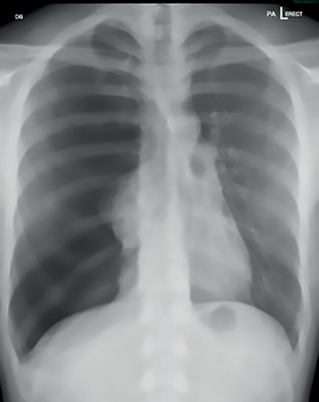
- On re-examination, check again for haemothorax, pneumothorax and cardiac tamponade, taking particular care to look for small pneumothoraces and an increase in the width of the mediastinum, which may be the only indication of an aortic dissection; CT of the chest is highly sensitive and specific in detecting cardiothoracic trauma [7].
- Browse's also flags the neck and clavicle, which sit at the boundary of the thoracic examination. Penetrating descending wounds of the root of the neck can be very dangerous, because they may damage the carotid, vertebral and subclavian arteries as well as the trachea, larynx, pharynx and oesophagus, and major structures within the upper chest can also be damaged.
- Neurological signs or ischaemia of the upper limb suggest a major arterial injury, as does a rapidly expanding haematoma or a machinery murmur [7]. Severe compound clavicular injuries are often associated with injuries to the subclavian or axillary vessels, the brachial plexus and the apex of the lung, so the vascular supply and peripheral nerves of both upper limbs should be examined [7].
- Gentle palpation should detect any subcutaneous surgical emphysema in the neck or supraclavicular fossae [7].
Etiology
- Rib fractures occur in approximately 10% of trauma admissions and are a marker of severe associated injury, with nearly 50% of multiply injured patients having a rib fracture; mortality with chest wall injury is 6–12% [4].
- Massive haemothorax in blunt trauma most commonly arises from torn intercostal or internal mammary vessels secondary to rib fractures; in penetrating trauma, thoracic and abdominal viscera may be involved [1].
- Tracheobronchial injury is most common with blunt trauma, and bronchial injuries are more common on the right; 90% occur within 1 cm of the carina [5].
- Diaphragmatic injuries are more common on the left and more often result from blunt trauma [1][5].
- Cardiac tamponade most commonly results from penetrating injury, but blood can also accumulate in the pericardial sac after blunt trauma [2].
- Blunt cardiac injury is associated with chest wall contusion and with sternal or rib fractures [2].
- Aortic disruption should be suspected wherever there is a history of a decelerating force together with a widened mediastinum on chest radiograph [2].
Diagnosis
- Chest radiography is the first-line investigation and is almost universally obtained in the primary survey [1][4].
- Pitfalls include failure to clinically assess tracheal shift, failure to examine both front and back in a supine patient, and confusing a supine haemothorax's homogeneous opacity for the contralateral, normal side [1].
- Thoracic CT and CT angiography have become the standard for chest wall, vascular, pleural, and parenchymal evaluation; erect chest radiography best reveals a small pneumothorax or air-fluid level, and up to 300 mL of blood may pool behind the diaphragm domes without becoming visible [1][4].
- Oxford's rule for the secondary survey is that in stab injuries the patient is exposed fully and positioned to assess the front, back and sides of the chest for wounds missed in the primary survey, and that an erect chest radiograph will aid in identifying simple pneumothorax and haemothorax, pulmonary contusion, tracheobronchial rupture, blunt cardiac injury and aortic disruption [2].
- Penetrating wounds within the "cardiac box", bounded by the sternal notch superiorly, the costal margins inferiorly and the nipples laterally, mandate evaluation for cardiac and great vessel injury with pericardial ultrasound, CT angiography, bronchoscopy, and oesophagoscopy or oesophagography as indicated [4][5].
- Diagnostic sensitivity for penetrating aerodigestive injury approaches 100% when multiple complementary studies are performed [4].
- For tracheobronchial injury, bronchoscopy confirms the diagnosis; for aortic injury, CT angiography of the chest is diagnostic [1][2][5].
- Diaphragmatic injury is difficult to detect; a nasogastric tube seen coiled in the chest on chest radiograph or an air-fluid level from herniated stomach is suggestive, but video-assisted thoracoscopy or laparoscopy is the most accurate evaluation [1][5].
- Blunt cardiac injury is excluded by a normal ECG and normal troponin; abnormal findings or haemodynamic instability warrant echocardiography and 24–48 hours of telemetry [5].
- Oxford's threshold for suspicion is significant abnormality on ECG or echocardiography, prompting cardiology or cardiothoracic advice [2].
NICE's imaging pathway for chest trauma splits three ways, on respiratory compromise and on age. All imaging for suspected chest trauma should be performed urgently, and the images interpreted immediately by a healthcare professional with training and skills in this area [3].
| Patient | First-line imaging |
|---|---|
| Adults 16 or over with severe respiratory compromise | Consider immediate chest X-ray and/or eFAST as part of the primary survey [3] |
| Adults 16 or over without severe respiratory compromise, who are responding to resuscitation or whose haemodynamic status is normal | Consider immediate CT [3] |
| Children under 16 | Consider chest X-ray and/or ultrasound; do not routinely use CT for first-line imaging [3] |
Table reformats the NG39 chest imaging pathway [3]. The CT referred to for the adult without severe respiratory compromise is the whole-body scan of recommendation 1.5.34, a vertex-to-toes scanogram followed by CT from vertex to mid-thigh, with the patient not repositioned during the scan [3].
Note the shape of this pathway relative to how chest trauma imaging is usually taught. The sicker the adult, the more NICE moves back toward the chest X-ray and eFAST; the more stable the adult, the more it moves forward to CT. In the child, CT is not the first-line test at all, whatever the physiology.
Imaging the chest after injury in Schwartz's account
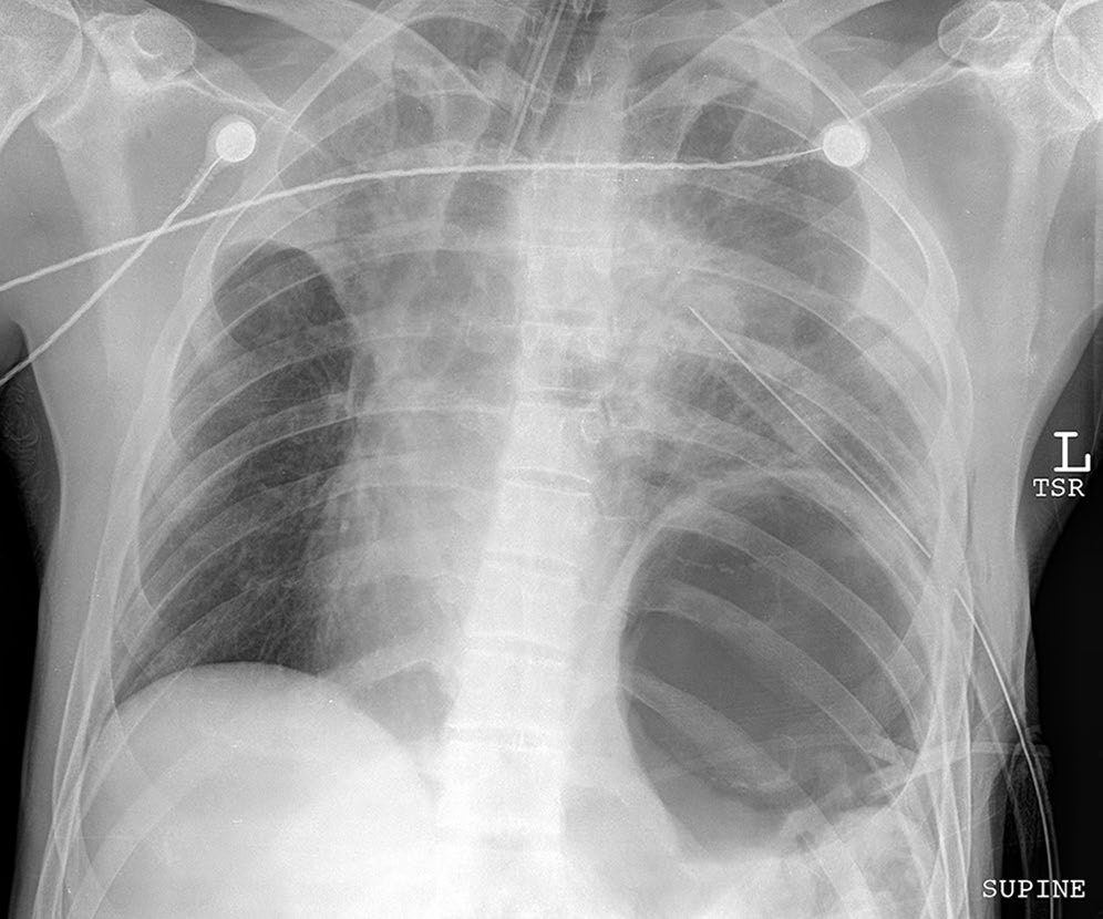
- Every emergency intervention (intubation, central line, chest tube) needs a repeat chest film; persistent pneumothorax, large air leak or difficult ventilation calls for bronchoscopy to exclude tracheobronchial injury or foreign body; a haemothorax not drained by two chest tubes is a caked haemothorax mandating thoracotomy; pneumomediastinum on CT after blunt trauma predicts aerodigestive injury poorly and is worked up selectively [8].
- Mediastinal widening from a pleura-contained haematoma suggests great vessel injury, left-sided with descending aortic and right-sided with innominate injury, though posterior rib and sternal fractures and venous bleeding mimic it, and the other radiographic signs of a descending tear are abnormal aortic contour, tracheal and nasogastric tube shift, left apical cap, paraspinal stripe thickening, depression of the left main bronchus, obliteration of the aortopulmonary window and left hilar haematoma; since at least 7% of descending injuries have a normal film, screening CTA is triggered by mechanism (frontal impact over 30 mph or lateral over 23 mph, ejection, falls over 25 ft, horse kick, snowmobile or ski impact); over 95% of survivors' injuries lie just beyond the left subclavian at the ligamentum arteriosum, 2–5% in the ascending aorta, arch or at the diaphragm, and multislice CTA reconstructions have replaced arteriography [8].
- Penetrating chest wounds are mostly defined by examination, PA and lateral films with wound markers and pericardial ultrasound, but oesophageal and tracheal injuries need bronchoscopy (persistent leak or mediastinal air) and bedside oesophagoscopy or soluble-contrast then barium oesophagography; stable transmediastinal gunshot wounds get CT for trajectory; over 60% of subclavian injuries have no pulse deficit, so CTA is done on proximity rather than brachial–brachial indices, and plain films screen contiguous cavities for retained fragments [8].
Scoring and Severity
- Traditional indications for emergent thoracotomy after chest tube placement for haemothorax include more than 1,500 mL drained immediately, more than 200 mL/h for 4 consecutive hours, more than 2,500 mL over 24 hours, or ongoing bleeding with haemodynamic instability [1][4][5].
- Sabiston qualifies those figures as deriving from Vietnam War-era data rather than contemporary trials [4].
- Occult pneumothorax is defined as identification on CT without evidence on plain radiography; observation may be considered for pneumothoraces with a CT radial diameter up to 35 mm in the absence of instability or respiratory compromise [4].
- Resuscitative or emergency department thoracotomy is considered futile after CPR for more than 15 minutes in penetrating thoracic trauma, more than 10 minutes in blunt thoracic trauma (each despite endotracheal intubation) or in blunt trauma with no signs of life at the scene [1].
- Oxford supplies the corresponding thresholds for the ward and for the drain rather than for theatre.
- A haemodynamically unstable postoperative patient, or one draining more than 200 mL of blood per hour, should be discussed urgently with the thoracic surgeons [9].
- Drains inserted for pleural effusion can be removed when they drain less than 250 mL in 24 hours; drains for empyema can be removed when they stop draining; and postoperative drains are normally removed when they drain nothing for 2 consecutive hours, unless there is an air leak [9].
- For a chest drain inserted for pneumothorax, do not remove the drain while there is an air leak, or a pneumothorax will rapidly re-form; when the air leak stops, take the drain off suction for 12 hours and repeat the chest radiograph, and if the lung is fully expanded the drain can be removed [9].
- A chest radiograph is always requested and reviewed after drain removal to check for pneumothorax [9].
Indications for thoracotomy in Schwartz's table
Over 85% of chest injuries are treated definitively by a chest tube; operation is indicated by initial drainage over 1000 mL (penetrating) or 1500 mL (blunt), ongoing drainage over 200 mL/h for 3 consecutive hours in a non-coagulopathic patient, caked haemothorax despite two tubes, great vessel injury (endovascular options in selected cases), tamponade, cardiac herniation, massive air leak with inadequate ventilation, tracheal or main bronchial injury on endoscopy or imaging, open pneumothorax, oesophageal perforation and air embolism, with the caveat that a delayed presenter whose 1.5 L output stops with a re-expanded lung may be observed if stable [8].
Treatment and Management
Tension pneumothorax: immediate decompression without waiting for radiographic confirmation, via needle or finger decompression in the "safe triangle", bordered by latissimus dorsi posteriorly, pectoralis major laterally, and a line at the nipple level inferiorly, followed by formal tube thoracostomy [1][5]. Oxford's technique is a 12G cannula into the second intercostal space in the mid-clavicular line, followed by insertion of an underwater seal chest drain into the fifth intercostal space between the anterior and mid-axillary lines [2]. Open pneumothorax: cover with a sterile occlusive dressing taped on three sides to act as a flutter valve, followed by chest tube placement remote from the wound, through a separate incision, before definitive closure [1][2][5].
Pericardial tamponade: pericardiocentesis has no role in tamponade from penetrating myocardial injury because clot typically prevents aspiration; definitive treatment is operative via subxiphoid window, sternotomy, or left anterolateral thoracotomy [1]. Oxford takes a different line for the critically ill patient, advising "blind" pericardiocentesis with a simultaneous call to cardiothoracic or general surgeons to consider emergency thoracotomy, and reserving urgent transthoracic echocardiography or focused ultrasound for the patient who is unwell but responding to treatment [2], a divergence worth noting, since the two books disagree on whether needle decompression of the pericardium is ever worth attempting.
- Massive haemothorax: correct hypovolaemic shock, insert an intercostal drain, and proceed to thoracotomy if the drainage criteria above are met; there is no role for clamping a chest tube to tamponade bleeding, with the exception of suspected tracheobronchial injury [1][5].
- Oxford adds two practical points: blood volume restoration and pleural decompression by wide-bore chest drain are done simultaneously, not sequentially, and blood from the chest tube should be collected in a device appropriate for autotransfusion [2].
- Where there is continued brisk bleeding and a need for persistent transfusion, urgent thoracotomy is considered in consultation with a regional thoracic centre [2].
- Retained haemothorax after two well-placed chest tubes is treated with VATS drainage, and is the most important risk factor for empyema; all blood should be evacuated within 48 hours to prevent fibrothorax, entrapment and empyema [5].
- Flail chest and pulmonary contusion: oxygen, adequate analgesia including epidural or intrapleural analgesia, and physiotherapy, with mechanical ventilation reserved for respiratory failure; fluid restriction is important because contused lung is very fluid-sensitive; surgical rib fixation may benefit selected patients [1][5].
- Oxford stratifies by segment size: if the segment is small and respiration is not compromised, nurse the patient in HDU with adequate analgesia, encourage early ambulation and vigorous physiotherapy, and perform regular blood gas analysis; in more severe cases endotracheal intubation with positive-pressure ventilation is required [2].
- Oxford calls pulmonary contusion the commonest potentially lethal chest injury, and its distinguishing feature is that respiratory failure develops over a period of time rather than immediately, so treatment is analgesia, physiotherapy and oxygenation, with respiratory support considered for significant hypoxia [2].
Tracheobronchial injury: intubate with a long single-lumen tube directed to the unaffected side, avoiding dual-lumen tubes; operative repair is indicated for large air leak with respiratory compromise, persistence beyond 2 weeks, inability to re-expand the lung, or injury involving more than one-third of the tracheal diameter [5]. Aortic injury: control systolic blood pressure at 100–120 mmHg with esmolol then nitroprusside until definitive repair, addressing other life-threatening injuries first. Endovascular covered stent grafting is preferred for distal transections, with open repair by left thoracotomy and partial left heart bypass if endovascular repair fails or is unsuitable, and significant intracerebral haemorrhage is a contraindication to open repair [1][5]. Diaphragmatic injury: operative repair is recommended in all cases; a transabdominal approach is used if diagnosed within a week and a transthoracic approach with lysis of adhesions if diagnosed later; penetrating diaphragmatic injuries must be repaired via the abdomen rather than the chest, to exclude concurrent hollow viscus injury [1][5].
Two NICE recommendations change the standard teaching on chest decompression, and both restrict it.
The first restricts when. Only perform chest decompression in a patient with suspected tension pneumothorax if there is haemodynamic instability or severe respiratory compromise [3]; the same rule is repeated for the hospital setting as a condition on decompressing before imaging [3]. Suspicion of tension pneumothorax is not, on its own, sufficient, one of those two physiological criteria must also be present.
- The second restricts how.
- Pre-hospital, use open thoracostomy instead of needle decompression if the expertise is available, followed by a chest drain via the thoracostomy in patients who are breathing spontaneously [3].
- In hospital the same instruction is unconditional: perform chest decompression using open thoracostomy followed by a chest drain in patients with tension pneumothorax [3].
- That is a direct substitution for the 12G-cannula-in-the-second-intercostal-space step taught in ATLS and in the textbooks above, and it applies wherever the expertise exists.
- After any decompression, observe patients for signs of recurrence of the tension pneumothorax [3].
For open pneumothorax, NICE agrees with the textbooks on the dressing but not on its description: cover the open pneumothorax with a simple occlusive dressing and observe for the development of a tension pneumothorax [3], the observation clause being the reason the three-sided technique is taught in the first place.
For blunt thoracic aortic injury, NICE is unequivocal and does not offer open repair as an equal alternative: use an endovascular stent graft [3].
Surgeries
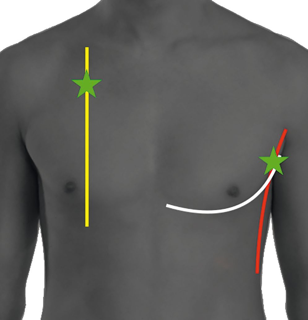
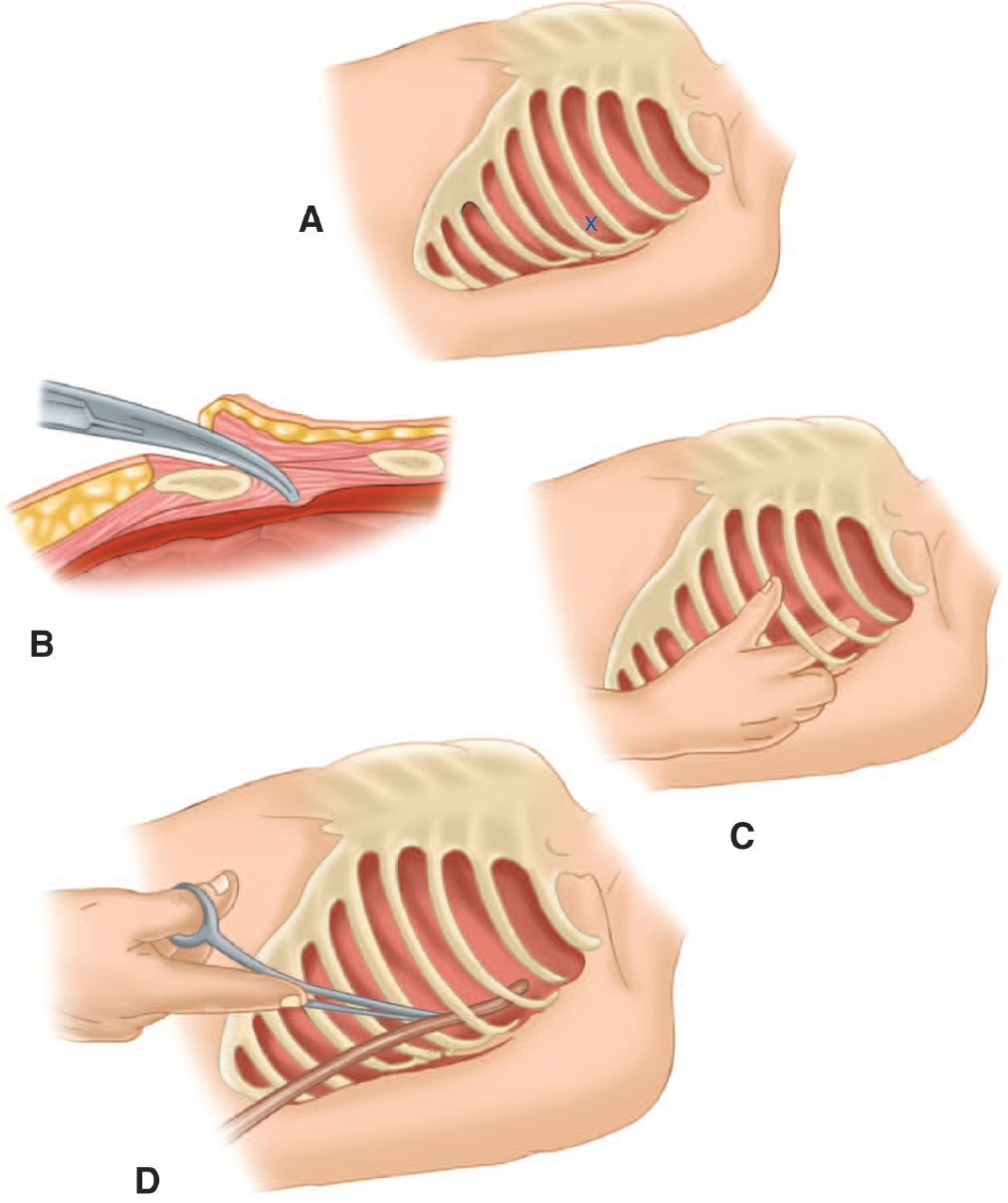
- Tube thoracostomy is performed via the "triangle of safety", nipple or inframammary fold inferiorly, midaxillary line posteriorly, lateral pectoralis major border medially, at the 4th or 5th intercostal space; an open technique is preferred in the unstable patient; chest tube size was historically 32–36 Fr, though 14 Fr percutaneous catheters show equivalent success with less morbidity; the tube is secured and connected to −20 cm H₂O suction [4].
- Oxford defines the same triangle by the anterior border of latissimus dorsi, the lateral border of pectoralis major, and a horizontal line at the level of the nipple, with the patient positioned semi-decubitus at 45° with the arm behind the head to expose the axilla, and notes that for pneumothorax the second intercostal space in the mid-clavicular line is an alternative, particularly in emergencies such as tension pneumothorax [10].
- Its three device options are needle thoracentesis for first-time treatment of simple effusions or pneumothoraces with low likelihood of recurrence; a pigtail chest drain, a 16G tube inserted by modified Seldinger technique, for simple effusions or pneumothoraces; and a large-bore chest tube, inserted bluntly or with a trocar, for tension pneumothorax, recurrent pneumothorax, haemothorax or empyema [10].
- Coagulopathy and local infection are contraindications [10].
Chest drain management
- Oxford sets out four principles that govern every chest drain [9].
- Drainage should always be into an underwater sealed container Swinging of the air–fluid meniscus with respiration demonstrates continuity with the intrapleural space, meaning the drain is working. Bubbling in the underwater seal, either continuously or on coughing, indicates an air leak from the lung parenchyma.
- And the safest mode is an unclamped drain connected to an underwater seal kept below the level of the patient at all times
- For a drain inserted for pneumothorax, use low-pressure, high-volume wall suction at −3 to −5 kPa initially (not the high-pressure wall suction used for tracheal toilet) and request and review daily chest radiographs [9].
- For drains inserted to drain collections, there is no evidence that suction improves outcome [9].
- Clamping should only be done under specialist supervision, because clamping a thoracic drain in a patient with an air leak may rapidly result in a tension pneumothorax, and clamping a mediastinal drain in a patient who is bleeding may rapidly result in cardiac tamponade [9].
- Oxford adds the trap that catches the unwary: connecting the drain to wall suction with the suction switched off, or pressing in the one-way valve on top of the underwater seal too tightly, effectively clamps the drain [9].
- Its general caution on drains applies here as much as anywhere: drains do not always drain the substance expected and may give a false sense of security, and there is no place for their routine use without a clear indication, "Better no drainage than ignorant use of it" [9].
Operative approaches
Thoracotomy approach: posterolateral thoracotomy at the 5th interspace provides access to the lungs, pulmonary vasculature and hemidiaphragm; a right-sided approach exposes the proximal and mid-oesophagus, trachea and bilateral mainstem bronchi; left thoracotomy best exposes the distal oesophagus, left lung, left ventricle, descending aorta and left subclavian artery; median sternotomy exposes the right heart, ascending aorta, aortic arch with right-sided vessels, and pulmonary vasculature [4][5]. Emergency department or resuscitative thoracotomy is reserved for penetrating injury with signs of life present: left anterolateral thoracotomy with a rib spreader, pericardiotomy anterior to the phrenic nerve, release of tamponade, control of the cardiac injury, and cross-clamping of the descending thoracic aorta [1][5]. Vascular approaches: median sternotomy, with a left 2nd-intercostal-space trap-door extension, for the ascending aorta, innominate artery and vein, and proximal subclavian and carotid injuries; left thoracotomy for the distal left subclavian artery and descending aorta; a midclavicular incision with medial clavicle resection for the distal right subclavian artery [5]. For bronchial injuries, right thoracotomy is used for right mainstem, tracheal and proximal left mainstem injuries because it avoids the aorta, and left thoracotomy for distal left mainstem injuries [5].
Repair of specific thoracic injuries in Schwartz's detail
- Over 90% of great vessel injuries are penetrating; simple ascending or arch lacerations take lateral aortorrhaphy while posterior, complex or interposition arch repairs need full cardiopulmonary bypass; innominate injuries use the bypass-exclusion technique, a 12 mm PTFE graft end-to-side from undamaged proximal aorta, tunnelled under the vein, end-to-end to the distal innominate before the haematoma is entered, then the origin oversewn, avoiding bypass; subclavian injuries take lateral arteriorrhaphy or PTFE interposition, not end-to-end anastomosis when segment is lost; descending blunt aortic injury yields to intracranial, abdominal or pelvic bleeding in priority, gets esmolol in the trauma bay to a systolic under 100 mmHg and heart rate under 100, is now mainly stented (durability in the young the open question), is repaired open on partial left heart bypass via the left superior pulmonary vein (less prone to tearing than the atrium) to protect cord and viscera and unload the ventricle, and grade I intimal injuries are managed with antiplatelets and pressure control [8].
- Penetrating cardiac wounds, mostly stabs, are controlled by a Satinsky clamp on atria and a finger on ventricles (Foley occlusion enlarges wounds with traction), temporarily or definitively stapled in the left ventricle when the edges coapt in diastole, and repaired with running 3-0 or pledgeted interrupted 2-0 polypropylene (pledgets protecting the thin right ventricle), horizontal mattress sutures beside coronary arteries to avoid occlusion, and BioGlue for friable stellate gunshot wounds; valvular or septal damage is diagnosed by auscultation, haemodynamics and echo, rarely repaired acutely, and followed by echo because it progresses; blunt cardiac injury shows tachycardia or conduction disturbance and occasionally atrial or right ventricular rupture with tamponade, has no pathognomonic ECG and enzymes do not predict complications, so stable suspects get 24 hours of telemetry and unstable ones echo for right ventricular hypokinesis, effusion, valve or chordal injury or low ejection fraction with pulmonary artery catheter and serial echo if pressors are needed [8].
- Intrathoracic tracheobronchial injury affects under 1%, blunt tears clustering within 2.5 cm of the carina; massive leaks are controlled by passing the tube beyond the injury or into the other main bronchus, repair is debridement and end-to-end 3-0 PDS with dissection limited to preserve bronchial blood supply and suture lines wrapped in pericardium, intercostal muscle or pleura, injuries under a third of the circumference without persistent leak are watched, and peripheral leaks may be sealed bronchoscopically with fibrin glue; parenchymal injuries found at thoracotomy are managed without resection where possible, stapled wedge for bleeding peripheral lacerations and pulmonary tractotomy for central tracks, the longest stapler (GIA-100) laid along the thinnest overlying parenchyma to fillet the track open for selective ligation of bronchioles and vessels, leaving the defect open, lobectomy only for a proximal injury revealed, and pneumonectomy-level injuries usually fatal from right heart failure, with bronchovenous fistula minimised by prompt control of major leaks; traumatic pneumatoceles follow a benign course with analgesia, toilet and serial films, but persistent fever prompts CT for abscess, 25% failing antibiotics alone and needing CT-guided drainage, and unresolving or refractory lesions resection [8].
- Oesophageal injuries, often with tracheobronchial wounds after penetrating trauma, are debrided and repaired end-to-end in a single layer with a vascularised pedicle between adjacent suture lines, gastro-oesophageal junction perforations repaired with fundoplication or resected with gastric pull-up, small injuries stented, and large or delayed injuries excluded with wide drainage, loop oesophagostomy and gastrostomy [8].
- Chest wall injuries are almost all non-operative: pre-emptive rib blocks with 0.25% bupivacaine in the trauma bay then thoracic wall catheters, epidurals for multiple segmental fractures, thoracotomy with ligation or angioembolisation for unusual persistent intercostal bleeding, plate fixation for extensive flail segments, markedly displaced bicortical fractures or loss of 20% of thoracic volume, local tissue or flap closure of open pneumothorax defects; scapular and sternal fractures rarely need surgery (displaced sternums may be plated) but mark force sufficient for blunt cardiac and aortic injury, clavicles are immobilised except posterior dislocation threatening the subclavian vessels; blunt diaphragmatic tears are usually large and linear, penetrating ones variable, both repaired abdominally to treat visceral injury after evacuating blood and debris from the chest and placing a tube, edges held with Allis clamps and closed with running No. 1 polypropylene, polypropylene or biological mesh or cephalad transposition by one or two interspaces bridging large defects [8].
Complications
- Retained haemothorax increases morbidity and is the most important risk factor for empyema; persistent pneumothorax despite two well-placed chest tubes warrants bronchoscopy to exclude mucus plug or tracheobronchial injury [4][5].
- Undiagnosed diaphragmatic rupture may present later with strangulation of herniated abdominal contents and high associated mortality [1].
- Myocardial contusion carries a risk of ventricular tachycardia or fibrillation, highest in the first 24 hours; supraventricular tachycardia is the most common arrhythmia overall [5].
- Endovascular stenting that covers the left subclavian artery origin can cause left hand ischaemia, treated with carotid-to-subclavian bypass [5].
- Pulmonary contusion causes progressively worsening hypoxaemia over the first 24–48 hours [1][5].
- The drain itself is a source of complications.
- Oxford lists them as damage to structures during insertion even under CT or ultrasound guidance; a potential route for the introduction of infection, especially in external drains left longer than a few days; damage to structures close to the drain, such as pressure injury from high-pressure suction; and failure to drain the substance expected, giving a false sense of security [9].
- Vasovagal reactions can occur during insertion, so patients must be monitored appropriately [10].
Post-traumatic empyema in Schwartz's account
Empyema is the commonest complication of chest injury and is managed on CT criteria: percutaneous drainage for a single loculation without appreciable rind, early VATS decortication for multiple loculations or a rind over 1 cm, fibrinolytics widely used but poorly supported, and presumptive antibiotics covering MRSA in the surgical ICU until cultures return [8].
Prognosis
- Aortic disruption is a common cause of sudden death after vehicle collision or fall from height; complete transection is rarely survived to hospital arrival, and hypotension in a patient with a widened mediastinum should prompt a search for another bleeding source rather than being attributed to the aortic injury [1].
- Delayed treatment of oesophageal injury is associated with exponentially rising mortality [1].
- More than 80% of chest injuries overall are managed successfully without thoracotomy [1], a figure consistent with Oxford's statement that fewer than 10% of blunt and fewer than 30% of penetrating chest injuries require an operation at all [2].
The service requirement behind the operative figures above is that hospital trust boards must ensure interventional radiology and definitive open surgery are equally and immediately available for haemorrhage control in all patients with active bleeding [11], so that a patient with a bleeding chest injury is not routed by what happens to be staffed. Where transfer to a major trauma centre is needed for a critical intervention, the patient must leave the sending emergency department within 30 minutes of the decision to transfer [11].
- Two NG39 recommendations bear on outcome after the acute episode.
- Heat loss must be minimised throughout [3], relevant in chest trauma because the pleural cavity is opened.
- And pain must be assessed regularly with a scale suited to the patient's age, developmental stage and cognitive function, using the same scale in hospital that was used pre-hospital, with intravenous morphine as the first-line analgesic [3].
- Analgesia is not incidental in chest trauma: it is what makes the physiotherapy and early ambulation that prevent respiratory failure after rib fractures possible at all.
References
- Bailey & Love's Short Practice of Surgery, 28th ed., Ch. 29 Torso and pelvic trauma
- Oxford Handbook of Clinical Surgery, 5th ed., Ch. 15 Major trauma
- NICE Guideline NG39: Major trauma — assessment and initial management (2016), 1.3.1; 1.3.2; 1.3.3; 1.3.4; 1.3.5; 1.3.6; 1.3.7; 1.4.1; 1.4.2; 1.4.3; 1.4.3 to 1.4.7; 1.4.4; 1.4.5; 1.4.6; 1.4.7; 1.5.34; 1.5.43; 1.6.1; 1.7.2; 1.7.3; 1.7.4 www.nice.org.uk
- Sabiston Textbook of Surgery, 22nd ed., Ch. 36 Management of Acute Trauma
- The ABSITE Review, 2022, Ch. 15 Trauma
- Schwartz's Principles of Surgery: ABSITE and Board Review, Ch. 7 Trauma
- Browse's Introduction to the Symptoms and Signs of Surgical Disease, 6th ed., Ch. 5 Major injuries
- Schwartz's Principles of Surgery, 11th ed., Ch. 7, Table 7-5, Figs. 7-22 to 7-24
- Oxford Handbook of Clinical Surgery, 5th ed., Ch. 2 Principles of surgery
- Oxford Handbook of Clinical Surgery, 5th ed., Ch. 4 Practical procedures, Figure 4.3
- NICE Guideline NG40: Major trauma — service delivery (2016), 1.5.4; 1.11.3 www.nice.org.uk