



Neck Trauma
Summary
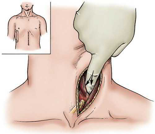
- The neck is divided into three zones because the zone determines the incision, not the diagnosis. Zone I requires a median sternotomy to reach; zones II and III use a lateral neck incision [1].
- The decision to explore is made on symptoms: any symptomatic blunt or penetrating neck injury needs exploration [1].
- This page covers the zones and their approaches, blunt cerebrovascular injury and when to image for it, oesophageal injury (the hardest neck injury to find) laryngeal and tracheal injury, and the vessels that can and cannot be ligated.
Definition
The three zones are defined by bony landmarks [1]:
| Zone | Boundaries | Approach |
|---|---|---|
| I | Clavicle to cricoid cartilage | Median sternotomy needed to reach these lesions |
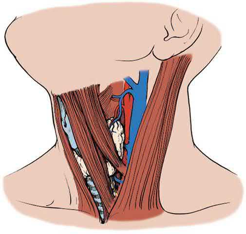
| II | Cricoid to angle of mandible | Lateral neck incision |
| III | Angle of mandible to base of skull | Lateral neck incision; may need jaw subluxation, digastric and sternocleidomastoid release, or mastoid sinus resection to reach vascular injuries |
The important implication of a zone I injury is the greater potential for intrathoracic great vessel injury [1].
Pathophysiology
The distal internal carotid artery is the commonest site of blunt cerebrovascular injury, which may take the form of dissection, transection, arteriovenous fistula or pseudoaneurysm [1].
An expanding neck haematoma can compromise the airway [1], which is why it appears among the symptoms mandating exploration rather than among the findings to observe.
Clinical features
The symptoms that mandate exploration are shock, bleeding, an expanding haematoma, losing or having lost the airway, subcutaneous air, stridor, dysphagia, haemoptysis, and a neurological deficit [1].
Laryngeal fracture and tracheal injury present with crepitus, stridor and respiratory compromise, and are airway emergencies addressed in the primary survey [1].
Recurrent laryngeal nerve injury presents with hoarseness [1].
Etiology
The indications for imaging in suspected blunt cerebrovascular injury are broad, and the threshold is deliberately low [1]: any significant blunt head or neck injury such as a Le Fort II or III fracture, basilar skull fracture, diffuse axonal injury or cervical spine fracture; any hyperflexion, hyperextension or rotation mechanism such as attempted hanging; a neurological finding not explained by brain imaging; epistaxis from a suspected arterial source; or a GCS below 8 with head injury.
An isolated neck skin seatbelt sign without soft tissue injury is not a risk factor and is not on its own enough to warrant a neck CT angiogram [1].
Diagnosis
Asymptomatic blunt injury gets a neck CT angiogram including assessment of the cervical spine [1].
Asymptomatic penetrating injury gets a neck CT angiogram, plus endoscopy or an oesophagogram for zone I and II injuries, and the chest included in the angiogram for zone I injuries [1].
Oesophageal injury is the hardest neck injury to find, and the best approach is combined: oesophagoscopy with oesophagogram finds essentially 95% of injuries when both are used [1].
Shotgun injuries to the neck need angiography and neck CT, with evaluation of the oesophagus and trachea [1].
Zones, symptoms and screening for blunt cerebrovascular injury in Schwartz's account
Penetrating wounds violating the platysma are potentially lethal; immediate operation is for instability, significant external bleeding or aerodigestive injury, and stable patients are sorted by hard signs (massive haemoptysis, rapidly expanding haematoma → exploration), soft signs (dysphagia, venous bleeding, subcutaneous emphysema, haematoma, hoarseness, stridor, odynophagia → CTA of neck and chest, with angiography, oesophagram and bronchoscopy as needed) or no symptoms (observation for 6–12 hours, except transcervical gunshot wounds, which get CTA for trajectory); zone I lies below the clavicles at the thoracic outlet, zone II between outlet and angle of the mandible and zone III above the angle, precise preoperative imaging being desirable for symptomatic zone I and III injuries, zone III injuries being managed by angioembolisation, and under 15% of penetrating neck trauma needs exploration; a blunt laryngeal fracture shows hoarseness, subcutaneous emphysema tracking in the prevertebral space or a palpable fracture [2]. Blunt carotid or vertebral injury, dissection, thrombosis or pseudoaneurysm usually in the inaccessible distal internal carotid, is sought during the latent period with multislice (ideally 64-channel) CTA in patients with signs (arterial bleeding from neck, nose or mouth, cervical bruit under 50, expanding haematoma, TIA, hemiparesis, vertebrobasilar symptoms or Horner's, deficit inconsistent with head CT, stroke on imaging) or risk factors (high-energy mechanism, Le Fort II or III, mandible, complex or basal skull or occipital condyle fracture, GCS under 6, any cervical spine fracture or ligamentous injury, near-hanging or strangulation, clothesline or seat-belt sign with swelling, scalp degloving, thoracic vascular injury, blunt cardiac rupture, upper rib fractures), graded by the Denver scale, I irregularity or dissection with under 25% narrowing, II thrombus, flap or ≥25% narrowing, III pseudoaneurysm, IV occlusion, V transection [2].
Thresholds and severity
Ligation of the common or internal carotid artery causes stroke in 20% [1].
Vertebral artery bleeding (posterior neck arterial bleeding) can be embolised or ligated, with no sequelae in the majority [1]. The external carotid can be ligated for extensive bleeding from facial fractures [1].
All oesophageal and hypopharyngeal repairs are drained, because the leak rate is 20% [1].
Bailey & Love treats the neck and spine together in its trauma section, and the practical UK emphasis is the same as the ABSITE approach above: the zone determines the exposure, and symptoms rather than imaging determine whether to explore [3].
Cricothyroidotomy is the airway of last resort, indicated where usual intubation cannot be accomplished (severe maxillofacial trauma, an airway foreign body, severe laryngospasm) and the patient has impending loss of the airway [1].
A cricothyroidotomy must later be converted to a tracheostomy [1], and tracheostomy is necessary for most laryngeal and tracheal injuries in any case, to allow oedema to subside and to check for stricture [1].
Treatment and Management
Blunt cerebrovascular injury is managed by degree of occlusion and by symptoms [1]:
- Completely occluded carotid, clopidogrel or heparin, to prevent clot propagation.
- Partially occluded and symptomatic, covered stent, with open repair if that fails.
- Partially occluded and asymptomatic, clopidogrel or heparin, with repeat CT angiography before discharge.
- Carotid AV fistula, pseudoaneurysm, or symptomatic partially occluded dissection, covered stent.
- Oesophageal injury is managed by whether it is contained.
- Contained injuries can be observed.
- For non-contained injuries, a small injury with minimal contamination gets primary closure; where the injury is extensive or contamination significant, a neck oesophageal injury is simply drained widely and will heal on its own (the same approach used when the injury cannot be found) while a chest oesophageal injury gets chest tubes and a spit fistula in the neck, and will eventually need oesophagectomy [1].
Laryngeal and tracheal injuries are repaired primarily, with strap muscle available for airway support [1].
Thyroid gland injuries are controlled by suture ligation and drainage, not thyroidectomy [1].
Recurrent laryngeal nerve injury can be repaired, or the nerve reimplanted into the cricoarytenoid muscle [1].
Vascular and aerodigestive repair in Schwartz's account
Antithrombotics cut the stroke rate of blunt cerebrovascular injury from 20% untreated to under 1%: heparin without loading dose at 15 units/kg/h to a PTT of 40–50 seconds, or aspirin 325 mg or clopidogrel 75 mg daily, equivalent in published series, for an empirical 6 months with repeat CTA at 7–10 days (grade I injuries that heal stop treatment), contraindicated by intracranial haemorrhage or falling haemoglobin with solid organ or complex pelvic injury; stenting of grade II–III carotid injuries is reserved for symptoms or a markedly enlarging pseudoaneurysm, and grade V injuries are repaired operatively if accessible or endovascularly [2]. Penetrating carotid injuries are repaired end-to-end after mobilising the common carotid, by interposition or transposition, in every patient except the comatose with delayed transport, with a Pruitt–Inahara shunt for profound shock, selective shunting otherwise and systemic heparin to an ACT of 250 seconds; tangential jugular wounds are closed by lateral venorrhaphy and extensive ones ligated, never both jugulars; vertebral injuries in the foramen transversarium are best angioembolised, Fogarty balloon occlusion controlling bleeding met at exploration; bilateral blunt jugular thrombosis aggravating cerebral oedema may warrant stenting; laryngeal and tracheal fractures shown on CT are repaired, tracheal injuries end-to-end with single-layer interrupted absorbable sutures after debridement, and coexisting oesophageal wounds are repaired with sternocleidomastoid or strap muscle interposed and a closed suction drain to prevent fistula [2].
Procedural interventions
The surgical approach to an oesophageal injury depends on its level [1]:
| Level | Approach |
|---|---|
| Neck | Left side |
| Upper half of thoracic oesophagus | Right thoracotomy, avoids the aorta |
| Lower third of thoracic oesophagus | Left thoracotomy, follows its left-sided course |
Zone III vascular injuries may require jaw subluxation, release of digastric and sternocleidomastoid, or mastoid sinus resection to gain access [1].
Complications
The 20% leak rate after oesophageal and hypopharyngeal repair is the reason drainage is mandatory [1].
Carotid ligation carries a 20% stroke rate [1], which is why stenting has displaced it wherever the anatomy allows.
Stricture is the reason a tracheostomy is left in place after airway repair rather than closed early [1].
Outcomes
The two numbers to carry from this page are both 20%: the stroke rate after carotid ligation, and the leak rate after oesophageal repair [1].
The diagnostic figure is 95%, the yield of combined oesophagoscopy and oesophagogram for the injury that is hardest to find [1].
References
- The ABSITE Review, 2022, Ch. 15 Trauma
- Schwartz's Principles of Surgery, 11th ed., Ch. 7, Trauma, Figs. 7-18 to 7-20
- Bailey & Love's Short Practice of Surgery, 28th ed., Ch. 30 The neck and spine
- Maingot's Abdominal Operations, 13th ed., Ch. 27