



Head Injury (Surgical Aspects)
Summary
- Head injury accounts for 3–4% of emergency department attendances, with an annual incidence of around 1500 per 100,000 population in the UK, and remains the leading cause of death and disability from childhood to early middle age [1].
- Traumatic brain injury (TBI) comprises a primary injury sustained at the moment of impact, which is not medically modifiable, and a secondary injury developing over subsequent hours to days, which is the principal target of medical and surgical management [1][2].
- Head injury is the most common cause of death after reaching the emergency room alive [3].
- Roughly 75% of head injuries are mild, 15% moderate and 10% severe, and up to half of all trauma deaths under the age of 45 are due to a head injury, with sequelae common in survivors [4].
- UK practice is governed by NICE NG232, Head injury: assessment and early management, published in 2023 to replace CG176.
- For its purposes a head injury is any trauma to the head other than superficial injuries to the face, including both closed and penetrating head injuries; babies are defined as under 1 year and children and young people as 1 year to under 16 [5].
- The guideline covers ten areas, from pre-hospital assessment through emergency department triage, CT criteria for the head and cervical spine, transfer to a neuroscience unit, admission and observation, and discharge and follow-up [5].
- The guideline endorses the standard courses rather than replacing them: people aged 16 and over should be initially assessed and managed according to Advanced Trauma Life Support or the European Trauma Course, International Trauma Life Support, Pre-hospital Trauma Life Support, the Advanced Trauma Nurse Course, the Trauma Nursing Core Course, or the JRCALC Clinical Practice Guidelines for Head Trauma.
- People under 16 according to Advanced or European Paediatric Life Support, Pre-hospital Paediatric Life Support, or Paediatric Education for Pre-hospital Professionals [5].
- Its own first principle is identical to the ATLS one: when administering immediate care, first treat the greatest threat to life and avoid further harm [5].
Definition
- Head injury severity is classified by the post-resuscitation Glasgow Coma Scale (GCS): minor (GCS 15, no loss of consciousness), mild (GCS 14–15 with loss of consciousness), moderate (GCS 9–13), and severe (GCS 3–8) [1].
- The GCS motor score is the best predictor of neurological outcome [1][3].
- Sabiston and Oxford both use the three-band version, mild TBI at GCS 13–15, moderate at 9–12 and severe at 3–8 [4][6].
Sabiston makes the point that this classification is increasingly recognised as inadequate. The term traumatic brain injury encompasses a heterogeneous group of diseases with distinct means of primary prevention, targets for treatment, and trajectories of recovery, and there is growing awareness of the need to classify TBI beyond mild, moderate and severe by incorporating neuroimaging detail, TBI-specific biomarkers such as GFAP and UCH-L1, and disease modifiers including social determinants of health [6]. The chapter also notes that TBI and criminal offences are bidirectionally linked, with a high prevalence of self-reported TBI history in the incarcerated population, and that TBI overlapping with intimate partner violence has disproportionately worse outcomes than other forms of intimate partner violence [6].
Pathophysiology
- Cerebral perfusion pressure (CPP) equals mean arterial pressure (MAP) minus intracranial pressure (ICP); normal cerebral blood flow of about 55 mL/min per 100 g of brain tissue is preserved across a MAP range of 50–150 mmHg through cerebral autoregulation, which can be disrupted after trauma [1].
- The Monro–Kellie doctrine holds that the cranium is a rigid box containing a nearly incompressible brain; expanding mass lesions are initially compensated by exclusion of venous blood and CSF, but further expansion produces an exponential rise in ICP [1].
- Uncontrolled ICP rise causes herniation syndromes: subfalcine, where the cingulate gyrus passes under the falx; uncal, where the temporal lobe passes over the tentorium compressing CN III; and central or tonsillar herniation compressing the brainstem, producing Cushing's triad of hypertension, bradycardia and irregular respiration as a late sign of impending herniation, or "coning" [1][3].
- Sabiston expresses the same doctrine as the reason mass lesions matter surgically: any increase in intracranial blood displaces brain and CSF within the fixed space of the skull, and the building pressure ultimately seeks an exodus by pushing the brain downward through the foramen magnum or across the tentorium or falx [6].
The three primary injuries
Sabiston divides primary brain injury into three components, which can occur in isolation or in any combination [6].
Fractures of the cranial vault and skull base are classified as open versus closed by violation of the overlying skin and soft tissue, depressed versus non-depressed, and linear versus comminuted. Skull base fractures should always prompt assessment for associated cranial nerve injuries, cerebrovascular injuries and CSF fistulas because of the close anatomical relationships, and they frequently accompany intracranial haemorrhage because disruption of the bone commonly disrupts the underlying meningeal arteries or dural venous sinuses [6].
- Intracranial haemorrhage is subdivided by compartment: epidural haemorrhage between dura and skull, typically from a meningeal artery tear; subdural haemorrhage between dura and arachnoid, from injury to the bridging veins running between the brain and the dural venous sinuses; subarachnoid haemorrhage into the spinal fluid spaces around the cortical vessels; and intraparenchymal or intracerebral haemorrhage, sometimes by a coup–contrecoup mechanism [6].
- Small intraparenchymal bleeds are also called contusions, and Sabiston's caution about them is that although initially underwhelming on imaging and physical examination, contusions can blossom and become life threatening within hours to days, earning them the label "talk and die" [6].
- Intraventricular haemorrhage is seen but rarely in isolation, because the blood must originate somewhere [6].
- Epidural and subdural collections occupy the space outside the brain and are therefore both the most likely to place external pressure on it and cause herniation, and the most amenable to urgent surgical decompression [6].
- Traumatic subarachnoid haemorrhage must be distinguished early from aneurysmal rupture, since aneurysmal bleeds are managed differently, though both carry a risk of cerebral vasospasm [6].
- Diffuse axonal injury (DAI), also called traumatic axonal injury or shear injury, is caused by rotational acceleration–deceleration injury to the white matter tracts, producing primary axotomy or functional or anatomical disruption of those pathways; in severe cases the primary axotomy is followed by a secondary axotomy of apoptosis and degeneration over the subsequent 6 to 12 hours [6].
- Visible lesions on MRI and CT are believed to be just the tip of the iceberg of the actual injury, so it is virtually impossible to know the true incidence and prevalence of DAI with current clinical neuroimaging [6].
- DAI should be on the differential in a patient who fails to improve after evacuation of a subdural or epidural haematoma, or who remains unconscious despite the absence of haemorrhage on CT, although the utility of pursuing clinical MRI is uncertain given its weak correlation with outcomes [6].
Secondary brain injury
- Secondary brain injury is driven by a self-perpetuating cycle of raised ICP, reduced cerebral perfusion, hypoxia, deranged autoregulation, and increased metabolic demand from seizure, pyrexia, and inflammation [1].
- Sabiston's account adds the mechanism and the epidemiology: at least half of all patients hospitalised with TBI have polytrauma or other associated injuries, which both raises their risk of secondary injury and forces competing life-threatening injuries to be managed simultaneously [6].
- Severe TBI disrupts cerebral vessel autoregulation, and adding systemic hypotension to impaired autoregulation reduces cerebral blood flow and produces relative ischaemia; that cycle is exacerbated by systemic hypoxia, intracranial hypertension and cerebral inflammation, leading to excitotoxicity, calcium influx and Na⁺/K⁺-ATPase dysfunction, and culminating in neuronal dysfunction and death [6].
- Elevated ICP is a global measure that may reflect oedema, cerebral venous outflow obstruction, hyperaemia from loss of autoregulation, mass effect or disturbed CSF circulation, but crucially, secondary brain injury may occur even in the absence of intracranial hypertension [6].
- At least 3% of dementia cases in the general population are attributable to TBI, and Sabiston regards this as likely an underestimate [6].
Clinical features
- Extradural haematoma classically produces transient loss of consciousness followed by a lucid interval, then rapid deterioration with contralateral hemiparesis, reduced conscious level, and ipsilateral pupillary dilatation; this "talk and die" pattern occurs in only about one-third of cases but is critical to recognise [1][3].
- Chronic subdural haematoma, common in older adults with cerebral atrophy and stretched bridging veins, presents with gradual deterioration over days to weeks, often after a trivial or unrecalled impact, especially on antiplatelet or anticoagulant therapy [1].
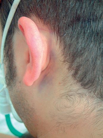
- Signs of basal skull fracture include Battle's sign, mastoid bruising associated with a middle fossa fracture and facial nerve injury; "raccoon eyes", periorbital bruising indicating an anterior fossa fracture; haemotympanum; and CSF rhinorrhoea or otorrhoea [1][3][4].
- A dilated or "blown" pupil suggests ipsilateral uncal herniation compressing the oculomotor nerve [1][3].
- Concussion presents with confusion and amnesia, without necessarily any loss of consciousness; post-concussive syndrome comprises persistent headache, dizziness, and neurocognitive disturbance [1].
- Cushing's reflex of bradycardia, hypertension and altered or Cheyne-Stokes respiration is a late sign of impending herniation [3].
The pupil examination
- Sabiston treats the pupils as a formal part of the assessment rather than an incidental sign, to be performed at the same time as the GCS once ABC has been stabilised [6].
- The normal light reflex requires a functioning lens, retina, optic nerve, brainstem and oculomotor nerve, and the consensual response assesses the function of the contralateral oculomotor nerve [6].
- Absence or asymmetry of these reflexes may indicate herniation or brainstem ischaemia; new pupillary changes or anisocoria, defined as unequal pupils with more than 1 mm of difference, may indicate a rise in ICP requiring reimaging or intervention [6].
- A unilateral fixed and dilated pupil may represent herniation, whereas bilateral fixed and dilated pupils may reflect global anoxia and a dismal prognosis [6].
- The examination may be unreliable after traumatic globe rupture or other ophthalmological trauma, and metabolic, pharmacological, toxic, surgical or genetic causes can all produce pupillary abnormality, but Sabiston's rule is that pupillary abnormalities should be presumed to indicate a primary brain injury until proven otherwise [6].
Etiology
- Road traffic accidents are the leading cause of head injury, up to 50% of cases, followed by falls and assault; firearms are the third leading cause in the USA [1].
- Oxford gives the same three as the commonest reasons in the UK: falls, road traffic accidents and assaults [4].
- Non-accidental injury must be considered in children and vulnerable adults, suggested by delayed presentation, injuries of disparate age, retinal haemorrhages, bilateral chronic subdural haematomas, multiple skull fractures, and neurological injury without external signs of trauma [1].
- Intracerebral or intraparenchymal haematoma, usually frontal or temporal, is the most common brain injury in blunt trauma [3].
- Diffuse axonal injury is a form of primary injury seen in high-energy accidents, usually rendering the patient comatose, and is associated with poor outcomes [1][3].
Diagnosis
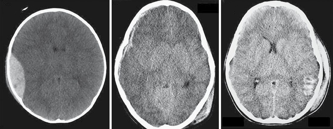
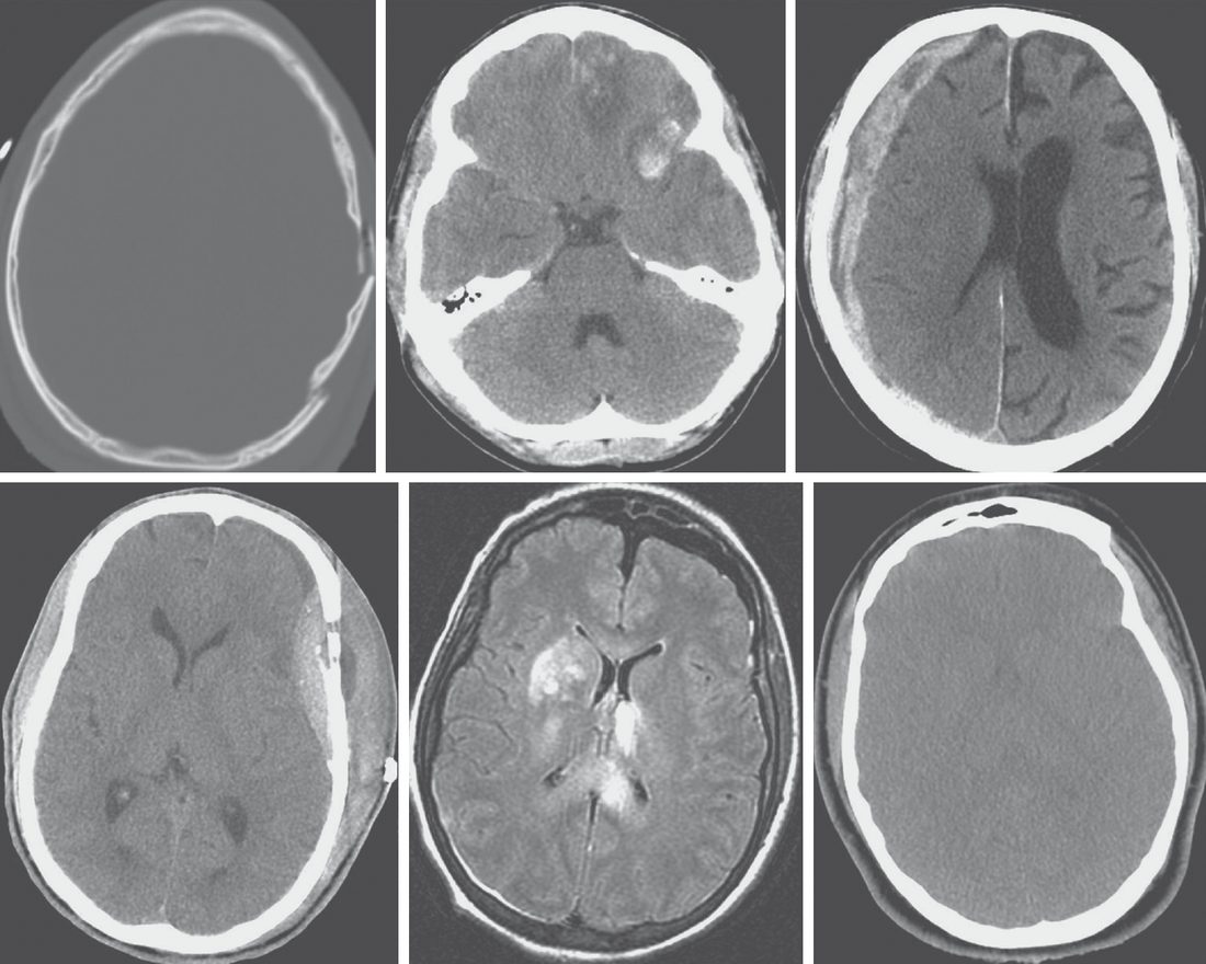
- On CT, extradural haematoma appears as a lentiform, biconvex hyperdense collection constrained by dural attachments to the skull; acute subdural haematoma appears as a diffuse concave, crescent-shaped collection free to spread over the brain surface because the dura is not adherent to the brain; chronic subdural haematoma appears diffusely hypodense [1][3].
- Diffuse axonal injury shows haemorrhagic foci in the corpus callosum and dorsolateral rostral brainstem on CT, but MRI is more sensitive, showing blurring of grey-white matter and multiple small punctate haemorrhages, classically as multifocal hyperintense lesions at the grey-white matter interfaces, graded in increasing severity with involvement of the cerebral lobes, corpus callosum and brainstem [1][3][6].
- A cervical spine fracture is found in up to 10% of moderate-to-severe TBI and must be presumed until excluded [1].
- Indications for head CT in the ABSITE account include suspected skull penetration, CSF or blood discharge from nose or ear, haemotympanum, intoxication, altered consciousness, focal signs, any loss of consciousness, and head injury with additional trauma; GCS 14 or less warrants head CT, and GCS 8 or less warrants intubation and ICP monitoring [3].
- Sabiston sets out what the non-contrast head CT is being read for: presence or absence of fracture, intracranial haemorrhage, midline shift, and the appearance of the basal and perimesencephalic cisterns, with routine scanning of the cervical spine at the same time to exclude acute fractures or traumatic dislocations [6].
- If life-threatening injuries in a polytrauma patient mandate immediate operative exploration for haemorrhage control, CT should be delayed until after stabilisation, and an intraoperatively placed ICP monitor can be a useful adjunct where there is concern about undiagnosed intracranial injury or an extended operating time [6].
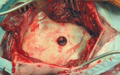
- Where a polytrauma patient has an impaired GCS and a suspected unilateral intracranial mass lesion evidenced by a unilateral fixed and dilated pupil, an exploratory burr hole may be performed in theatre concurrently with laparotomy or thoracotomy, but Sabiston stresses this situation should rarely occur, because CT neuroimaging is best used to guide emergent evacuation of epidural and subdural haematomas [6].
- The same scenario is the one ABSITE Review describes as an unstable patient with a blown pupil, in whom hypotension from abdominal or pelvic bleeding should be addressed before head CT, with a burr hole considered only if CT access will be significantly delayed [3].
NG232's CT criteria are the ones a UK trainee is examined on, and the 2023 update changed the wording of the first trigger. The primary investigation of choice for detecting an acute clinically important traumatic brain injury is CT imaging of the head; MRI should not be done as the primary investigation for safety, logistic and resource reasons, although it can sometimes add prognostic information [5]. Plain skull X-rays must not be used to diagnose important traumatic brain injury before a discussion with a neuroscience unit, though people under 16 presenting with suspected non-accidental injury may need a skeletal survey [5].
For people aged 16 and over, do a CT head scan within 1 hour of any of these being identified [5]:
| Risk factor (16 and over, CT within 1 hour) |
|---|
| --- |
| A GCS score of 12 or less on initial assessment in the emergency department |
| A GCS score of less than 15 at 2 hours after the injury on assessment in the emergency department |
| Suspected open or depressed skull fracture |
| Any sign of basal skull fracture, haemotympanum, "panda" eyes, CSF leakage from the ear or nose, Battle's sign |
| Post-traumatic seizure |
| Focal neurological deficit |
| More than 1 episode of vomiting |
- Table reformats the 1-hour CT criteria [5].
- For people 16 and over who have had some loss of consciousness or amnesia, do a CT head scan within 8 hours of the injury (or within the hour if they present more than 8 hours after it) if they have age 65 or over, any current bleeding or clotting disorder, a dangerous mechanism (a pedestrian or cyclist struck by a motor vehicle, an occupant ejected from a motor vehicle, or a fall from a height of more than 1 m or 5 stairs), or more than 30 minutes' retrograde amnesia of events immediately before the injury [5].
- A provisional written radiology report must be available within 1 hour of a CT scan [5].
- The paediatric thresholds are separate and lower.
- For people under 16, CT within 1 hour is indicated for suspicion of non-accidental injury, post-traumatic seizure, a GCS score of less than 14 on initial assessment (or less than 15 for babies under 1 year), a GCS score of less than 15 at 2 hours, suspected open or depressed skull fracture or a tense fontanelle, any sign of basal skull fracture, focal neurological deficit, or (for babies under 1 year) a bruise, swelling or laceration of more than 5 cm on the head [5].
- A second list of risk factors triggers CT within 1 hour only if more than one is present: witnessed loss of consciousness lasting more than 5 minutes, abnormal drowsiness, 3 or more discrete episodes of vomiting, a dangerous mechanism, amnesia lasting more than 5 minutes, or any current bleeding or clotting disorder [5].
- A child with only one of those is observed for a minimum of 4 hours from the time of injury, and scanned within 1 hour if the GCS falls below 15, there is further vomiting, or there is a further episode of abnormal drowsiness [5].
Anticoagulation is its own indication. For a person with no other indication for a CT head scan who is on anticoagulant treatment (vitamin K antagonists, DOACs, heparin or low molecular weight heparins) or on antiplatelet treatment excluding aspirin monotherapy, consider a CT head scan within 8 hours of injury, or within the hour if they present more than 8 hours after it [5].
For the cervical spine, do a CT scan within 1 hour in a person 16 or over if the GCS is 12 or less on initial assessment, the person has been intubated, a definitive diagnosis of cervical spine injury is urgently needed, there has been blunt polytrauma involving the head and the chest, abdomen or pelvis in someone alert and stable, or there is clinical suspicion of cervical spine injury together with age 65 or over, a dangerous mechanism, focal peripheral neurological deficit, or paraesthesia in the upper or lower limbs [5]. Where there is neck pain or tenderness without those high-risk indications, CT within 1 hour is still indicated if it is not thought safe to assess the range of neck movement, if safe assessment shows the person cannot actively rotate the neck 45 degrees to left and right, or if they have a condition predisposing to cervical spine injury such as axial spondyloarthritis [5]. MRI is added to CT if there are neurological signs and symptoms suggesting cervical spine injury, and CT or MR angiography of the neck vessels is done if vascular injury is suspected because of vertebral malalignment, a high-grade or complex facial fracture or base of skull fracture likely to involve the internal carotid or vertebral artery, or a posterior circulation syndrome [5].
Two further rules have no textbook counterpart. Do not refer people who have had a head injury for neuroimaging by direct access from the community [5]. And only assume a depressed conscious level is due to intoxication after an important traumatic brain injury has been excluded [5].
Scoring and Severity
- The Glasgow Coma Scale sums eye-opening (1–4), verbal (1–5), and motor (1–6) responses; intubated patients receive a verbal score of "1T", so the best possible score for an intubated patient is 11T [1][3][4][6].
- Of the three components, the motor score carries the most prognostic weight [1][6].
- Sustained ICP above 20–25 mmHg is associated with poor outcome; target parameters in neurointensive care include PaCO₂ 4.5–5.0 kPa, PaO₂ above 11 kPa, MAP 80–90 mmHg, ICP below 20 mmHg, CPP above 60 mmHg, and sodium above 140 mmol/L [1][3].
- Craniotomy is generally indicated for significant neurological deterioration or midline shift greater than 5 mm on imaging [3].
- Skull fracture surgical indications include depression greater than 1 cm, contamination in an open fracture, or persistent CSF leak unresponsive to conservative treatment [3].
- Peak ICP and brain swelling typically occur 48–72 hours after injury [3].
- The Glasgow Outcome Scale (good recovery, moderate disability, severe disability, persistent vegetative state, death) quantifies long-term recovery [1].
Physiological targets and how the GCS is used over time
Sabiston tabulates the Brain Trauma Foundation and American College of Surgeons TQIP targets separately for the prehospital and emergency department phase and for the intensive care unit [6]:
| Parameter | Prehospital and ED | Intensive care unit |
|---|---|---|
| Oxygen saturation | ≥90% | ≥94% |
| PaO₂ | Not stated | 80–100 mmHg |
| Systolic blood pressure | 100–150 mmHg | ≥100 mmHg |
| End-tidal CO₂ | 35–40 mmHg (30–35 mmHg accepted for imminent herniation) | Not stated |
| PaCO₂ | Not stated | 35–45 mmHg |
| Intracranial pressure | Not stated | <22 mmHg |
| Cerebral perfusion pressure | Not stated | 60–70 mmHg |
| Brain tissue oxygen tension | Not stated | ≥15 mmHg |
| Temperature | 36.0–37.0 °C | 36.0–37.9 °C |
| Glucose | Not stated | 100–180 mg/dL |
| Serum sodium | Not stated | 135–145 mEq/L |
| Serum osmolality | Not stated | 300–320 mOsm |
| INR / platelets / haemoglobin | Not stated | ≤1.4 / ≥75 × 10³/mm³ / ≥7 g/dL |
Table reformats the goals of treatment for traumatic brain injury [6]. Two thresholds in that table differ from the numbers taught elsewhere: hypotension is defined at a systolic below 100 mmHg, not 90 mmHg, and intracranial hypertension is defined as persistent elevation above 22 mmHg rather than 20 [6].
- The GCS is a repeated measure, not a single number: it should be determined at least every 30 minutes in the prehospital setting and whenever there is an apparent change in mental status, and should be measured before sedative or paralytic drugs are given if the patient's stability allows [6].
- An initially depressed GCS may reflect true injury severity, but may equally reflect haemorrhagic shock with poor cerebral perfusion, hypoglycaemia, seizure activity, sedatives or other ingestions, all of which may improve the score once addressed [6].
- The Simplified Motor Score and Simplified Verbal Score are also in some prehospital use [6].
Sabiston uses the Glasgow Outcome Scale-Extended (GOSE) as its main outcome metric, an eight-point scale running from 1 dead, 2 vegetative state, 3 lower severe disability with complete dependence, 4 upper severe disability with dependence for some activities, 5 lower moderate disability with inability to return to work or social activities, 6 upper moderate disability with return to work or social function at reduced capacity, 7 lower good recovery with minor social or mental deficits, to 8 upper good recovery [6]. Acute-phase scales include the Rancho de Los Amigos Scale and the Galveston Orientation and Amnesia Test [6].
- NG232 does not set physiological targets for intensive care, but it is prescriptive about how the GCS is recorded and communicated, which is where UK practice diverges most visibly from the international literature.
- Monitoring and exchange of information must be based on the 3 separate responses, so that a person with a GCS score of 13 based on 4 on eye opening, 4 on verbal response and 5 on motor response is described as E4, V4, M5 [5].
- When recording or passing on a total score it is given out of 15, for example "13 out of 15" [5], and the individual components must be described in all communications and every patient record and must always accompany the total score [5].
- The paediatric version of the GCS includes a "grimace" alternative to the verbal score so that preverbal children can be scored [5].
- A fifth recommendation removes a common trap: in some people, for example those with dementia, underlying chronic neurological disorders or learning disabilities, the pre-injury baseline GCS score may be less than 15, and this should be established where possible and taken into account during assessment [5].
- Ambulance crews and paramedics should be fully trained in the adult and paediatric versions of the GCS and its derived score [5].
- A pre-alert call must be made to the destination emergency department for anyone with a GCS score of 8 or less [5], and a trained member of staff must assess anyone presenting with a head injury within a maximum of 15 minutes of arrival at hospital, establishing whether they are at high or low risk for clinically important brain or cervical spine injury [5].
- Anyone presenting with a GCS score of less than 15 must be assessed immediately by a trained member of staff [5].
Treatment and Management
- Resuscitation follows ATLS principles, beginning with airway and cervical spine control, with hypoxia and hypotension avoided to prevent secondary injury; blood glucose is checked early to exclude reversible hypoglycaemia [1].
- ICP is controlled first by simple measures, raising the head of the bed, loosening a tight collar, and controlling seizures and pyrexia, before escalating to sedation, analgesia, and paralysis [1][3].
- Mannitol, loaded at 1 g/kg then 0.25 g/kg every 4 hours, or hypertonic saline, can be used to temporise or treat raised ICP by an osmotic effect [1][3].
- Modest relative hyperventilation to a PaCO₂ of 30–35 mmHg or 4.5–5.0 kPa produces cerebral vasoconstriction to lower ICP, but excessive hyperventilation risks cerebral ischaemia [1][3].
- Routine, non-directed corticosteroids in severe head injury increase mortality and are not recommended [1].
- Prophylactic antiepileptics such as phenytoin, fosphenytoin or levetiracetam for about a week are given to patients at high risk of early seizures with moderate-to-severe TBI [1][3].
- Blind nasogastric or nasotracheal tube placement is contraindicated with suspected skull base fracture [1][3].
- Anticoagulation should be reversed with vitamin K, prothrombin complex concentrate or DOAC-specific reversal agents in patients on anticoagulants with an abnormal head CT; a normal CT still warrants a repeat scan at 8 hours because of the risk of delayed bleeding [1][3].
- Enteral nutrition should begin within 72 hours of injury [1].
- Second impact syndrome, malignant brain swelling after a repeat and apparently trivial injury while still symptomatic, means symptomatic athletes must not return to play [1].
- Oxford adds that systemic analgesia should be avoided until full neurological assessment is made, and that an anaesthetist should be involved for airway management in a patient with a GCS below 8 or an AVPU score of P or U [4].
The three prehospital pitfalls
- Sabiston frames prehospital management around three things to be anticipated, prevented and avoided in any patient with suspected TBI: hypoxia (saturation below 90%), hypotension (systolic below 100 mmHg), and hyperventilation (end-tidal CO₂ below 35), with monitoring at least every 5 minutes, or continuously where available [6].
- Preventing those three pitfalls is what the Excellence in Prehospital Injury Care Study implemented across more than 130 EMS systems in Arizona, improving TBI outcomes [6].
- Ventilation of patients with altered consciousness should target an end-tidal CO₂ of 35 to 45 mmHg, and both hypothermia and hyperthermia should be avoided [6].
- The single exception to the ban on hyperventilation is a patient with a prehospital GCS below 9 and signs of imminent herniation, Cushing's triad, posturing or lateralising findings, or unilateral or bilateral fixed dilated pupils, in whom hyperventilation should temporarily target an end-tidal CO₂ of 30 to 35 mmHg [6].
- Two other prehospital points are stated as negatives: hyperosmolar therapy with mannitol or hypertonic saline should not be given for the prophylactic treatment of suspected elevated ICP in the prehospital setting, having shown no benefit in large randomised trials, and the evidence for hypertonic fluids prehospital is weak [6].
- Prehospital tranexamic acid is described only as used empirically in some practice settings for clot stabilisation in patients with suspected intracranial haemorrhage or elevated ICP [6].
- Patients with suspected moderate to severe TBI should be transported directly from the scene to the highest-level trauma centre available, and any patient with suspected TBI of any type, particularly with polytrauma, should go directly to the nearest trauma centre rather than being stabilised at a non-trauma centre first [6].
- Tranexamic acid for isolated head injury is the clearest divergence between UK guidance and the textbooks, because NICE gives a different dose, a different window and a different indication from the major-trauma regimen.
- For people with a head injury and a GCS score of 12 or less who are not thought to have active extracranial bleeding, NICE says to consider a 2 g intravenous bolus injection of tranexamic acid for people 16 and over, or 15 mg/kg to 30 mg/kg up to a maximum of 2 g for people under 16, given as soon as possible within 2 hours of the injury, in the pre-hospital or hospital setting, and before imaging [5].
- In March 2023 these were off-label uses of tranexamic acid [5].
- Where extracranial bleeding is suspected or confirmed, the major-trauma regimen applies instead [5]: intravenous tranexamic acid as soon as possible in patients with major trauma and active or suspected active bleeding [8], and not more than 3 hours after injury unless there is evidence of hyperfibrinolysis [8].
- So the same drug carries a 2-hour window and a single 2 g bolus for isolated head injury, and a 3-hour window under the major-trauma protocol when the patient is also bleeding elsewhere.
- Two further prehospital and resuscitation points come from NG39 by cross-reference.
- For a patient with both haemorrhagic shock and traumatic brain injury, if haemorrhagic shock is the dominant condition, continue restrictive volume resuscitation; if the brain injury is dominant, use a less restrictive volume resuscitation approach to maintain cerebral perfusion [8].
- And pain must be managed effectively because it can lead to a rise in intracranial pressure, provide reassurance, splint limb fractures and catheterise a full bladder when needed [5].
Critical care management
- Goal-directed treatment extends from the prehospital and emergency department phases into the ICU.
- Systolic and mean arterial pressure should be tracked closely to avoid hypotension and optimise CPP; body temperature, glucose and electrolytes should be kept in the normal range; and hyponatraemia should specifically be avoided because of the risk of cerebral oedema [6].
- Patients with severe TBI merit frequent monitoring of serum sodium and osmolality because they are at risk of developing diabetes insipidus or SIADH [6]. Paroxysmal sympathetic hyperactivity, or "storming", can occur after severe TBI, is associated with elevated catecholamines producing hypertension and other negative effects, and is commonly treated with beta-blockade, though evidence for benefit is variable [6].
- ICP monitoring is indicated for comatose patients with a GCS of 8 or less and evidence of structural brain injury on CT, or in patients judged at high risk of progression because of large contusions or coagulopathy, and may also be considered in moderate TBI undergoing urgent extracranial surgery [6].
- The external ventricular drain's advantage over an intraparenchymal bolt is the ability to drain CSF and thereby lower ICP; subdural and epidural monitors tend to be less accurate and are used less [6].
- Sabiston's caveat is important: monitoring should not replace careful, repeated neurological assessments and prompt repeat CT imaging in cases of neurological deterioration, and in under-resourced settings reasonably good outcomes from severe TBI can still be obtained using institutional protocols of empiric treatment of elevated ICP, repeat imaging and meticulous clinical examination [6].
Treatment of intracranial hypertension follows a three-tier approach [6]. Tier 1 elevates the head of the bed to 30 degrees with the head neutral to improve cerebral venous outflow, taking care that cervical immobilisation devices do not obstruct jugular venous flow, adds short-acting sedation such as propofol or fentanyl in intubated patients to allow frequent reassessment off sedation, and uses intermittent CSF drainage where an EVD is in place. Tier 2 adds hyperosmolar therapy with mannitol or hypertonic saline, with serum sodium and osmolality checked every 6 hours and treatment held if levels reach 160 mEq/L or 320 mOsm/L respectively; a single test dose of neuromuscular paralysis is given, with continuous infusion only if the test dose reduces ICP; and hyperventilation to a PaCO₂ of 30–35 mmHg may be considered although brain hypoxia may result from the reduced cerebral blood flow. Tier 3 comprises decompressive bilateral craniectomy, continuous neuromuscular blockade if the tier 2 test dose worked, or barbiturate coma with continuous EEG titrated to burst suppression; hypothermia is not recommended as salvage treatment for intracranial hypertension [6]. In a deeply sedated patient, the pupillary examination and the ICP measurement are all that remain, and a persistently elevated ICP or a new blown pupil necessitates emergent CT neuroimaging [6].
Sabiston tabulates eleven treatments that should be avoided in severe TBI: mannitol by non-bolus continuous infusion; scheduled infusion of hyperosmolar therapy every 4–6 hours; lumbar drainage of CSF; furosemide; routine use of steroids; routine therapeutic hypothermia to below 35 °C; high-dose propofol to attempt EEG burst suppression; hyperventilation to a PaCO₂ below 30 mmHg; routinely raising CPP above 90 mmHg; platelet transfusion in adults on antiplatelet therapy; and antibiotic prophylaxis for external ventricular drain placement [6].
- Other supportive measures: nutritional support should begin within 24 to 48 hours of injury with full nutrition by 7 days at the latest, enterally by preference, because the hypermetabolic and hypercatabolic state lasts from 1 week to several months [6].
- Anticonvulsant prophylaxis is given to prevent early post-traumatic seizures within the first week [6]. Once there is no evidence of ongoing intracranial haemorrhage, pharmacological VTE prophylaxis should be started as soon as 24 to 48 hours after injury, because of high VTE rates in this population [6].
- The dopamine agonist amantadine may improve cognitive function, especially when given within the first week [6].
- Early tracheostomy within the first 5 to 7 days should be considered in patients requiring mechanical ventilation, to reduce ventilator-associated pneumonia and other complications [6].
- Coagulopathy should be reversed when apparent on laboratory testing or from a history of anticoagulant use, to limit expansion of intracranial bleeding, using prothrombin complex concentrate for anticoagulants and desmopressin for non-aspirin antiplatelet agents [6].
- Transfer to a neuroscience unit.
- Local transfer guidelines drawn up between referring trusts, the neuroscience unit and the ambulance service must recognise that transfer would benefit anyone with serious head injuries, meaning a GCS score of 8 or less, irrespective of the need for neurosurgery; where transfer of patients who do not need neurosurgery is not possible, ongoing liaison with the neuroscience unit over clinical management is essential [5].
- Occult extracranial injuries must be considered in patients with multiple injuries, who must not be transferred to a service unable to deal with other aspects of trauma [5].
- Initial resuscitation and stabilisation must be completed and comprehensive monitoring established before transfer, and a person with persistent hypotension despite resuscitation must not be transported until the cause has been identified and they are stabilised [5].
- Intubate and ventilate anyone with a GCS of 8 or less needing transfer [5], and immediately when there is coma, loss of protective laryngeal reflexes, ventilatory insufficiency judged by blood gases as hypoxaemia with PaO₂ less than 13 kPa on oxygen, or hypercarbia with PaCO₂ more than 6 kPa, or irregular respirations [5].
- Intubation before the journey is also indicated for a significantly deteriorating conscious level of one or more points on the motor score even if not coma, unstable fractures of the facial skeleton, copious bleeding into the mouth, or seizures [5].
- Once intubated, aim for a PaO₂ of more than 13 kPa and a PaCO₂ of 4.5 kPa to 5.0 kPa, unless there is clinical or radiological evidence of raised intracranial pressure, in which case more aggressive hyperventilation is justified with an increased inspired oxygen concentration; maintain the mean arterial pressure at 80 mmHg or more by infusion of fluid and vasopressors as indicated [5].
- Anticoagulant reversal.
- Offer immediate prothrombin complex concentrate transfusion for the emergency reversal of warfarin anticoagulation in people with either severe bleeding or head injury with suspected intracerebral haemorrhage [9], monitoring the INR to confirm adequate reversal and considering further prothrombin complex concentrate [9].
- Under the major trauma guideline, anticoagulation in a patient with major trauma and haemorrhage must be rapidly reversed, trusts must have a protocol for rapid identification of anticoagulated patients, plasma must not be used to reverse a vitamin K antagonist, and a haematologist must be consulted immediately for advice on any anticoagulant other than a vitamin K antagonist [8]. Do not reverse anticoagulation in patients who do not have active or suspected bleeding [8].
Surgeries
- Significant extradural haematoma requires urgent transfer for surgical evacuation, typically via craniotomy, in deteriorating or comatose patients or those with large bleeds; smaller bleeds may be observed with serial imaging [1].
- Acute subdural haematoma from high-energy injury with significant midline shift or deteriorating neurology requires urgent evacuation, typically by craniotomy or craniectomy [1].
- Chronic subdural haematoma is generally drained through burr holes; if clinically stable, a 7–10 day delay may be used to allow platelet function to normalise after stopping antiplatelet agents, and liquefaction of clot over 7–10 days can allow less invasive burr-hole evacuation even of some acute collections [1].
- ICP monitoring uses an intraparenchymal bolt or an external ventricular drain, which also allows therapeutic CSF drainage, indicated for GCS 8 or less with head injury, suspected raised ICP, or inability to follow the clinical examination, as when the patient is intubated [1][3].
- Depressed skull fractures, where bone is displaced inward by at least the thickness of the skull and which are usually compound, require exploration and elevation, especially with intracranial air suggesting a dural breach; fractures through air sinuses are managed as open fractures with broad-spectrum antibiotics with or without exploration [1].
- Traumatic intraventricular haemorrhage causing hydrocephalus requires ventriculostomy [3].
Operative thresholds and the craniectomy question
- Sabiston gives explicit volumetric criteria. An epidural haematoma of more than 30 mL, in association with neurological deficits or radiographic signs of midline shift or effacement of the basal cisterns, is commonly accepted as the criterion for emergent craniotomy
- A subdural haematoma of at least 1 cm in size, or with any midline shift, in combination with a decline in neurological examination should also prompt evacuation [6].
- Regardless of examination findings, a large mass lesion should be evacuated before neurological deterioration develops, if that accords with the patient's goals of care [6].
- Both types are treated with a formal craniotomy centred on the clot, rather than a burr hole, and depending on brain swelling the neurosurgeon may elect to leave the bone flap off (a craniectomy) to allow the brain to heal before coverage.
- A recent international trial reached equipoise on routine craniectomy versus craniotomy [6].
- ICP monitors are often placed at the time of craniotomy, particularly for a preoperative GCS below 8, at the operating surgeon's discretion [6].
- Decompressive craniectomy is considered when ICP cannot be controlled medically, though evidence for long-term outcome benefit is limited [1][3].
- Sabiston states the trial evidence behind that caution: decompressive craniectomy for intracranial hypertension in the absence of mass lesions is performed at some institutions as tier 3 management but is not well supported by the literature
- The 2011 randomised DECRA trial showed that the successful reduction in ICP achieved by decompressive craniectomy was associated with worse neurological outcomes at 6 months, with similar findings in the 2016 RESCUEicp trial [6].
- NICE makes no recommendation on decompressive craniectomy, and that silence is itself the finding.
- NG232 is a guideline on assessment and early management: it covers pre-hospital care, emergency department triage, imaging criteria, transfer, observation and discharge, but it contains no recommendations on operative thresholds for evacuating an extradural or subdural haematoma, on ICP or CPP targets, or on decompressive craniectomy [5].
- A search of NICE guidance for decompressive craniectomy returns no product of any type, no guideline, no technology appraisal and no HealthTech guidance.
- UK operative practice therefore runs on the Brain Trauma Foundation criteria and on the DECRA and RESCUEicp trial evidence, with NICE contributing only the decision about who reaches a neurosurgeon and when
- What NG232 does specify is the referral trigger. Discuss with a neurosurgeon the care of anyone with new and surgically significant abnormalities on imaging, with the definition of "surgically significant" developed by local neurosurgical centres and agreed with referring hospitals along with the referral procedures [5].
- And regardless of imaging, discuss a person's care plan with a neurosurgeon if they have persisting coma with a GCS of 8 or less after initial resuscitation, unexplained confusion persisting for more than 4 hours, deterioration in GCS after admission (with more attention paid to motor response deterioration) progressive focal neurological signs, a seizure without full recovery, a definite or suspected penetrating injury, or a cerebrospinal fluid leak [5].
- Oxford's list of indications for neurosurgical referral matches it: major intracranial injury such as extradural haematoma, moderate or larger subdural haematoma or intracerebral haematoma; progressive focal neurological signs; definite or suspected penetrating head injury; CSF leak or base of skull fracture; and persisting coma after initial resuscitation or deterioration in GCS after admission.
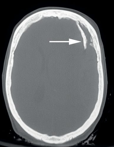
Burr holes, maxillofacial and spinal injury in Schwartz's account
- Schwartz keeps ICP under 20 mmHg (normal upper limit 10) with cerebral perfusion pressure above 60, evacuates haematomas on clot volume, shift over 5 mm (not absolute), location, GCS and ICP, small posterior fossa clots for brainstem compression, small clots to relieve refractory ICP, and treats open or depressed fractures and penetrating wounds operatively; the general surgeon without neurosurgical cover must know burr-hole placement for a life-threatening epidural haematoma, whose course is loss of consciousness, a lucid interval and recurrent coma with an ipsilateral fixed dilated pupil as the temporal lobe compresses the third nerve and brainstem: epidurals need evacuation within 70 minutes while subdurals may wait, the hole is made on the side of the dilated pupil, external carotid branches are usually ligated for access, no attempt is made to control intracranial bleeding through the hole, the head is wrapped in a bulky dressing and the patient transferred for craniotomy [11].
- Secondary injury is limited by avoiding systolic under 100 mmHg, PaO₂ under 60 or saturation under 90%, keeping PCO₂ 35–40 (hyperventilation below 30 only briefly for acute hypertension), using sedation, osmotic diuresis, paralysis, ventricular drainage and barbiturate coma in sequence, noting that moderate hypothermia (32–33°C) helped experimentally but not clinically and decompressive craniectomy remains controversial after a multicentre trial, and giving 7 days of prophylactic anticonvulsants [11].
- Maxillofacial injury is divided into upper (frontal sinus and brain), mid (orbits, nose, zygomaticomaxillary complex) and lower (mandible) face, high energy fracturing frontal sinus, orbital rims and mandible and low energy nose and zygoma; bleeding from facial fractures is controlled by nasal packing, Foley tamponade of the posterior nose and oropharyngeal packing with prompt angioembolisation for exsanguination, tooth-bearing fractures are open fractures needing antibiotics and semi-urgent repair for airway, occlusion and aesthetics, orbital fractures threaten vision, cause diplopia and enophthalmos, nasoethmoid fractures threaten the lacrimal system and cribriform plate with CSF rhinorrhoea, and three-dimensional CT follows a cranial nerve examination [11].
- Cervical spine injuries are reduced and stabilised by supervised axial traction with tongs or, more often, a halo vest, or braced; fusion is performed for neurological deficit, angulation over 11° or translation over 3.5 mm, or persistent instability, immediate surgery for deterioration or fracture-dislocation with incomplete deficit, and urgent decompression for bilateral locked facets with incomplete tetraplegia or deterioration, surgery within 24 hours shortening stay and complications; methylprednisolone (24-hour infusion within 3 hours, 48-hour within 3–8 hours) is no longer recommended, complete injuries are essentially untreatable, and about 3% of flaccid quadriplegics have concussive injury and recover [11].
Complications
- Depressed skull fractures carry a high incidence of infection, neurological deficit, and late-onset epilepsy [1].
- Skull base fractures may be complicated by pituitary dysfunction, arterial dissection, and cranial nerve deficits including anosmia, facial palsy and hearing loss; persistent CSF leak can result in meningitis [1].
- Electrolyte disturbance is common in TBI: cerebral salt wasting and SIADH both cause hyponatraemia, which worsens brain swelling, so hypotonic fluids are avoided, while impaired ADH secretion causes diabetes insipidus with hypernatraemia [1].
- Post-traumatic seizure cumulative probability ranges from 2% in mild TBI to 60% in severe TBI, with risk factors including intracerebral haemorrhage, depressed skull fracture and dural tear [1].
- Coagulopathy after TBI results from release of tissue thromboplastin [3].
- Cervical collars, especially if ill-fitting, may increase ICP and impair airway management [2].
- Observation after admission is the part of UK head injury practice that is specified in the most detail, and it is where most examinable numbers sit.
- People are admitted after a head injury for new, clinically important abnormalities on imaging, noting that an isolated simple linear non-displaced skull fracture is unlikely to be a clinically important abnormality unless the person is taking anticoagulant or antiplatelet medication; for a GCS that has not returned to 15 or the pre-injury baseline after imaging, regardless of the imaging result; when there are indications for CT that cannot be met in time; for continuing worrying symptoms such as persistent vomiting, severe headaches or seizures; or for other sources of concern including intoxication, other injuries, shock, suspected non-accidental injury, meningism, CSF leak, or suspicion of ongoing post-traumatic amnesia [5].
- Patients with multiple injuries are admitted under the team trained to deal with their most severe and urgent problem [5].
- The minimum acceptable documented neurological observations are GCS score, pupil size and reactivity, limb movements, respiratory rate, heart rate, blood pressure, temperature and blood oxygen saturation [5].
- Observations are carried out and recorded half-hourly until the GCS is 15; for a person whose GCS is 15, observations start after the initial emergency department assessment and run half-hourly for 2 hours, then 1-hourly for 4 hours, then 2-hourly [5].
- A person with a GCS of 15 who deteriorates at any time after the initial 2-hour period reverts to half-hourly observations and follows the original schedule again [5].
Urgent reassessment by a supervising doctor is required for any of these signs of neurological deterioration [5]:
| Sign of neurological deterioration |
|---|
| --- |
| Agitation or abnormal behaviour |
| A sustained drop (for at least 30 minutes) of 1 point in GCS score, giving more weight to a drop of 1 point in the motor response score |
| Any drop of 3 or more points in the eye opening or verbal response scores, or 2 or more points in the motor response score |
| Severe or increasing headache, or persistent vomiting |
| New or evolving neurological symptoms or signs, such as pupil inequality or asymmetry of limb or facial movement |
- Table reformats the deterioration triggers [5].
- To reduce interobserver variability and unnecessary referrals, a second competent member of staff should confirm deterioration before the supervising doctor is involved, immediately if possible, but if no second observer is available, the supervising doctor is contacted without confirmation [5].
- If deterioration is confirmed, consider an immediate CT scan [5], and if a person has had a normal CT scan but does not have a GCS score of 15 after 24 hours of observation, consider a further CT or MRI scan in discussion with the radiology department [5].
- Hypopituitarism is the complication NG232 added in 2023, and it is easy to miss because it can present months later.
- Be aware that any severity of head injury can cause pituitary dysfunction, which may present immediately, or hours, weeks or months after the injury, with a variety of symptoms [5].
- In people admitted to hospital with a head injury who have persistently abnormal low sodium levels or low blood pressure, consider investigations for hypopituitarism [5]; and in people presenting to primary or community care with persistent symptoms consistent with hypopituitarism in the weeks or months after a head injury, consider investigations or referral [5].
- That extends the textbook association between pituitary dysfunction and skull base fracture to head injury of any severity.
Prognosis
- Prompt evacuation of extradural haematoma without associated primary brain injury carries an excellent prognosis [1].
- Chronic subdural haematoma in the elderly is a common and treatable cause of acute neurological deterioration [1].
- Diffuse axonal injury carries a very poor prognosis [3].
- Penetrating head injury has the worst survival of all head injury types [3].
- UK trauma audit data show higher mortality when severe TBI is managed at non-neurosurgical centres, supporting early transfer regardless of the ultimate need for surgery [1].
- Reduced CPP results in secondary traumatic brain injury and worsens outcomes; hypotension and hypoxia are the principal preventable drivers of secondary injury [2][3].
Prognostication and the 72-hour rule
- Sabiston is unusually direct about the limits of prognostication and its consequences.
- After discharge, TBI recovery is varied and poorly understood, with predictions limited to 6 months; it is unknown at what point recovery plateaus, and in some patients post-TBI neuroplasticity continues for multiple years [6]. Currently available prognostic models do not adequately predict long-term outcomes for individual patients and should be used with caution when making treatment-limiting decisions: despite broad acceptance in research, the IMPACT and CRASH-TBI models are infrequently used clinically and predict only 35% of the variance in patient outcomes [6].
- The one exception is the Baylor score for penetrating intracranial gunshot wounds, which does adequately predict mortality and functional outcomes to inform goals-of-care discussions acutely [6].
- From that follows the practical rule.
- Clinical fluctuations during the first 72 hours after injury may help inform prognosis and trajectory, and patients with severe TBI should receive full treatment for at least 72 hours unless their goals of care do not align with a trial of full treatment [6].
- There is a growing consensus that premature withdrawal of life-sustaining measures, meaning within 72 hours, can lead to a self-fulfilling prophecy of fatalism or inappropriate therapeutic nihilism, and that a substantial minority of patients with seemingly devastating TBI can recover to independent functional status [6].
- The chapter closes on Hippocrates: no head injury is too severe to despair of, nor too trivial to ignore [6].
- Discharge is gated on the GCS and on supervision, not on the scan alone.
- If CT is not indicated on history and examination and there is no suspicion of clinically important traumatic brain injury, discharge is appropriate provided there are no other factors warranting admission and there are appropriate support structures for safe discharge, for example competent supervision at home [5].
- Where imaging of the head is normal and the risk of clinically important brain injury is low, transfer to the community requires the GCS score to have returned to 15 or the pre-injury baseline, no other factors warranting admission, and appropriate support structures [5].
- The rule is stated again as an absolute: do not discharge people presenting with a head injury until their GCS score is 15, or, in preverbal and non-verbal children, until consciousness is normal on the paediatric GCS, and in people with pre-injury cognitive impairment until the GCS is back to the documented pre-injury value [5]. Only transfer people with any degree of head injury to their home if there is somebody suitable at home to supervise them; discharge a person with no carer at home only if suitable supervision has been organised or the risk of late complications is thought negligible [5].
- People with pre-injury cognitive impairment such as dementia or a learning disability, and people returning to a custodial setting, must be supervised and monitored with arrangements in place should there be signs of deterioration [5].
- Verbal and printed discharge advice must be given to people with any degree of head injury and to the person responsible for their care after discharge, whether family, carers, social workers or custodial staff [5].
- Beyond the acute episode, rehabilitation after traumatic injury is covered by its own guideline [12].
References
- Bailey & Love's Short Practice of Surgery, 28th ed., Ch. 28 Traumatic brain injury
- Sabiston Textbook of Surgery, 22nd ed., Ch. 36 Management of Acute Trauma
- The ABSITE Review, 2022, Ch. 15 Trauma
- Oxford Handbook of Clinical Surgery, 5th ed., Ch. 15 Major trauma
- NICE Guideline NG232: Head injury — assessment and early management (2023; replaces CG176), 1.3.1; 1.3.2; 1.3.3; 1.3.4; 1.3.5; 1.3.6; 1.3.7; 1.3.8; 1.3.10; 1.3.11; 1.3.15; 1.3.17; 1.3.18; 1.3.19; 1.4.2; 1.4.4; 1.4.7; 1.4.11; 1.4.15; 1.4.16; 1.5.1; 1.5.2; 1.5.4; 1.5.8; 1.5.9; 1.5.10; 1.5.11; 1.5.12; 1.5.13; 1.5.14; 1.6.2; 1.6.3; 1.6.9; 1.6.10; 1.8.1; 1.8.2; 1.8.6; 1.8.7; 1.8.8; 1.8.9; 1.8.10; 1.9.1; 1.9.3; 1.9.6; 1.9.7; 1.9.8; 1.9.10; 1.9.11; 1.9.12; 1.9.13; 1.9.14; 1.9.15; 1.9.16; 1.10.1; 1.10.2; 1.10.4; 1.10.5; 1.10.6; 1.10.8; Recommendations www.nice.org.uk
- Sabiston Textbook of Surgery, 22nd ed., Ch. 41 Emergency Care of Neurologic Injuries
- Sabiston Textbook of Surgery, 22nd ed., Ch. 42
- NICE Guideline NG39: Major trauma — assessment and initial management (2016), 1.5.4; 1.5.5; 1.5.6; 1.5.7; 1.5.9; 1.5.10; 1.5.12; 1.5.21 www.nice.org.uk
- NICE Guideline NG24: Blood transfusion (2015, last updated February 2026), 1.12.3; 1.12.6 www.nice.org.uk
- Bailey & Love's Short Practice of Surgery, 28th ed., Ch. 8
- Schwartz's Principles of Surgery, 11th ed., Ch. 7, Trauma, Fig. 7-52
- NICE Guideline NG211: Rehabilitation after traumatic injury (2022), Recommendations www.nice.org.uk