



Kidney and Pancreas Transplantation
Summary
- Kidney transplantation is the commonest form of organ transplant [1], and it is the operation against which the rest of transplant surgery is measured: all patients with end-stage renal failure should be considered for renal transplant unless there are specific contraindications [1].
- Pancreas transplantation is grouped with it because 90% of pancreas transplants are performed simultaneously with a kidney.
- This page covers matching, the operation, postoperative course, the specific complications (urine leak, lymphocele, vascular thrombosis) and the results. Transplantation extends life by 15 years, and the commonest cause of death afterwards is myocardial infarction [2].
Definition
Three types of pancreas transplant are performed [1]:
- SPK, simultaneous pancreas and kidney, 90% of all pancreas transplants, for type 1 diabetes with end-stage renal failure.
- PAK, pancreas after kidney, in diabetic patients who have already had a kidney transplant, usually from a living donor.
- PTA, pancreas transplant alone, for type 1 diabetes with normal kidney function but life-threatening complications such as recurrent hypoglycaemic unawareness.
Historical and policy landmarks in Schwartz's account
- Ullman attempted the first human kidney transplant in 1902 and half a century of failures followed until Murray's identical-twin transplant of 1954 (Nobel Prize 1990, shared with E.
- Donnall Thomas); azathioprine from 1960 with steroids gave 1-year graft survival near 80% for living related and 65% for deceased donor kidneys, polyclonal antilymphocyte globulin arrived in the 1970s, cyclosporine in the 1980s, tacrolimus in the 1990s and biologics in the 2000s; the 1972 Social Security amendments gave Medicare cover to end-stage renal disease and the National Organ Transplant Act of 1984 created what became UNOS, making transplantation the most transparent field in medicine, with centre performance public and under-performing programmes closed [3].
- Kidney transplantation remains the most definitive and durable renal replacement (better survival, quality of life and cost-effectiveness than dialysis) with nearly 100,000 adults waiting and nearly 20,000 transplanted in 2016, 1-year graft survival of nearly 98% for living and about 95% for deceased donor kidneys in 2015, and the elective nature of living donation permitting desensitisation for ABO-incompatible or cross-match-positive pairs [3].
- Pancreas transplantation is the only definitive long-term treatment that restores normoglycaemia without severe hypoglycaemia and prevents, halts or reverses secondary diabetic complications; diabetes affects 10–15% of the US population (10% early-onset), is the leading cause of end-stage renal disease, blindness, impotence, major amputation and bypass surgery, and intensive insulin therapy in the DCCT slowed complications at the price of iatrogenic hypoglycaemia with annual mortality up to 2–3%; since Kelly and Lillehei's first case in December 1966 over 35,000 pancreas transplants have been reported to the International Pancreas Transplant Registry [3].
Pathophysiology
The transplanted kidney does not replace the native kidneys' position or their vasculature. It is placed extraperitoneally in the iliac fossa and anastomosed to the iliac vessels, which is why the complications are those of an iliac-fossa organ (lymphocele, ureteric problems and iliac vascular thrombosis) rather than those of the renal bed [1][2].
In a living donor the remaining kidney hypertrophies [2].
Pancreas transplantation corrects some diabetic complications and not others. Successful pancreas and kidney transplantation stabilises retinopathy, decreases neuropathy, increases nerve conduction velocity, and reduces autonomic dysfunction including gastroparesis and orthostatic hypotension, but there is no reversal of vascular disease [2].
Clinical features
Postoperative oliguria is usually acute tubular necrosis, whose pathology shows dilation and loss of tubules; postoperative diuresis is usually due to urea and glucose [2]. The Oxford Handbook frames the same period differently: delayed graft function with oliguria or anuria is common early, especially after ischaemic injury before retrieval or a prolonged cold ischaemic time, and intermittent haemodialysis may be needed; polyuria is then common once the kidney starts working, until tubular function recovers, and fluid must be replaced to prevent pre-renal failure of the graft [1].
New proteinuria suggests renal vein thrombosis [2].
Postoperative diabetes is a side effect of ciclosporin, tacrolimus and steroids rather than a graft problem [2].
Acute rejection usually occurs in the first 6 months and shows tubulitis on pathology, with vasculitis in the more severe form; chronic rejection is usually not seen until after 1 year and has no good treatment [2].
Pancreas rejection is hard to diagnose if the patient does not also have a kidney transplant, one of the practical arguments for SPK; when it can be detected it shows as a rising glucose or amylase, with fever and leucocytosis [2].
Etiology
A surprising number of donor findings do not disqualify a kidney. A urinary tract infection still allows the kidney to be used; an acute rise in creatinine from 1.0 to 3.0 still allows the kidney to be used; and HIV is not a contraindication [2]. In living donors, a dual collecting system is not a contraindication either [2].
- Pancreas transplantation carries greater morbidity and mortality than kidney transplantation because of the increased risk of cardiovascular complications in the diabetic population, so donor and recipient selection criteria are fairly stringent [1].
- It is predominantly performed in type 1 diabetes, though it has recently been shown to benefit a select group with type 2 [1].
- The ABSITE Review gives the commonest indication as diabetes with renal failure [2].
Diagnosis
Kidney rejection workup is triggered by a rising creatinine or poor urine output [2]. It comprises ultrasound with duplex (to exclude a vascular problem and ureteric obstruction) and biopsy, together with an empirical reduction in ciclosporin or tacrolimus, since both are nephrotoxic, empirical pulse steroids, and an empirical fluid and furosemide challenge [2].
Graft function is monitored by serial creatinine measurements, and early graft failure is usually lack of perfusion from arterial or venous thrombosis, so perfusion should be assessed by Doppler ultrasound if there is no immediate graft function [1]. Biopsy to confirm suspected rejection is done percutaneously under ultrasound guidance [1].
Renal artery stenosis is diagnosed on ultrasound, where flow acceleration occurs at the level of the stenosis [2].
Pretransplant evaluation in Schwartz's detail
- Active infection or malignancy, active substance abuse and poorly controlled psychiatric illness are the few absolute contraindications; group education meetings precede evaluation [3].
- Cardiovascular disease causes 30–42% of deaths with a functioning graft, so all candidates have a resting ECG, echocardiography for ventricular function and pulmonary hypertension, and stress testing when diabetic, dialysed over a year, over 60, hypertensive, dyslipidaemic, smoking or with ventricular hypertrophy, troponin T adding prognostic value; untreated malignancy is an absolute bar except non-melanoma skin cancer and incidental renal cell carcinoma found at concurrent nephrectomy for polycystic disease, low-risk treated tumours wait at least 2 years and high-risk tumours (melanoma, lymphoma, renal cell, breast, colon) at least 5, since the Israel Penn registry records post-transplant recurrence in 67% of myeloma, 53% of non-melanoma skin cancer, 29% of bladder and 23% of breast cancers [3].
- Vaccinations are completed 4–6 weeks beforehand (attenuated vaccines are unsafe afterwards) with encapsulated-organism cover before any planned splenectomy for ABO-incompatible or cross-match-positive transplantation; tuberculosis screening is routine, fungal serology and chest film are taken for endemic exposure, chronic osteomyelitis or endocarditis is fully treated, acute viral hepatitis contraindicates but chronic HBV or HCV does not once liver biopsy grades severity, HBV carriers get antivirals such as lamivudine (enzymes do not predict damage because the virus is non-cytopathic), HCV often brings glomerulonephritis and cirrhotics are considered for combined liver–kidney transplantation, and interferon is avoided after transplantation because it provokes rejection; HIV is acceptable with undetectable viral load, CD4 above 200/mm³ and no opportunistic infection for a year; CMV and EBV serostatus of donor and recipient guide prophylaxis [3].
- Recurrent glomerular disease (FSGS, IgA nephropathy, haemolytic uraemic syndrome, lupus, membranoproliferative GN) is the third commonest cause of graft loss, FSGS recurring in 30–40% with up to half of those losing the graft, so nephrotic proteinuria after transplantation prompts biopsy and immediate plasmapheresis with rituximab as adjunct; a history of thrombosis, recurrent miscarriage or familial thrombophilia triggers screening for activated protein C resistance, factor V Leiden, prothrombin 20210, antiphospholipid antibody and lupus anticoagulant, protein C, S and antithrombin III deficiency and hyperhomocysteinaemia, such patients avoiding paediatric or vascularly complex grafts and receiving perioperative anticoagulation [3].
- Congenital or genitourinary causes call for voiding cystourethrography and lower tract assessment, native nephrectomy is indicated for chronic pyelonephritis, huge polycystic kidneys, significant reflux or uncontrollable renovascular hypertension, and a pulsatile abdominal mass, absent pulses, claudication, rest pain or tissue loss lead to CT, Doppler or angiography of the iliac inflow [3].
- Immunological work-up requires ABO and HLA-A, -B and -DR typing; the panel-reactive antibody assay tests candidate serum against a panel of HLA-typed lymphocytes, the percentage predicting a positive cross-match and the difficulty of finding a donor, while Luminex HLA-coated fluorescent microbeads with flow cytometry (the gold standard) identify specific donor-specific antibodies so that unacceptable antigens can be excluded in a virtual cross-match, and candidates with a historically negative PRA who have been recently transfused need a prospective cross-match [3].
- Pancreas rejection is diagnosed by rising serum amylase and lipase and, with bladder drainage, a sustained fall in urinary amylase of more than 25% from baseline prompting biopsy; graft tenderness and fever precede hyperglycaemia, a late sign after which fewer than 5% of episodes reverse, and percutaneous biopsy confirms the diagnosis [3].
Thresholds and severity
Matching requires ABO compatibility and a negative crossmatch [2]. Donor and recipient must normally be ABO-compatible, since hyperacute rejection occurs in ABO-incompatible patients unless desensitisation has been performed preoperatively [1].
Graft survival is better with no more than one mismatch for HLA-A and/or HLA-B and no mismatches for HLA-DR [1].
Children are given priority, and even small children can receive adult kidneys [1].
UK kidney transplant results are reported as graft and patient survival at 1 and 5 years, and the living-donor advantage is consistent across all four [1].
| Measure | Deceased donor | Living donor |
|---|---|---|
| Graft survival, 1 year | 94% | 98% |
| Graft survival, 5 years | 86% | 92% |
| Patient survival, 1 year | 96% | 99% |
| Patient survival, 5 years | 88% | 95% |
UK activity in 2016-17 was 3042 kidney transplants (1218 DBD, 887 DCD and 937 living donor) against around 6000 patients on the waiting list [1].
Pancreas activity is far smaller and overwhelmingly combined. A total of 179 pancreas transplants were performed in the UK in 2016-17, of which 162 (91%) were SPK and 17 (9%) were either PTA or PAK [1]. One-year graft survival for SPK is 87% with patient survival 97%, substantially improved in recent years through advances in immunosuppression and surgical technique and refinement of donor and recipient selection [1].
Preoperative native nephrectomy is only occasionally needed, for continued or recurrent urinary infection, tuberculosis of the kidney, or massive polycystic kidney disease [1].
Treatment and Management
Early postoperative management centres on balancing adequate renal perfusion against blood pressure control [1].
Complications are managed by cause [1][2]:
- Urine leak, drainage and stenting is best. The Oxford Handbook notes it can often be managed by urinary catheterisation for 6 weeks, followed by cystogram to confirm healing, with re-implantation of the ureter if required.
- Renal artery stenosis, percutaneous angioplasty with a stent.
- Ureteric stenosis, ureteroplasty and a stent, or surgery.
- Lymphocele, percutaneous drainage first; if that fails, a peritoneal window, making a hole in the peritoneum so lymphatic fluid drains into it and is reabsorbed, which is 95% successful. The Oxford Handbook describes the same as laparoscopic or open marsupialisation into the peritoneum.
- Viral infection, CMV treated with ganciclovir, HSV with aciclovir.
- Renal vein or artery thrombosis, may result in loss of the kidney.
Recipient categories and islet transplantation in Schwartz's account
- Almost 80% of pancreas transplants are simultaneous pancreas–kidney in uraemic diabetics (already committed to immunosuppression, so only surgical risk is added, and rendered dialysis-free and insulin-independent), about 15% pancreas-after-kidney for poor control or progressing complications including nephropathy in the graft, and about 5% pancreas alone for brittle non-uraemic diabetes with hypoglycaemic unawareness where surgical risk is weighed against the disease; SPK recipients show improvement in nephropathy, autonomic and peripheral neuropathy, micro- and macrovascular disease, retinopathy and gastroparesis, and pancreas transplantation also treats endocrine and exocrine failure after total pancreatectomy for benign disease [3].
- Islet transplantation isolates islets enzymatically at a specialised facility, purifies them on density gradients, cultures and tests them and infuses them percutaneously into a portal vein branch as an outpatient; about half the pancreas's million islets are lost in isolation and roughly 10,000 islet equivalents per kilogram are needed, so two to four donor pancreases are commonly used; complications are portal hypertension, portal vein thrombosis, hepatic abscess and bacteraemia [3].
- Watson-Williams and Harsant implanted minced sheep pancreas subcutaneously in a ketoacidotic boy in 1893, Sutherland and Najarian introduced islet autotransplantation after pancreatectomy for chronic pancreatitis in the 1970s, only 5% of 270 allograft recipients were insulin-independent at 1 year in the 1995 registry, and the 2000 Edmonton protocol (over 10,000 islet equivalents/kg of fresh islets with steroid-free low-dose tacrolimus, sirolimus and IL-2 receptor antibody induction) gave consistent short-term reversal; long-term independence remained poor (15% at 9 years, though 73% kept hypoglycaemia awareness and corrected HbA1c) until single-donor protocols with T-cell-depleting induction (anti-CD3, alemtuzumab or antithymocyte globulin) plus a TNF-α inhibitor (etanercept or infliximab) achieved about 50% diabetes reversal at 5 years in expert centres; islet transplantation is standard of care and reimbursed in Canada since 2001 and in the UK, Sweden, Switzerland, France and Italy but still experimental and unreimbursed in the United States, its trial goal now being fewer hypoglycaemic events, lower insulin needs and better HbA1c rather than independence, so centres favour islets for high-surgical-risk and whole pancreas for low-risk patients, whole-pancreas remaining the β-cell replacement of choice [3].
- Living donor pancreas transplantation (about 150 cases since 1979, over 85% 1-year graft survival in the last decade) takes the body and tail on the splenic vessels, exploiting the fact that distal hemipancreatectomy leaves endocrine function intact, suits identical twins, other relatives and highly sensitised patients, and drains exocrine secretions by ductocystostomy or enteric anastomosis [3].
Procedural interventions
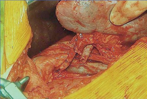
The kidney is placed extraperitoneally into the iliac fossa, and either kidney can go into either iliac fossa [1]. The renal vessels are anastomosed to the external iliac vessels, with the common or internal iliac artery used if the external is diseased, and the ureter is anastomosed to the bladder, usually over a stent [1].
The pancreas is retrieved as the whole organ with the duodenum attached, and transplanted either intraperitoneally or extraperitoneally into the right iliac fossa using techniques similar to renal transplantation [1]. Its dual arterial supply, from splenic artery and SMA branches, is reconstructed as an arterial Y-graft [1]. The ABSITE Review states the requirement as needing both the donor coeliac artery and SMA for arterial supply, and the donor portal vein for venous drainage [2].
Most units use enteric drainage for the pancreatic duct: the second part of the donor duodenum is taken along with the ampulla of Vater and the pancreas, and the donor duodenum is anastomosed to recipient bowel [2].
Operative technique in Schwartz's detail
- The kidney is placed heterotopically in the iliac fossa, retroperitoneal for biopsy and ureteric access, the right side preferred for shallower vessels unless a future pancreas or a previous failed graft dictates the left; a three-lumen catheter is clamped under the drapes to fill the bladder (or saline instilled if anuric), a curvilinear incision one to two fingers above the pubis along the lateral rectus sheath opens the anterior sheath, the absent posterior sheath below the arcuate line gives extraperitoneal access without dividing rectus, the peritoneum is swept medially, inferior epigastric vessels and round ligament divided and the cord looped, iliac artery dissection is limited and perivascular lymphatics ligated to prevent lymphocele, renal artery and vein are anastomosed end-to-side to the external iliac vessels (internal iliac or endarterectomy for a calcified artery, a Carrel patch or caval extension conduit for a short right renal vein), and urinary continuity is by Leadbetter–Politano (large dome cystotomy, 1 cm submucosal tunnel, spatulated ureter sewn from inside with interrupted absorbable sutures) or extravesical Lich ureteroneocystostomy (1 cm of anterolateral detrusor divided to a mucosal bubble, diamond-spatulated ureter sewn mucosa-to-mucosa with running absorbable sutures over a temporary stent, muscle closed over it against reflux), stents generally reserved for ureteroureterostomy or paediatric transplants and fixation only for small or en bloc kidneys [3].
- Multiple renal arteries occur in 10–30% of donor kidneys and fare as well as single vessels except in hypercoagulable recipients, implanted separately, joined into a common channel or combined on one Carrel patch; kidneys from donors under 5 years or 20 kg may be transplanted en bloc on the donor aorta and cava with suprarenal ends oversewn, lumbar branches ligated, hilum untouched, orientation marked against torsion, colour compared after reperfusion, retroperitoneal fixation and two ureters implanted separately or as a common patch, with encouraging long-term results from the few centres doing it [3].
- Perioperatively electrolytes are checked with emergency dialysis for hyperkalaemia or overload, catheter sites inspected, a non-nephrotoxic antibiotic covering skin flora and Gram-negatives given, heparin given before clamping, the recipient kept well hydrated with a central venous pressure near 10 mmHg and systolic above 120 mmHg before reperfusion (supraphysiological in children receiving adult kidneys), mannitol given as radical scavenger and diuretic with furosemide, and afterwards urine output replaced hourly with equal or reduced fluid, electrolytes (calcium, magnesium, potassium) replaced during brisk diuresis, and urea and creatinine trusted over urine volume when native kidneys still make urine [3].
- The pancreas donor is excluded by type 1 diabetes and relatively by previous pancreatic surgery, chronic pancreatitis or IPMN but not by hyperglycaemia (insulin resistance of brain death); a no-touch technique takes gland, spleen, duodenum and surrounding tissue together, the pancreas is not flushed extensively (splenic artery and SMA temporarily clamped), the coeliac axis on an aortic patch stays with the liver while the splenic artery and the SMA on its own patch go with the pancreas, a replaced right hepatic artery is dissected from the posterior pancreas so that the proximal SMA and patch go with the liver and the distal SMA with the inferior pancreaticoduodenal artery with the pancreas, and pancreas, liver and intestine can be procured together for three recipients [3].
- Back-table preparation in chilled solution removes the spleen, shortens and reinforces the mesenteric root, trims and reinforces the duodenum and reconstructs inflow as a Y-graft from the donor iliac bifurcation (external iliac to SMA, internal iliac to splenic artery) so the common iliac is sewn as a single vessel to the recipient common iliac, with the portal vein kept short to avoid kinking; the graft lies intra-abdominally on the right with head and duodenum caudal, recipient internal iliac vein branches divided to relieve tension, anastomoses end-to-side, and exocrine drainage to bladder (antimesenteric duodenum to bladder dome, hand-sewn or circular-stapled) or, in over 80% of US cases, side-to-side to jejunum or a Roux loop, with systemic venous drainage in over 90% [3].
- Bladder drainage lets urinary amylase detect exocrine rejection 5–7 days before endocrine rejection, when over 90% of episodes reverse if treated while normoglycaemic, and localises any leak to the right lower quadrant; enteric drainage is more physiological but carries bacterial contamination [3].
Complications
Urine leak is the commonest complication of kidney transplantation [2].
Lymphocele is the most common cause of external ureteric compression, most often occurring 3 weeks after transplantation and presenting as late reduction in urine output with hydronephrosis and a fluid collection [2].
Venous thrombosis is the commonest complication of pancreas transplantation, and is hard to treat [2].
For living kidney donors, the commonest complication is wound infection at 1%, and the commonest cause of death is fatal pulmonary embolism [2].
Complication profile in Schwartz's account
After kidney transplantation hypotension (hypovolaemia, vasodilatation, infarction) and hypertension (catecholamines, overload, drugs) are managed promptly; a sudden fall in urine output is most often volume change but also catheter blockage, urine leak, vascular thrombosis, nephrotoxicity, ATN or rejection, investigated by Doppler, nuclear renography or biopsy; bleeding is uncommon, tamponaded by haematoma and explored only for ongoing transfusion, instability or graft compression (lower threshold on anticoagulants), usually from small hilar or retroperitoneal vessels; graft thrombosis in under 1% follows hypercoagulability, severe vascular disease, paediatric or en bloc grafts, procurement injury, intimal dissection, torsion or hyperacute rejection, presents in the first days with abrupt anuria, haematuria or graft pain, and thrombectomy rarely salvages the graft; urological complications in up to 5% arise from ureteric ischaemia, distal ureter damage or technique, leaks (fever, pain, swelling, rising creatinine, cutaneous drainage; fluid-to-serum creatinine comparison) are repaired or stented via nephrostomy, early obstruction from oedema, clot, torsion or haematoma and late ischaemic strictures show as hydronephrosis and are treated by nephrostomy and stenting, then reimplantation or ureteropyelostomy if that fails [3]. Pancreas transplantation carries the highest technical complication rate of any solid organ because of organ-specific pancreatitis, abscess, necrosis, fistula and pseudocyst with low blood flow, hollow-viscus anastomosis, high immunogenicity and diabetic comorbidity: thrombosis 5–15% (usually in the first week, venous commoner than arterial with swollen tender graft, haematuria, ipsilateral leg oedema and DVT, arterial thrombosis silent and Doppler-diagnosed, usually ending in graft pancreatectomy), abscess 5–10% (increasingly drained radiologically, with 7 days of broad-spectrum antimicrobials), bleeding 6–8% often needing relaparotomy, leaks not always requiring pancreatectomy but pseudoaneurysm, AV fistula and dehiscence often doing so; rejection affects about 30% in the first year, intra-abdominal infection is the second cause of graft loss after thrombosis, and bladder drainage causes enzymatic cystitis, haematuria, dysuria, bicarbonate loss with dehydration and acidosis, so 20–30% convert to enteric drainage within 5 years [3].
Outcomes
Five-year graft survival overall is 70% (65% for cadaveric and 75% for living donor grafts) and transplantation extends life by 15 years [2]. The UK figures above are higher and more recent, and are broken down by graft against patient survival.
Mortality after kidney transplantation is primarily from stroke and myocardial infarction, with myocardial infarction the commonest single cause [2], which is the reason cardiovascular risk dominates the long-term follow-up of a patient whose kidney is working perfectly well.
Registry outcomes in Schwartz's account
One-year deceased donor kidney graft failure fell from about 20% in 1989 to under 7% in 2009 and 4.8% in 2015, and living donor failure from 8.5% to under 3%, with steroid-free and calcineurin-free protocols validated; the commonest cause of graft loss is now death with a functioning graft (usually cardiovascular), then chronic allograft nephropathy of mixed immunological and non-immunological cause, technical failure holding at 1–2% [3]. US pancreas transplants fell over 20% between 2005–09 and 2010–14 (PAK down 50%, SPK 10%, PTA 20%) while numbers rose elsewhere, donor criteria tightened towards young trauma donors with short preservation, and protocols moved to antibody induction with tacrolimus and MMF and growing steroid avoidance; 1-year SPK patient survival rose from 95.7% to 97.4%, pancreas graft function from 88.3% to 91.3% and kidney function from 93.6% to 95.5%, PAK patient survival from 96.4% to 97.9% and graft function from 81.0% to 86.0%, and PTA graft survival from 81.0% to 85.7% with patient survival steady at 97%; islet recipients are mostly C-peptide positive with retained hypoglycaemia awareness even when insulin dependence returns, and pancreas-alone recipients achieve higher insulin independence than islet-alone recipients despite up to three donor pancreases each [3].
References
- Oxford Handbook of Clinical Surgery, 5th ed., Ch. 20 Transplantation
- The ABSITE Review, 2022, Ch. 12 Transplantation
- Schwartz's Principles of Surgery, 11th ed., Ch. 11, Figs. 11-11 to 11-13
- Bailey & Love's Short Practice of Surgery, 28th ed., Ch. 88 Kidney transplantation and the principles of transplantation