



Immunosuppression
Summary
- The goal of immunosuppression is to inhibit the immune response to alloantigen while preserving the immune response to infection and malignancy [1].
- That balance is never fully achieved, which is why the complications of immunosuppression (opportunistic infection, skin cancer and post-transplant lymphoproliferative disorder) are as much a part of transplant practice as rejection itself. Skin cancer is the commonest malignancy following any transplant, squamous cell carcinoma most of all [2].
- This page covers the drug classes and their mechanisms, the toxicity that distinguishes them, and the infections and tumours that follow from the dampened response.
Definition
Immunosuppression consists of a short, intense induction phase followed by a maintenance phase in which the dosage is tapered [1]. A careful balance is kept between therapeutic and toxic doses, generally achieved by combining drugs with different mechanisms of action rather than pushing any single agent [1].
Phases and a conventional protocol in Schwartz's account
Immunosuppression evolved from azathioprine and steroids in the 1960s–70s through cyclosporine in the 1980s to tacrolimus and mycophenolate mofetil in the 1990s, and is now given as multidrug regimens that hit several pathways to raise efficacy while lowering each agent's toxicity, some protocols withdrawing, avoiding or minimising a class; induction begins immediately after transplantation when rejection risk peaks and maintenance follows within days for the life of the graft, so immunosuppression is heaviest in the first 3–6 months, when antibacterial, antiviral and antifungal prophylaxis is given; a conventional protocol is antibody induction (depleting or non-depleting) then a calcineurin inhibitor, an antiproliferative and corticosteroids [3]. Schwartz groups the drugs as immunophilin binders (calcineurin inhibitors cyclosporine and tacrolimus; the non-calcineurin sirolimus), antimetabolites blocking de novo purine synthesis (azathioprine, mycophenolate), biologics (polyclonal Atgam and antithymocyte globulin; monoclonal muromonab-CD3, basiliximab, belatacept, alemtuzumab, rituximab, bortezomib, eculizumab) and corticosteroids [3].
Pathophysiology
Each class interrupts a different step of the alloresponse.
Corticosteroids inhibit the immune response at many levels: they decrease production of gamma-interferon and the interleukins that would upregulate the lymphocyte response, and reduce macrophage function [1]. The ABSITE Review describes the same action as inhibiting inflammatory cells (macrophages) and the genes for cytokine synthesis, IL-2 most importantly [2].
- Calcineurin inhibitors work one step further along.
- Ciclosporin binds cyclophilin protein, and the resulting complex inhibits calcineurin, decreasing cytokine synthesis with IL-2 most important [2].
- Tacrolimus binds FK-binding protein instead, with actions similar to ciclosporin but more potent [2].
- The Oxford Handbook states the shared endpoint: both inhibit production of IL-2 by T helper cells, selectively reducing the cytotoxic T-cell response [1].
mTOR inhibitors bind the same protein as tacrolimus but act elsewhere. Sirolimus binds FK-binding protein like tacrolimus but inhibits the mammalian target of rapamycin, so that it inhibits the T- and B-cell response to IL-2 rather than its production [2]. Everolimus acts likewise [1].
Antiproliferative agents starve lymphocytes of purines. Mycophenolate inhibits de novo purine synthesis, which inhibits growth of T cells; azathioprine has a similar action [2]. Mycophenolic acid inhibits purine synthesis in lymphocytes, reducing clonal expansion and lymphocyte counts [1].
Clinical features
- The side effects separate the calcineurin inhibitors from each other and from everything else.
- Ciclosporin causes nephrotoxicity, hepatotoxicity, tremor, seizures and haemolytic uraemic syndrome [2].
- Tacrolimus causes nephrotoxicity, and more gastrointestinal symptoms, mood changes and diabetes than ciclosporin, with much less enterohepatic recirculation [2].
- The Oxford Handbook adds that both cause a usually dose-dependent tremor and impair glucose tolerance, and that ciclosporin is associated with gingival hypertrophy [1].
Sirolimus is not nephrotoxic, unlike ciclosporin and tacrolimus [2], but the Oxford Handbook adds the qualifier that matters in combination therapy: mTOR inhibitors are not themselves nephrotoxic, yet they appear to potentiate the nephrotoxicity of calcineurin inhibitors if given together [1]. Sirolimus has its own signature toxicity: interstitial lung disease [2].
Mycophenolate's main side effect is gastrointestinal intolerance (nausea, vomiting and diarrhoea) with myelosuppression second [2]. It comes in two forms: mycophenolate mofetil, which can cause severe gastrointestinal effects, and mycophenolate sodium, which has fewer [1]. Azathioprine causes bone marrow suppression and pancreatitis, and allopurinol inhibits its metabolism, a drug interaction with obvious consequences [1].
Anti-thymocyte globulin causes cytokine release syndrome (fever, chills, pulmonary oedema and shock) which is why steroids and an antihistamine are given before the drug; it also causes PTLD and myelosuppression [2].
Corticosteroid side effects are the familiar Cushingoid features: centripetal obesity, thin skin and proximal myopathy among them [1].
Etiology
The agents
| Agent | Class and mechanism | Role |
|---|---|---|
| Prednisolone, methylprednisolone | Corticosteroid; blocks cytokine genes and macrophage function | Induction, maintenance, acute rejection |
| Ciclosporin | Calcineurin inhibitor via cyclophilin | Maintenance |
| Tacrolimus | Calcineurin inhibitor via FK-binding protein; more potent | Maintenance; preferred CNI |
| Sirolimus, everolimus | mTOR inhibitor; blocks response to IL-2 | Maintenance |
| Mycophenolate, azathioprine | Antiproliferative; blocks purine synthesis | Maintenance |
| Basiliximab | Monoclonal antibody to the IL-2 receptor | Induction |
| Anti-thymocyte globulin | Polyclonal anti-T-cell antibody; cytolytic | Induction and severe rejection |
| Alemtuzumab | Monoclonal antibody to CD52 | Induction |
| Belatacept | Blocks co-stimulation of T-cells by APCs | Newer agent, trials ongoing |
Tacrolimus is the preferred calcineurin inhibitor, as it has a better side effect profile [1], and there are fewer rejection episodes in kidney transplantation with tacrolimus than with ciclosporin [2]. Mycophenolic acid is preferred to azathioprine [1].
Anti-thymocyte globulin is equine (ATGAM) or rabbit (thymoglobulin) polyclonal antibody against T-cell antigens CD2, CD3 and CD4, used for induction and for acute rejection, and is cytolytic and complement dependent [2]. The Oxford Handbook describes it as derived from rabbits or horses immunised with T-cells, primarily directed against the T-cell receptor [1].
Alemtuzumab binds CD52, depleting T-cells, B-cells, NK cells, lymphocyte precursors, dendritic cells and macrophages, so it reduces every cell involved in antigen presentation and in both cellular and humoral rejection; side effects are bleeding and sepsis [1].
Belatacept blocks the co-stimulatory signal and is neither nephrotoxic nor diabetogenic, though its long-term effects are unknown and trials are still under way [1].
- Schwartz's dosing and pharmacology: rabbit antithymocyte globulin (Thymoglobulin, largely replacing equine Atgam) carries antibodies to T and B cells, integrins and adhesion molecules, a total of about 6 mg/kg giving adequate depletion and better graft survival while 3 mg/kg may not prevent rejection and more prolongs infection and lymphoma risk, premedicated with paracetamol and diphenhydramine, causing fever, chills, arthralgia, thrombocytopenia and leukopenia; basiliximab blocks the IL-2 receptor α-chain (Tac/CD25) found only on activated T cells, depletes nothing (so cannot treat rejection) and adds no infection or malignancy risk, being followed by a calcineurin inhibitor, steroids and MMF; a single 30 mg dose of alemtuzumab (anti-CD52, originally for chronic lymphocytic leukaemia) depletes 99% of lymphocytes by complement, antibody-mediated cytotoxicity and apoptosis, monocytes recovering at 3 months, B cells at 12 and T cells to only 50% at 36 months, with cytokine release needing steroid and antihistamine premedication and lasting risk of infection and PTLD [3].
- Steroids bind glucocorticoid-responsive DNA elements to block transcription of cytokine and cytokine-receptor genes, are first-line for acute rejection, are given in large perioperative doses then tapered to 5–15 mg daily or stopped, and are CYP3A4/3A5 and P-glycoprotein substrates; azathioprine is converted to 6-mercaptopurine, inhibits de novo and salvage purine synthesis, has been relegated to an adjunct since cyclosporine but is preferred over teratogenic MMF in recipients planning a pregnancy and in those intolerant of MMF's gut effects, at 1–3 mg/kg/day, and allopurinol blocks its metabolism so the pair is avoided or the azathioprine dose cut by 75%; mycophenolate mofetil (FDA-approved May 1995), the prodrug of Penicillium-derived mycophenolic acid inhibiting inosine monophosphate dehydrogenase, is started at 1 g twice daily, the enteric-coated Myfortic has unproven benefit beyond less reported GI intolerance, MPA levels do not correlate with rejection and are not routinely measured, oesophagitis or gastritis in about 5% may signal CMV or herpes infection, and leukopenia may reverse on lowering the dose or stopping valganciclovir [3].
- Sirolimus, the first mTOR inhibitor, binds FKBP and the complex blocks mTOR's switch from G1 to S phase in response to IL-2, also inhibiting vascular smooth muscle proliferation that may ease allograft vasculopathy; dosed 2–4 mg/day to trough, it is used to withdraw or avoid steroids or spare calcineurin inhibitors, and causes statin- and fibrate-resistant hypertriglyceridaemia, impaired wound healing (worst immediately post-transplant), thrombocytopenia, leukopenia and anaemia, all worse with MMF [3].
- Cyclosporine binds cyclophilin to inhibit calcineurin and IL-2 gene expression; oil-based Sandimmune has poor and variable bioavailability whereas microemulsified Gengraf and Neoral absorb better; troughs are held at 250–350 ng/mL for 3 months then 150–250, the oral dose about 5 mg/kg/day in two doses; it causes hirsutism, gingival hyperplasia and more hypertension and hyperlipidaemia than tacrolimus [3].
- Tacrolimus, the backbone of most regimens, binds FKBPs and inhibits IL-2 production 10–100 times more potently than cyclosporine, is given intravenously (0.015 mg/kg/day infusion), orally (0.05 mg/kg/day in two doses) or sublingually to troughs of 8–12 ng/mL for 3 months then 6–10, and causes more new-onset diabetes than cyclosporine plus alopecia, nephrotoxicity, neurotoxicity, hypertension, hyperkalaemia and hypomagnesaemia [3].
- Belatacept (LEA29Y) is abatacept (CTLA4-Ig) with two amino-acid substitutions giving higher avidity for CD80/CD86, infused monthly (5–10 mg/kg); against cyclosporine it was non-inferior for patient and graft survival but had more biopsy-proven acute cellular rejection and more PTLD, greatest in EBV-seronegative recipients so the FDA restricts it to seropositive patients, liver trials stopped for excess mortality, and it spares lipids, blood pressure, glucose, nerves and appearance [3].
Diagnosis
Monitoring is by trough level for the calcineurin inhibitors and by white cell count for the cytolytic and antiproliferative agents. Ciclosporin trough should be kept at 200 to 300; tacrolimus trough at 10 to 15 [2]. White cells should be kept above 3 on mycophenolate and on anti-thymocyte globulin [2].
Ciclosporin undergoes hepatic metabolism and biliary excretion and is reabsorbed in the gut, giving enterohepatic recirculation, which tacrolimus largely avoids [2].
CMV is diagnosed by detecting CMV-PCR DNA copies in serum, or by evidence of CMV in biopsy of the affected organ [1]. Pneumocystis jiroveci is diagnosed by bronchoalveolar lavage [1].
Drug interactions in Schwartz's tables
Calcineurin inhibitor and sirolimus levels rise with verapamil, diltiazem, nicardipine, amiodarone, clarithromycin, erythromycin, azithromycin, azole antifungals, protease inhibitors including ritonavir and grapefruit juice, and fall with isoniazid, nevirapine, rifampicin, carbamazepine, phenobarbital, phenytoin, caspofungin and St John's wort; calcineurin nephrotoxicity (a dose-dependent, reversible constriction of the afferent arteriole) is potentiated by ganciclovir, aminoglycosides, amphotericin, NSAIDs, ACE inhibitors and ARBs, and hyperkalaemia by potassium-sparing diuretics, ACE inhibitors, ARBs, β-blockers and co-trimoxazole; MMF levels fall with cholestyramine and antacids and its marrow suppression is additive with valganciclovir, ganciclovir and co-trimoxazole; azathioprine marrow suppression is potentiated by allopurinol and sulphonamides [3].
Thresholds and severity
| Drug | Target |
|---|---|
| Ciclosporin | Trough 200–300 |
| Tacrolimus | Trough 10–15 |
| Mycophenolate | Keep white cells above 3 |
| Anti-thymocyte globulin | Keep white cells above 3 |
The risks of long-term immunosuppression are cancer, cardiovascular disease, infection and osteopenia [2].
UK prophylaxis against opportunistic infection runs to a defined schedule with defined drugs.
CMV prophylaxis is given to transplant recipients based on their own and the donor's CMV status, for the first 3 to 6 months after transplantation (or following a rejection episode) as oral valganciclovir, dosed by eGFR [1]. Established CMV infection is treated with oral valganciclovir, or intravenous ganciclovir for severe infection [1].
Pneumocystis jiroveci prophylaxis is oral co-trimoxazole for the first 6 months after transplantation [1]. Established infection is treated with high-dose co-trimoxazole, occasionally with second- or third-line agents [1].
The clinical trap with Pneumocystis is the mismatch between symptoms and film. Patients often present with a dry cough and shortness of breath with minimal chest radiograph signs at presentation, but deteriorate rapidly [1].
BK virus infection is generally asymptomatic but is a possible cause of renal transplant failure [1], a reason a failing kidney graft is not automatically assumed to be rejecting.
Treatment and Management
Steroids serve three roles, induction after transplantation, maintenance, and treatment of acute rejection episodes [2]. Ciclosporin, tacrolimus, sirolimus and mycophenolate are maintenance agents; basiliximab, alemtuzumab and anti-thymocyte globulin are induction agents, with ATG also used for severe rejection [1][2].
Post-transplant lymphoproliferative disorder is treated first by withdrawing immunosuppression, then with rituximab (anti-CD20, which decreases B cells) with chemotherapy and radiotherapy needed for aggressive tumours [2].
Steroids and an antihistamine are given before anti-thymocyte globulin to try to prevent cytokine release syndrome [2].
Procedural interventions
There is no procedure specific to immunosuppression itself; the procedural burden falls on monitoring (trough levels, blood counts, CMV PCR) and on the biopsies used to separate rejection from drug toxicity from infection, since all three can present as a failing graft [1].
Where humoral rejection is diagnosed, plasmapheresis is added to remove the antibodies [1].
Complications
Infection
Dampening the immune response leads to more severe infection and to opportunistic infection, which may be viral, bacterial or fungal [1]. The ABSITE Review lists the viral opportunists as CMV, HSV and VZV, and the fungal as Pneumocystis jiroveci, Aspergillus, Candida and Cryptococcus [2].
CMV is one of the commonest infections seen post-transplant. It presents non-specifically with malaise, lethargy and fever, or affects a specific organ as CMV colitis or CMV pneumonitis, and can be life-threatening [1].
- Schwartz separates early infections (within a month), mostly surgical (peritonitis, intra-abdominal abscess, wound infection), most severe in liver and pancreas recipients where intra-abdominal infection is the second cause of pancreas graft loss after thrombosis, favoured by long operations, blood loss, prolonged ischaemia, spillage of bile, urine or bowel content, heavy induction and anastomotic leaks, presenting with fever, hypotension, ileus and pain (often masked), polymicrobial with E. coli, Enterococcus, Klebsiella, Pseudomonas and Candida albicans, krusei and glabrata, treated by prompt reoperation or percutaneous drainage, while medical respiratory, urinary and bloodstream infections get empirical antibiotics and antifungals before cultures and donor-culture-directed therapy, from late infections of depressed cell-mediated immunity [3].
- Herpesviruses lead: CMV, transmitted by seropositive donor organs or reactivated, strikes at 3–6 months or during rejection treatment, has been greatly reduced by 12-week antiviral prophylaxis, ranges from flu-like illness to pneumonitis, hepatitis and GI ulceration, and symptomatic or tissue-invasive disease gets intravenous ganciclovir and reduced immunosuppression; EBV ranges from mononucleosis to hepatitis and PTLD, a proliferation of EBV-positive B cells from localised tumour to diffuse infiltration including brain, driven by heavy immunosuppression and a seronaive recipient with a seropositive donor, treated by reducing immunosuppression early and rituximab when advanced [3].
- After 6 months fungal infection follows environmental exposure, Blastomyces in Midwest and Southeast soil (biopsy, intravenous amphotericin), Coccidioides in the Southwest, Mexico and Latin America (resilient, high-dose amphotericin), Histoplasma in Ohio and Mississippi valley bird and bat droppings (disseminates, CNS in a quarter, 3–13 months of itraconazole), with rare Aspergillus, Cryptococcus, Mucor and Rhizopus, 20% mortality for invasive Candida or Aspergillus, fluconazole prophylaxis reducing fungal infection in liver recipients and lifelong daily co-trimoxazole virtually eliminating Pneumocystis [3].
Malignancy
Immunosuppression reduces immune surveillance of tumours, so some become commoner and others more aggressive; virally mediated tumours and squamous cell carcinoma of the skin are especially common [1].
- PTLD is the next most common malignancy after skin cancer, and is Epstein-Barr virus related [2].
- It presents with small bowel obstruction, a mass or adenopathy, and cytolytic drugs are the risk factor [2].
- The Oxford Handbook gives the mechanism: it is a B-cell proliferation that may extend to B-cell lymphoma, caused by drug inhibition of the IL-2-dependent mechanism by which T-cells regulate the B-cell proliferation of EBV infection [1].
Malignancy can also be transmitted from donor to recipient. The risk is very small for standard donors (of the order of 1 in 2000) but should be considered in unusual cases [1].
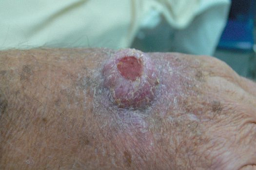
In a cohort of over 175,000 solid organ recipients 10,656 developed cancer, a standardised incidence ratio of about 2, with at least fivefold excess of Kaposi's sarcoma, non-melanoma skin cancer, non-Hodgkin's lymphoma and cancers of liver, anus, vulva and lip, and significant excess of melanoma, Hodgkin's lymphoma and cancers of lung, kidney, colon, rectum and pancreas [3].
Outcomes
The long-term price of a functioning graft is a defined list: cancer, cardiovascular disease, infection and osteopenia [2].
That list is why the maintenance phase tapers rather than holds, why drugs with different mechanisms are combined rather than any one pushed to its ceiling, and why the newer agents are judged on what they avoid, belatacept being neither nephrotoxic nor diabetogenic, sirolimus not nephrotoxic on its own [1][2].
References
- Oxford Handbook of Clinical Surgery, 5th ed., Ch. 20 Transplantation
- The ABSITE Review, 2022, Ch. 12 Transplantation
- Schwartz's Principles of Surgery, 11th ed., Ch. 11, Transplantation, Table 11-2
- Bailey & Love's Short Practice of Surgery, 28th ed., Ch. 45 Skin and subcutaneous tissue