



Mediastinum and Trachea
Summary
- Mediastinal masses are sorted first by compartment, because location predicts histology. The anterior mediastinum is the commonest site, and its differential is the four Ts, thymoma, thyroid, T-cell lymphoma and teratoma [1].
- Two figures set the index of suspicion: 50% of symptomatic mediastinal masses are malignant, and 90% of asymptomatic ones are benign [1].
- This page also covers tracheal tumours and stenosis, and the two fistulae that complicate a tracheostomy, including tracheo-innominate artery fistula, which can exsanguinate a patient in minutes
Definition
Neurogenic tumours are the most common mediastinal tumour in both adults and children, and usually sit in the posterior mediastinum [1].
The anterior mediastinum contains the thymus; the middle contains heart, trachea and ascending aorta; the posterior contains oesophagus and descending aorta [1].
Pathophysiology
The thoracic anatomy that matters at mediastinoscopy is what lies on each side of the scope [1]. Looking into the middle mediastinum, the left-sided structures are the recurrent laryngeal nerve, oesophagus, aorta and main pulmonary artery; the right-sided structures are the azygos vein and superior vena cava; and anteriorly lie the innominate vein, innominate artery and right pulmonary artery.
Two nerves relate to the hilum in opposite ways: the phrenic nerve runs anterior to it, the vagus posterior [1].
Tracheo-innominate fistula is a positional complication. It occurs after tracheostomy and is avoided by keeping the tracheostomy between the second and third tracheal rings [1].
Post-intubation stenosis occurs at different sites depending on the tube: at the stoma site with a tracheostomy, and at the cuff site with an endotracheal tube [1].
Compartments, contents and tumour distribution in Schwartz's account
- In the three-compartment model the anterior compartment (sternum to the front of heart and great vessels) holds thymus or its fatty remnant, internal mammary vessels, nodes and fat; the visceral or middle compartment (great vessels to trachea) holds pericardium, ascending and transverse aorta, both cavae, brachiocephalic vessels, phrenic and upper vagus nerves, trachea, main bronchi and nodes and the central pulmonary vessels; and the posterior compartment (paravertebral sulci and para-oesophageal area) holds descending aorta, oesophagus, thoracic duct, azygos and hemiazygos veins and nodes [2].
- In children neurogenic tumours (40%, posterior) lead, then lymphoma (18%), cysts (18%), germ cell tumours (11%) and mesenchymal tumours (9%) with thymoma rare; in adults neurogenic tumours (21%), cysts (20%), thymoma (19%), lymphoma (13%), germ cell (11%), mesenchymal (7%) and endocrine tumours (6%); about 25% are malignant in both groups, up to two-thirds of adult tumours are incidental radiological findings, and symptomatic tumours are significantly likelier to be malignant [2].
- Tracheal stenosis after tracheostomy once occurred in 3–12% (severe in 1–2%) and has fallen to 4.9 per million a year with low-pressure cuffs; intubation injury destroys cartilage by ischaemic necrosis and heals as a web-like stenosis at cuff level, whereas stomal stenosis reflects granulation tissue; risk factors are prolonged intubation, high tracheostomy through the first ring or cricothyroid membrane, transverse rather than vertical incision, oversized tubes, prior intubation, traumatic intubation, age, female sex, radiation, steroids, autoimmune disease, reflux, sleep apnoea and severe respiratory failure, with cuff pressure kept under 20 mmHg [2].
- Tracheo-innominate fistula follows tracheostomy below the fourth ring (the cannula's inner curve pressing the artery) or cuff hyperinflation, most cuff fistulas appearing within 2 weeks, so the tube is placed through rings two to four regardless of the sternal notch; tracheo-oesophageal fistula follows cuff compression of the membranous trachea against a nasogastric tube during prolonged ventilation [2].
Clinical features
Most mediastinal tumours are asymptomatic, and may present with chest pain, cough or dyspnoea [1].
Myasthenia gravis presents with fatigue, weakness, diplopia and ptosis, the ocular symptoms being commonest, and is caused by antibodies to acetylcholine receptors [1].
Neurogenic tumours present with pain and neurological deficit [1].
A tracheo-innominate fistula may announce itself with a sentinel bleed (a small amount of bleeding) before catastrophic haemorrhage [1].
Presentation of tracheal and mediastinal disease in Schwartz's account
- Tracheal stenosis causes stridor and exertional dyspnoea usually 2–12 weeks after extubation or decannulation (sometimes immediately or 1–2 years later) and is often mistreated as asthma or bronchitis; tracheo-innominate fistula presents with bleeding, a premonitory non-massive haemorrhage that must never be dismissed as wound bleeding; tracheo-oesophageal fistula reveals saliva, gastric contents or feed on suctioning with gastric insufflation from positive pressure; tracheal tumours present with cough, dyspnoea, haemoptysis, stridor or invasion of recurrent nerve or oesophagus, stenosis being visible radiologically in only 50% and symptoms of slow-growing non-squamous tumours persisting for months, with about 50% presenting at stage IV [2].
- Mediastinal masses compress the trachea to cause cough, dyspnoea or stridor, cause pain or dyspnoea from effusion, tamponade or phrenic involvement, and cause hoarseness from left recurrent nerve compression at the aortopulmonary window; systemic symptoms occur in 50% of lymphoproliferative masses against 29% of others, and raised ESR, CRP and leukocytosis in 86% against 58%; night sweats, weight loss, fatigue and extrathoracic adenopathy point to lymphoma, fluctuating weakness, ptosis and diplopia to thymoma with myasthenia, wheeze and haemoptysis to a visceral-compartment granuloma, and young male sex, a testicular mass and raised hCG or AFP to a germ cell tumour [2].
- Acute mediastinitis, from oesophageal perforation (iatrogenic dilatation, bougienage, endoscopy, sclerotherapy; Boerhaave's; foreign body; trauma; anastomotic leak; cancer erosion), deep sternotomy infection, Ludwig's angina, quinsy, retropharyngeal abscess, cervical lymphadenitis, lung, pleural or subphrenic infection, rib or vertebral osteomyelitis or haematogenous abscess, causes fever, chest pain, dysphagia, respiratory distress and cervical and upper thoracic crepitus and can deteriorate rapidly to sepsis and death [2].
Etiology
By compartment
| Compartment | Contents and masses |
|---|---|
| Anterior | Thymoma (commonest anterior mass in adults); thyroid cancer and goitres; T-cell lymphoma; teratoma and other germ cell tumours; parathyroid adenomas |
| Middle | Bronchogenic cysts; pericardial cysts; enteric cysts; lymphoma |
| Posterior | Enteric cysts; neurogenic tumours; lymphoma |
Lymphoma is the commonest cause of mediastinal adenopathy [1].
Thymoma and myasthenia
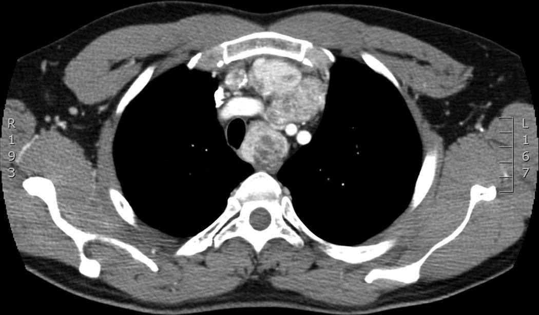
Thymoma runs on a series of 50s: 50% of thymomas are malignant, 50% of patients with thymoma have symptoms, and 50% of patients with thymoma have myasthenia gravis, but only 10% of patients with myasthenia gravis have a thymoma [1]. Thymomas are rare in children [1].
Germ cell tumours
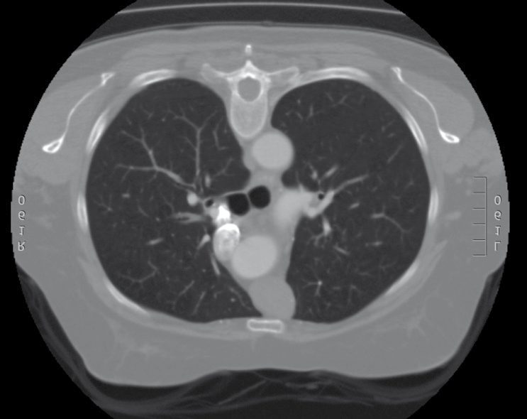
Teratoma is the commonest germ cell tumour in the mediastinum and may be benign or malignant [1]. Seminoma is the commonest malignant germ cell tumour: 10% are beta-HCG positive and it should not produce AFP [1]. Non-seminoma has elevated beta-HCG and AFP in 90% [1].
Tracheal tumours
The commonest benign tracheal tumour is papilloma in adults and haemangioma in children; the commonest malignant tumour is squamous cell carcinoma in adults and carcinoid in children [1].
Thymic, neurogenic, germ cell and cystic lesions in Schwartz's detail
- Thymoma, the commonest adult anterior mediastinal neoplasm at 40–60 years, is accompanied by myasthenic symptoms or acetylcholine-receptor antibodies in 10–50% though under 10% of myasthenics have a thymoma; thymectomy improves myasthenia in only about 25% of thymoma patients but gives complete remission in up to 50% and improvement in 90% without thymoma; 5% have red cell aplasia, hypogammaglobulinaemia, lupus, Cushing's or SIADH; thymomas are mixed epithelial cells and mature lymphocytes, usually encapsulated and cytologically bland even when invasive (hence "invasive thymoma" rather than "malignant"), whereas thymic carcinoma is microscopically malignant, classified by Suster and Rosai as low-grade (squamous, mucoepidermoid, basaloid) or high-grade (lymphoepithelial, small cell, sarcomatoid, clear cell, anaplastic), must be distinguished from metastatic lung cancer, invades and metastasises early and has 30–50% 5-year survival; rebound thymic hyperplasia appears about 9 months (2 weeks to 12 months) after chemotherapy for lymphoma or germ cell tumour as a bilobed retrosternal triangle with low PET uptake; thymolipoma is a benign fat-density mass dotted with thymic islands [2].
- Nerve sheath tumours are 20% of mediastinal tumours and over 95% benign: schwannomas (neurilemomas) from intercostal Schwann cells show compact palisading Antoni A and loose myxoid Antoni B regions, may extend through the foramen as a "dumbbell" needing MRI and a combined approach, and small asymptomatic ones may be watched in the elderly; neurofibromas (up to 25% of sheath tumours, 40% with von Recklinghausen's disease) are 70% benign but degenerate to neurofibrosarcoma in 25–30%, more with age, neurofibromatosis and radiation, with 53% 5-year survival falling to 16% with neurofibromatosis or tumours over 5 cm; ganglion cell tumours arise from the sympathetic chain, ganglioneuroma (benign, VIP diarrhoea, intraspinal extension, curable), ganglioneuroblastoma (infants under 3, nodular pattern metastasises, 80% 5-year survival) and neuroblastoma (14% thoracic, more resectable than other chest cancers); thoracic phaeochromocytoma (10% of extra-adrenal, 10% malignant, costovertebral sulcus) needs urinary catecholamines, MIBG and α- then β-blockade, and chemodectomas around arch, vagi or aorticosympathetics rarely secrete but are malignant in up to 30% [2].
- Lymphoma is the commonest mediastinal malignancy, primary there in about 50% of Hodgkin's and non-Hodgkin's cases, anterior then middle, rarely posterior, cured by chemoradiotherapy in up to 90% of early and 60% of advanced Hodgkin's disease [2].
- Primary mediastinal germ cell tumours are under 5% of germ cell tumours and under 1% of mediastinal tumours, arise from misplaced pluripotent cells rather than metastasis (autopsy series show no gonadal scar and no late testicular recurrence), a gonadal primary is always excluded, one-third are seminomas and two-thirds non-seminomatous or teratomas (teratomas 60–70% of the total, with ectodermal teeth, skin and hair, mesodermal cartilage and bone and endodermal bronchial, gut or pancreatic tissue), and teratocarcinoma is uniformly poor [2].
- Benign cysts are up to 25% of masses and the commonest middle-compartment lesion: pericardial cysts (right costophrenic angle, mesothelial-lined, observed), bronchogenic cysts (foregut budding, behind carina or main bronchus, 15% intrapulmonary, respiratory epithelium with cartilage and glands, half incidental, 78% asymptomatic in young military personnel but up to 67% eventually symptomatic, resected when over 6 cm or symptomatic before infection makes dissection difficult), enteric cysts (resected regardless because of haemorrhage, infection and perforation), thymic cysts (simple ones inconsequential but cystic thymoma and Hodgkin's excluded), thyroid extensions (up to 5% of masses, over 95% resectable cervically) and mediastinal parathyroids (10–20% of abnormal glands, most reachable from the neck) [2].
- Chronic sclerosing mediastinitis, usually from histoplasmosis or tuberculosis, entraps the SVC, innominate and azygos veins, oesophagus and pulmonary arteries; there is no definitive treatment, surgery serves diagnosis, obstruction relief or vascular reconstruction, stenting palliates, ketoconazole controlled progression in one 22-patient series and 30% of 71 patients died of complications, the disease resembling retroperitoneal fibrosis, sclerosing cholangitis and Riedel's thyroiditis [2].
Diagnosis
Mediastinoscopy will not reach an anterior or posterior mediastinal lesion [1]. Where open biopsy of a germ cell tumour is required, an anterior thoracotomy (parasternal mediastinotomy, the Chamberlain procedure) is used instead [1].
In a suspected mediastinal germ cell tumour, look for the primary elsewhere: check the testicles in men and perform pelvic ultrasound in women [1].
For a tracheo-innominate fistula with a small sentinel bleed, bronchoscopy is used to look for the fistula [1].
Imaging, markers and biopsy strategy in Schwartz's account
- Contrast CT delineates soft tissue from vessels, MRI is more accurate for vascular or spinal invasion, a thyroid scan precedes iodinated contrast for suspected goitre, octreotide or MIBG scans localise phaeochromocytoma and neuroblastoma, sestamibi finds mediastinal parathyroids, and PET separates benign from malignant but has no established staging role for resectable lesions [2].
- AFP or hCG is raised in over 90% of non-seminomatous tumours and a level above 500 ng/mL is nearly 100% specific, some centres treating on markers alone, whereas seminoma never raises AFP and raises hCG (usually under 100 ng/mL) in only 10%; intact PTH confirms a parathyroid adenoma and normalises rapidly after resection [2].
- Because up to 60% of anterior masses are ultimately treated non-surgically, tissue is obtained least invasively: combined FNA and core biopsy reach 98% accuracy against 79% for either alone, EBUS and EUS reach stations 4, 5, 7, 8 and 9, lymphoma yield is now 75–80% with multiple passes, on-site cytology and flow cytometry, and surgical biopsy is planned in the same session; thymoma is usually diagnosed at resection, biopsy is avoided when imaging is typical, lymphoma shows marked adenopathy where thymoma is a solitary encapsulated mass, SUV under 5 suggests Masaoka I–II and over 5 invasive thymoma or lymphoma, thymic carcinoma is highly FDG-avid, and if needed transmediastinal (not transpleural) CT-guided FNA is 87% sensitive and 95% specific; a CT showing water density in a typical site is virtually 100% diagnostic of a cyst; mature teratoma shows a multilocular encapsulated mass with fluid, soft tissue, calcium and fat [2].
- Tracheal stenosis needs full bronchoscopic documentation of length, location and distances to cords and carina, rigid bronchoscopic dilatation relieving severe obstruction; tracheo-oesophageal fistula is shown by bronchoscopy with the tube withdrawn or by oesophagoscopy showing the cuff in the oesophagus; tracheal tumours need neck and chest CT and rigid bronchoscopy, which also permits debridement or laser ablation [2].
Thresholds and severity
Fifty per cent of symptomatic mediastinal masses are malignant; 90% of asymptomatic masses are benign [1].
All thymomas require resection [1]. Resection is also indicated where the thymus is too big or is associated with refractory myasthenia gravis [1].
Ten per cent of neurogenic tumours have intraspinal involvement requiring simultaneous spinal surgery [1].
Masaoka staging and survival figures in Schwartz's account
Masaoka stage I is an encapsulated tumour without capsular invasion, II gross or microscopic capsular invasion or invasion of mediastinal fat or pleura, III gross invasion of pericardium, great vessels or lung, IVA pleural or pericardial dissemination and IVB lymphatic or haematogenous metastasis, with survival falling by stage; tracheal cancer overall has 40% 5-year survival (15% at stage IV), adenoid cystic carcinoma 73% and 57% at 5 and 10 years against 47% and 36% for other tracheal cancers, and squamous and adenoid cystic carcinoma together make up about 65% of tracheal tumours with small cell, mucoepidermoid, adenocarcinoma and lymphoma the rest [2]. Non-seminomatous germ cell tumours treated with cisplatin, bleomycin and etoposide then resection survive 67% at 2 and 60% at 5 years; residual masses contain viable tumour in up to 20%, mature teratoma in 40% and fibrosis in 40%, and post-resection marker elevation, post-chemotherapy pathology and persistent cancer predict survival [2].
Treatment and Management
Myasthenia gravis is treated with anticholinesterase inhibitors such as neostigmine, steroids and plasmapheresis, and 80% improve with thymectomy, including patients who do not have a thymoma [1].
- Germ cell tumours are treated by histology.
- Teratoma is resected, with possible chemotherapy. Seminoma is extremely radiosensitive and is treated with radiotherapy, chemotherapy being reserved for metastases or bulky nodal disease and surgery for residual disease thereafter.
- Non-seminoma is treated with chemotherapy (cisplatin, bleomycin and etoposide) with surgery for residual disease [1].
Cysts are treated differently by type: bronchogenic cysts, usually posterior to the carina, are resected; pericardial cysts, usually at the right costophrenic angle, can be left alone, being benign [1].
Neurogenic tumours are resected [1]. The commonest is neurolemmoma (schwannoma); paraganglioma can produce catecholamines and is associated with von Recklinghausen's disease; neuroblastomas and neurofibromas also occur [1].
Post-intubation tracheal stenosis is managed by serial dilatation, bronchoscopic resection or laser ablation if minor, and by tracheal resection with end-to-end anastomosis if severe or recurrent [1].
Resection principles and multimodality therapy in Schwartz's account
- Benign tracheal stenosis is resected through a collar incision (most lesions in the upper third) removing 2–4 cm with all scarred tissue, primary anastomosis being possible after resection of up to half the trachea; unfit patients or long stenoses receive silicone T-tubes (never wire mesh stents, which erode), balloon dilatation, laser or tracheoplasty having marginal efficacy, and decellularised donor or biosynthetic tracheal scaffolds seeded with recipient chondrocytes and epithelium remain experimental and controversial [2].
- A bleeding tracheo-innominate fistula is controlled by cuff hyperinflation, then by opening the tracheostomy wound and compressing the artery against the manubrium with a finger, oral intubation, suction and emergency resection of the arterial segment usually without reconstruction; tracheo-oesophageal fistula is managed by removing oesophageal tubes, weaning ventilation, positioning the cuff below the fistula, gastrostomy for decompression and jejunostomy for feeding, cervical oesophagostomy if aspiration persists, and once weaned a single-stage tracheal resection with primary anastomosis, single-layer tracheal and double-layer oesophageal closure and a sternohyoid muscle flap interposed [2].
- Resectable tracheal tumours are treated by resection and anastomosis of up to 50% of tracheal length with anterolateral mobilisation only, the chin sutured to the sternum for 7 days and laryngeal or right hilar release for tension, mortality up to 10% relating to length, release, resection type and histology; radiotherapy of 50 Gy or more follows resection of both adenoid cystic and squamous tumours and is primary but rarely curative for unresectable disease, nodal positivity not worsening survival, and stenting or laser treat recurrent obstruction; unresectability means probable gross positive margin, metastasis, length precluding reconstruction or invasion of unresectable organs [2].
- Minimally invasive resection (VATS, robotic, transcervical) is routine for middle and posterior tumours and anterior tumours under 5–6 cm, Chang's 2005 prospective comparison showing equal myasthenia outcomes and less pain than open thymectomy; larger or suspected malignant anterior tumours have median sternotomy, extended to hemi-clamshell for lateral extension, and anterior mediastinotomy (Chamberlain) suits parasternal tumours [2].
- Thymoma is treated by complete excision with total thymectomy including pericardium, pleura, adherent lung, phrenic nerve, vessels and pleural deposits, resecting and reconstructing SVC or innominate involvement but never both phrenic nerves, VATS being acceptable when the capsule is not breached; stage II has traditionally had adjuvant radiation without randomised proof and a single-institution series showed no benefit, advanced disease responds to platinum–doxorubicin chemotherapy (about 70%) and steroids, SEER's 476 stage III patients survived 127 versus 105 months with adjuvant radiation, guidelines recommend radiation for unresectable tumours failing induction chemotherapy and for incompletely resected invasive thymoma or carcinoma, and the surgeon clips areas of adherence to guide radiation; thymic carcinoma receives postoperative radiation to residual or margin-positive disease and carboplatin–paclitaxel [2].
- Seminoma is treated with cisplatin, bleomycin and etoposide or vinblastine (complete response over 75%), small incidental seminomas by resection and residual masses by excision; non-seminomatous tumours get chemotherapy then resection of residual masses, with perioperative oxygen restricted after bleomycin because oxygen toxicity can cause fatal respiratory failure; mature teratoma is resected with excellent prognosis [2].
- Acute mediastinitis is a surgical emergency: correct the source, debride and drain mediastinum, neck and pleura, give antibiotics and fluids, repeat debridement and CT as needed [2].
Procedural interventions
Massive bleeding from a tracheo-innominate fistula has an immediate manoeuvre: place a finger in the tracheostomy hole and hold pressure against the back of the sternum, then proceed to median sternotomy with ligation and resection of the innominate artery (ligation only, no graft) closing the tracheal hole primarily and interposing strap muscle between the ligated artery and the trachea [1].
Tracheo-oesophageal fistula from prolonged intubation is managed by placing a large-volume cuff endotracheal tube below the fistula, with a decompressing gastrostomy if needed, and attempting repair only after the patient is weaned from the ventilator; repair is tracheal resection and reanastomosis, closure of the oesophageal hole, and a sternohyoid flap between oesophagus and trachea [1].
Complications
The commonest early complication after tracheal surgery is laryngeal oedema, treated with reintubation, nebulised adrenaline and steroids; the commonest late complication is granulation tissue formation [1].
Tracheo-innominate artery fistula is the complication that can cause rapid exsanguination, and is prevented by tracheostomy placement between the second and third rings [1].
Outcomes
The prognostic split in mediastinal disease is captured by the two percentages at the top of this page: symptoms make malignancy roughly even odds, while their absence makes benign disease overwhelmingly likely [1].
For myasthenia gravis, thymectomy improves 80% of patients whether or not a thymoma is present [1], which is why the operation is offered for the disease and not only for the tumour.
References
- The ABSITE Review, 2022, Ch. 25 Thoracic
- Schwartz's Principles of Surgery, 11th ed., Ch. 19, Chest Wall, Lung, Mediastinum, and Pleura
- Sabiston Textbook of Surgery, 22nd ed., Ch. 110 Lung, Chest Wall, Pleura, and Mediastinum