



Lung Cancer
Summary
- Lung cancer is the most common cause of cancer-related death in the United States, and the decision that matters most is not whether the tumour can be removed but whether the patient can survive its removal [1].
- Two questions are asked separately: is the patient operable (the FEV1 and DLCO question) and is the tumour resectable, which turns on nodal and metastatic status [1]. Nodal involvement has the strongest influence on survival, and any N2 disease makes the tumour unresectable [1].
- This page also covers the solitary pulmonary nodule, carcinoid and bronchial adenomas, hamartoma, mesothelioma and SVC syndrome.
Definition
Non-small cell carcinoma accounts for 80% of lung cancer; small cell carcinoma for 20% [1].
A pulmonary lesion above 30 mm is considered a mass rather than a nodule [1].
Pathophysiology
The two non-small cell histologies sit in different places: squamous cell carcinoma is usually more central, adenocarcinoma more peripheral, and adenocarcinoma, not squamous, is the commonest lung cancer [1].
Small cell carcinoma is neuroendocrine in origin and usually central [1].
Paraneoplastic syndromes divide by cell type: squamous cell carcinoma produces PTH-related peptide; small cell produces ACTH and ADH, and small cell ACTH is the commonest paraneoplastic syndrome [1].
Bronchoalveolar carcinoma can look like pneumonia, growing along alveolar walls and being multifocal [1].
Preinvasive lesions and the 2011 adenocarcinoma classification in Schwartz's account
- The tracheobronchial tree branches about 23 times to the alveoli and is lined by pseudostratified ciliated columnar and goblet cells derived from basal cells, with submucosal salivary-type glands containing mucous, serous and neuroendocrine Kulchitsky cells that give rise to mucoepidermoid and adenoid cystic carcinoma; type I pneumocytes are 40% of alveolar cells but cover 95% of the surface and cannot regenerate, while type II pneumocytes are 60% of cells covering 3% of the surface [2].
- Three preinvasive lesions are recognised: squamous dysplasia and carcinoma in situ (smoke-induced squamous metaplasia progressing through mild, moderate and severe dysplasia with altered polarity, more layers and mitoses, to carcinoma confined by the basement membrane); atypical adenomatous hyperplasia (a lesion under 5 mm of type II–like cells, the first step towards adenocarcinoma in situ and invasive adenocarcinoma, seen as small multiple ground-glass foci on thin-section CT); and diffuse idiopathic pulmonary neuroendocrine cell hyperplasia, lesions over 5 mm or breaching the basement membrane being carcinoid tumours [2].
- The 2011 IASLC/ATS/ERS classification abolished "bronchioloalveolar carcinoma" and "mixed subtype": adenocarcinoma in situ is a solitary lesion of 3 cm or less with pure lepidic growth along alveolar walls, no stromal, vascular or pleural invasion, rarely mucinous, with 100% disease-specific survival after resection and staged Tis; minimally invasive adenocarcinoma is the same lesion with 5 mm or less of invasion (largest invasive focus), near 100% survival, staged T1mi, usually a part-solid nodule with a dominant ground-glass component; lepidic-predominant adenocarcinoma has lymphovascular or pleural invasion, necrosis or over 5 mm of invasion, and the invasive size sets the T stage; invasive adenocarcinoma is classified by predominant pattern (lepidic, acinar, papillary, micropapillary, solid) scored in 5% increments, with colloid, fetal and enteric variants and invasive mucinous adenocarcinoma (formerly mucinous BAC: consolidation with air bronchograms, multifocal, CK20 and KRAS positive, TTF-1 and EGFR mostly negative, against the ground-glass, TTF-1-positive, EGFR-mutant ~45% non-mucinous group) [2].
- Bubble-like lucency or extensive ground glass in tumours of 2 cm or less and intratumoral air bronchograms mark slow-growing well-differentiated tumours, whereas coarse thick spiculation (2 mm or more) and pleural retraction predict vascular invasion, nodal metastasis and worse survival, and signet-ring features in solid tumours flag the EML4–ALK fusion [2].
- Neuroendocrine carcinoma is graded I to IV: grade I (typical carcinoid, 80% central, younger patients, vascular stroma that can bleed dangerously at bronchoscopic biopsy, 15% nodal metastasis, rare death); grade II (atypical carcinoid, smoking-related, peripheral, necrosis and pleomorphism, 30–50% nodal and 25% distant metastasis at diagnosis); grade III (large cell neuroendocrine, heavy smokers, mid-to-peripheral, identified by at least one neuroendocrine marker); and grade IV (small cell, 25% of lung cancers, 10–20 µm cells with scant cytoplasm, central, early widespread metastasis, the leading producer of paraneoplastic syndromes, distinguished from crush-artefact NSCLC and lymphoma by immunohistochemistry or rebiopsy) [2].
Clinical features
Presentation may be an incidental finding on a routine chest radiograph, or cough, haemoptysis, atelectasis, pneumonia, pain and weight loss [1].
A Pancoast tumour invades the apex of the chest wall and produces either Horner's syndrome (ptosis, miosis and anhidrosis from invasion of the sympathetic chain) or ulnar nerve symptoms [1].
SVC syndrome gives severe venous swelling of head, neck and upper limbs, and can also cause laryngeal or tracheobronchial compression [1]. It is most commonly due to lung cancer invading the SVC (small cell most often) with lymphoma second; non-malignant causes are indwelling devices such as a pacemaker lead or haemodialysis catheter, sarcoid, substernal thyroid and fibrosing mediastinitis [1]. If it is due to lung cancer the tumour is unresectable, because it has invaded the mediastinum [1].
Carcinoid is usually central, 5% have metastases at diagnosis and 50% have symptoms, cough and haemoptysis [1].
Local invasion syndromes and paraneoplastic phenomena in Schwartz's detail
- Pulmonary symptoms in order of frequency are cough, dyspnoea, wheeze (central narrowing over 50%), haemoptysis (blood-streaking that signals a central tumour and is rarely massive), pneumonia and abscess; chest wall involvement produces pleuritic pain from parietal contact, localised pain from rib or intercostal muscle invasion and radicular pain from intercostal nerve invasion, mimicking renal colic when inferoposterior [2].
- Pancoast's syndrome (first rib and chest wall pain, Horner's enophthalmos, ptosis, miosis and anhidrosis from the stellate ganglion, and radicular arm pain from T1 and sometimes C8) is joined by phrenic palsy (referred shoulder pain, hiccups, exertional dyspnoea, elevated hemidiaphragm with paradoxical motion on the sniff test), left recurrent nerve palsy (breathy loss of tone and coughing on liquids from vagal invasion above the arch by a medial left upper lobe tumour or hilar and aortopulmonary nodes), SVC syndrome (head, neck and arm swelling, headache and conjunctival oedema, commonest with small cell cancer, from bulky nodes or a medial right upper lobe tumour), pericardial tamponade, localised severe back pain from vertebral invasion, dysphagia from nodal compression with lower lobe tumours, and diaphragmatic invasion causing effusion or shoulder pain [2].
- Paraneoplastic syndromes may precede local symptoms, do not alter resectability, abate with treatment and recur with relapse; most accompany small cell cancer: hypertrophic pulmonary osteoarthropathy (painful periostitis of fibula, tibia, radius, metacarpals and metatarsals, clubbing in up to 30% of small cell cases, symmetric long-bone uptake on bone scan, relieved by NSAIDs and cured by tumour eradication); hypercalcaemia in up to 10% (mostly metastatic, but ectopic PTH from squamous carcinoma in up to 15%, normalising after resection); SIADH in 10–45% of small cell cases (and ectopic atrial natriuretic peptide); Cushing's syndrome from an ACTH-like peptide with hypokalaemia below 3.0 mmol/L, alkalosis and hyperglycaemia but rarely the classic habitus, symptomatic in under 5% though immunoreactive ACTH is present in nearly all small cell extracts; immune-mediated neuropathies in up to 16% (half small cell, a quarter squamous), after excluding CNS metastases by CT or MRI; and Lambert–Eaton syndrome from IgG against presynaptic voltage-gated calcium channels, causing proximal weakness and gait disturbance that may precede the tumour, improves with tumour treatment, responds to guanidine, prednisone, azathioprine or plasma exchange and, unlike myasthenia, not to neostigmine [2].
- Metastases go to brain (10% at diagnosis, another 10–15% later; lung cancer is the commonest cause of cord compression), bone (25%, lytic and painful), liver and adrenal (asymptomatic, occasionally adrenal hypofunction), lung, skin and soft tissue (8% of those dying, painless masses sometimes eroding skin), and anorexia, weight loss, fatigue and malaise raise concern for metastasis [2].
Etiology
Asbestos exposure increases lung cancer risk 90-fold [1], and is the exposure behind mesothelioma, the most malignant lung tumour, in which aggressive local invasion, nodal involvement and distant metastases are common at diagnosis [1].
For a solitary pulmonary nodule, the probability of malignancy scales with both age and size [1]:
| Feature | Malignancy risk |
|---|---|
| Age under 50 | 5% |
| Age over 50 | 50% |
| Under 5 mm | 1% |
| 5–10 mm | 10% |
| 11–20 mm | 50% |
| 21–30 mm | 70% |
The commonest nodule is a granuloma, the commonest tumour a hamartoma, and the commonest cancer lung adenocarcinoma; non-calcified lesions are more likely to be malignant [1].
Hamartoma is the commonest benign adult lung tumour, composed of fat, cartilage and connective tissue, with calcification giving a popcorn appearance on CT; the diagnosis can be made on CT, it does not require resection, and a repeat CT at 6 months confirms it [1].
Risk factors and screening evidence in Schwartz's account
- Lung cancer caused 26% of US cancer deaths in 2017, more than breast, prostate, ovary and colorectal cancers combined, with 22% presenting with regional and 57% with distant metastasis; women (18.3% vs 13.8% 5-year survival), the young (22.8% under 45 vs 13.7% over 65) and whites (16.1% vs 12.2%) fare better, the racial gap vanishing with unrestricted military healthcare [2].
- Smoking causes about 90% of male and nearly 80% of female lung cancers, squamous and small cell carcinoma being extraordinarily rare without it; current smokers have a relative risk of 15.8–16.3, falling to 5.9–19.5 after 1–9 years, 2.0–6.1 after 10–19 and 1.9–3.7 beyond 20 years of abstinence but never to baseline; one large cigar equals 21 cigarettes of second-hand exposure, living with a smoker raises risk 24% and 3500 US deaths a year follow passive smoke; over 7000 chemicals and over 70 carcinogens (chiefly polycyclic aromatic hydrocarbons) are in smoke; radon causes most of the rest, asbestos, arsenic and chromium add risk (asbestos multiplicatively with smoking), and pre-existing COPD or tuberculous scarring raises risk up to 13% even in never-smokers [2].
- About 25% of lung cancers worldwide and 53% in women are unrelated to smoking, 62% of them adenocarcinomas, with never-smoker risks from spousal or workplace smoke (RR ~1.2), residential radon (8–11% per 100 Bq/m³), cooking-oil vapours (2.1) and indoor coal or wood burning (up to 2.7), family history (1.5–3.0) and HPV 16/18 in Taiwanese women (10.1); adenocarcinoma is 30% of male and 40% of female smokers' cancers and 60% and 80% of male and female never-smokers' cancers, squamous carcinoma is 30–40% (central airways, keratin pearls, cavitation), large cell 10–20% (30–50 µm cells), and any smoker has field cancerisation of the whole aerodigestive tract needing examination of mouth, pharynx, larynx and oesophagus [2].
- The National Lung Screening Trial randomised 53,353 people aged 55–74 with over 30 pack-years (quit within 15 years, no cancer within 5 years, no CT within 18 months) to three annual low-dose CTs or chest radiography, achieving a 20% relative reduction in lung cancer mortality, an absolute reduction of four deaths per 1000 screened and a 7% fall in all-cause mortality at the cost of a 7% false-positive rate; USPSTF recommends annual low-dose CT from 55 to 80 (averting 14% of deaths), Medicare covers 55–77 with mandatory registry submission, standardised reporting and shared decision-making, screening stops after 15 smoke-free years or when the patient could not tolerate resection, and screening from age 50 with 20 pack-years plus a validated risk score above 1.3% is also considered [2].
Diagnosis
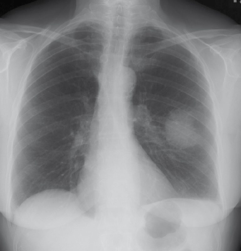
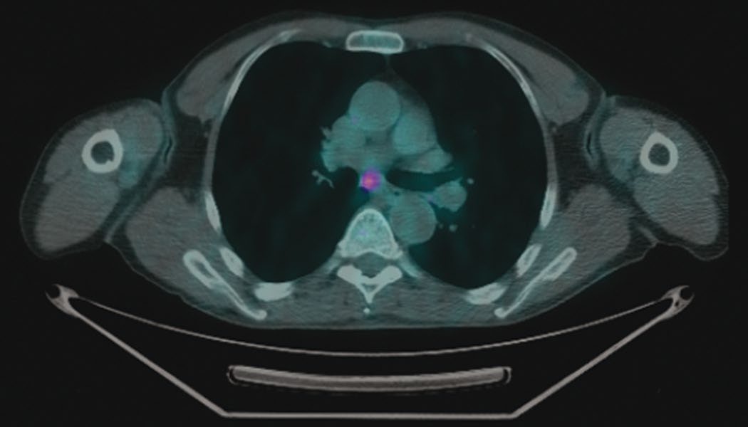
Chest and abdominal CT is the single best test for clinical assessment of T and N status, and the best test overall for resectability; PET is the best test for M status [1]. Brain MRI is indicated for neurological symptoms, stage III or IV disease, small cell carcinoma and Pancoast tumours [1].
Mediastinoscopy is used for centrally located tumours and suspicious adenopathy, nodes above 0.8 cm, or subcarinal nodes above 1.0 cm, on CT [1]. It assesses ipsilateral N2 and contralateral N3 mediastinal nodes, and if mediastinal nodes are positive the tumour is unresectable [1]. It does not assess the aortopulmonary window nodes, which drain the left lung [1].
The Chamberlain procedure (anterior thoracotomy or parasternal mediastinotomy through the left second rib cartilage) assesses enlarged AP window nodes [1].
Bronchoscopy is needed for centrally located tumours, to check for airway invasion [1].
Nodules, tissue acquisition and mediastinal staging in Schwartz's account
- A solitary pulmonary nodule is a well-circumscribed sphere of 3 cm or less surrounded by aerated lung without atelectasis, hilar enlargement or effusion; about 150,000 are found incidentally each year, up to 50% accompany one to six other subcentimetre nodules on screening CT, malignancy probability is 50% or more in smokers against 20–40% in never-smokers and higher with symptoms, age, male sex and occupational exposure [2].
- Thin-section spiral CT (1–2 mm collimation for parenchyma, 5–7 mm to hunt metastases, 3–5 mm for central airways) characterises number, location, size, margin, calcification and growth: diffuse, solid, central and laminated ("popcorn") calcification are benign, granulomas being 70–80% and hamartomas 10% of benign nodules; stippled, amorphous or eccentric calcification, growth, increasing density (40–50% of part-solid nodules are malignant against 15% of subcentimetre solid or non-solid ones), size over 3 cm, lobulated or spiculated edges and the corona radiata sign (fine 4–5 mm radial strands) favour cancer; volume-doubling times of 20–400 days fit lung cancer, shorter suggesting infection and longer benign or indolent tumours; FDG-PET is 97% sensitive and 78% specific with false negatives in AIS, MIA, lepidic tumours, carcinoids and tumours under 1 cm [2].
- For 8–30 mm nodules without benign calcification or 2-year stability, probability under 5% earns serial CT at 3, 6, 12 and 24 months, 5–60% earns PET, contrast CT, transthoracic FNA or bronchoscopy, and over 60% goes to VATS frozen section and resection, pure ground-glass lesions needing longer annual follow-up [2].
- Bronchoscopy is 20–80% sensitive depending on size and centrality, yielding brushings and washings, forceps biopsy, EBUS-FNA of external compression, or fluoroscopic or electromagnetic navigational transbronchial biopsy (up to 80% yield, 1–3.5% pneumothorax, also placing fiducials or tattoos), biopsy preceding brushings for peripheral lesions; CT- or ultrasound-guided transthoracic FNA diagnoses up to 95% of selected peripheral lesions but false negatives run 3–29% so an indeterminate result needs further work-up, with pneumothorax in up to 30%; VATS wedge or segmental excision (nodule under 3 cm) or core biopsy suits outer-third lesions, avoiding nodule manipulation and pleural breach and bagging the specimen to prevent chest wall seeding, proceeding to lobectomy on frozen section; thoracotomy is reserved for deep undiagnosed lesions or invasion unresolvable short of palpation, and pneumonectomy is never performed without tissue proof of cancer [2].
- Histology now drives therapy, pemetrexed or bevacizumab benefits adenocarcinoma but bevacizumab has caused fatal haemorrhage in squamous cancer, and EGFR mutation mandates first-line tyrosine kinase inhibition, so small biopsies are typed by morphology or one adenocarcinoma marker (TTF-1, mucin) and one squamous marker (p63, CK5/6), neuroendocrine stains are reserved for neuroendocrine morphology, adenocarcinomas are tested for EGFR, KRAS and EML4–ALK, and two or three extra passes are taken for cell block after on-site cytology confirms tumour [2].
- Contrast CT nodes over 1 cm predict metastasis in only about 70% (30% reactive), so enlargement must be confirmed histologically and never denies resection alone; false-negative CT is under 10% with normal nodes and T1 tumours but nearly 30% with central or T3 tumours, and T1 adenocarcinomas and large cell carcinomas micrometastasise early; PET is 79–88% sensitive and 91% specific for mediastinal nodes against CT's 63% and 76%, combined PET-CT reaching 93% sensitivity and 94% accuracy, with right upper lobe tumours most prone to occult N2 disease and PET-positive nodes still needing EBUS or mediastinoscopy because a false N3 label would wrongly stage IIIB [2].
- Invasive staging is indicated for enlarged mediastinal nodes (over 1 cm is absolute), central tumours, N1 enlargement, size over 3 cm and FDG-avid normal-size nodes in peripheral stage I: EBUS reaches stations 4R, 4L, 7, 10 and 11 (not 3, 5 or 6) with rapid on-site cytology; EUS reaches 4R, 4L, 7, 8 and 9 and lesions near the oesophagus but not the pretracheal mediastinum, though multi-station positive EUS may spare mediastinoscopy; cervical video-mediastinoscopy samples all paratracheal and subcarinal nodes, judges extracapsular spread and mediastinal invasion, and is recommended for central, T2 and T3 tumours and T1 adenocarcinoma or large cell tumours; a negative EBUS in a radiologically suspicious mediastinum is not enough, so the authors follow negative on-site EBUS with same-session mediastinoscopy but skip it when EBUS is positive, preserving mediastinoscopy for post-induction restaging; left VATS samples stations 5 and 6 for left upper lobe tumours after cervical mediastinoscopy, indicated when other stations are negative and induction would follow any positive station, when a protocol requires N2 proof, when CT shows bulky or extracapsular nodes, or for tissue from a hilar mass or nodes causing recurrent nerve palsy [2].
- A pleural effusion is not assumed malignant: cytology finds malignant cells in only 50% at first thoracentesis and five negative taps give 95% certainty of benignity, so thoracoscopy is used as a staging step; integrated PET-CT is standard and finds 10–15% more distant metastases than CT and bone scan, confirmed by MRI or biopsy in otherwise early disease; brain MRI is done for stage III or symptoms (head CT is negative in 95% of asymptomatic patients); indeterminate liver lesions and adrenal masses go to MRI, lipid-rich adenomas separating from lipid-poor metastases [2].
- New nodules after another cancer are more likely metastatic if multiple, smooth and round and close in time to the primary, but a solitary nodule is a new lung primary in 74% after uterine, 89% after bladder, 92% after lung and 94% after head and neck cancer [2].
Thresholds and severity
Operability
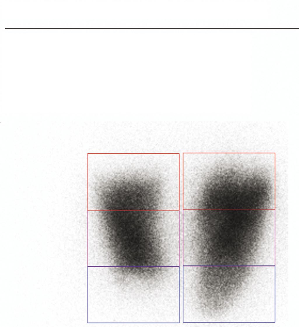
Predicted postoperative FEV1 must exceed 0.8 L, or 40% of the predicted postoperative value [1]. If it is borderline, a qualitative V/Q scan shows the contribution of the diseased lung to overall FEV1, and if that contribution is low resection may still be possible [1]. FEV1 is the best predictor of pulmonary complications and of being able to wean from the ventilator [1].
Predicted postoperative DLCO must exceed 10 mL/min/mmHg, or 40% of predicted; it measures carbon monoxide diffusion and represents oxygen exchange capacity, depending on pulmonary capillary surface area, haemoglobin content and alveolar architecture [1].
No resection if the preoperative pCO2 is above 50 or the pO2 below 60 at rest, or if VO2 max is below 10 to 12 mL/min/kg [1].
- Schwartz adds the functional shortcuts: a patient who can walk indefinitely on the flat without oxygen or rest will tolerate lobectomy, one who can climb two flights without stopping will tolerate pneumonectomy, and nearly all without CO₂ retention tolerate single-lung ventilation and wedge resection; raw FEV1 over 2.0 L suggests pneumonectomy and over 1.5 L lobectomy is tolerable, but percent predicted matters more, a raw FEV1 of 1.3 L is 30% predicted in a 190 cm man (normal 4.31 L) yet 59% in a 157 cm woman (normal 2.21 L) [2].
- Predicted postoperative FEV1 or DLCO is the percent predicted multiplied by the fraction of lung remaining (right upper lobectomy removes 3 of 20 segments, leaving 85%), values under 50% correlate with pulmonary complications rising stepwise with each 10% decline, quantitative perfusion scanning refines the estimate when partial central obstruction makes the contribution of a lobe or lung unclear (a right lung receiving 21% rather than the normal 55% means pneumonectomy costs little), and exercise testing resolves discordance between spirometry and function: VO₂max under 10 mL/kg/min carries 26% mortality after major resection against 8.3% at 10–15 and over 15 generally permits pneumonectomy; the ACCP algorithm adds cardiopulmonary exercise testing when predicted postoperative FEV1 or DLCO is under 40%, or FEV1 under 30% or their product under 1650 [2].
- Formal cardiac assessment (Thoracic Revised Cardiac Risk Index) is mandatory; current smokers and those over 60 pack-years have 2.5 times the pulmonary complications and three times the pneumonia, each 10% fall in DLCO raises complication risk 42%, cessation ideally precedes surgery by 8 weeks and at least 2 weeks (stopping on the day increases sputum retention), and chronic sputum producers merit preoperative culture, antibiotics and bronchodilators [2].
Staging
T stage is T1 for 3 cm or less; T2 for 3.1 to 5.0 cm but more than 2 cm from the carina; T3 for 5.1 to 7.0 cm, or invasion of chest wall, pericardium or diaphragm, or within 2 cm of the carina; and T4 for 7.1 cm or more (still possibly resectable) or invasion of mediastinum, oesophagus, trachea, vertebra, heart or great vessels, or a malignant effusion, which usually indicate unresectability [1].
N stage is N1 for ipsilateral hilar nodes; N2 for ipsilateral mediastinal, subcarinal or aortopulmonary window nodes, which is unresectable; and N3 for contralateral mediastinal or supraclavicular nodes, also unresectable [1]. Hilar nodal involvement does not preclude resection [1].
In the AJCC 8th edition (2018, from 77,156 patients) the T category advances per centimetre to 5 cm: Tis for AIS, T1mi, T1a ≤1 cm, T1b >1–2, T1c >2–3, T2a >3–4, T2b >4–5, T3 >5–7, T4 >7 cm; main bronchus involvement at any distance from the carina and total atelectasis are now T2, diaphragmatic invasion T4, visceral pleural invasion lifts a ≤3 cm tumour to T2, satellite nodules in the same lobe are T3 and in another ipsilateral lobe T4, and M is split into M1a (contralateral lobe nodule, pleural or pericardial nodules or malignant effusion), M1b (single extrathoracic metastasis in one organ) and M1c (multiple); stage groups run IA1–IA3 for T1a–c N0, IB T2a N0, IIA T2b N0, IIB for N1 or T3 N0, IIIA for N2 with T1–2 or T3–4 N1, IIIB for N3 with T1–2 or N2 with T3–4, IIIC for T3–4 N3, IVA for M1a–b and IVB for M1c; grade, lymphovascular invasion, margins, adequacy of nodal dissection and mutation status are also recorded [2]. Small cell cancer is "limited" when confined to one hemithorax within a tolerable radiation field (any T, any N, M0, unless nodules are scattered through the ipsilateral lung) and "disseminated" beyond it, pleural or pericardial effusions counting as disseminated [2].
Screening is by annual low-dose CT [1].
It is indicated for patients aged 50 to 80 with more than a 20 pack-year smoking history who currently smoke, or who quit within the last 15 years [1].
Screening stops when the patient has not smoked for 15 years, or becomes ineligible for surgery because of comorbidity or their own preference [1].
A benign nodule needs no further workup where there has been no growth over 2 years, the contour is smooth, and there is popcorn calcification [1]. Low-risk lesions get serial chest CT, at a frequency set by clinical suspicion, starting at 3 months where there is concern, with biopsy if it grows [1].
Biopsy is indicated for suspicious lesions above 10 mm, with growth over 2 years being worrisome: bronchoscopy-guided for central lesions, CT-guided for peripheral ones, and VATS wedge resection if those fail [1]. The full cancer workup must be completed before VATS, because a frozen section showing cancer means proceeding to formal lung resection at the same sitting [1].
Treatment and Management
Treatment by stage [1]:
- Stage I and II, resection, with definitive radiotherapy if not a surgical candidate; stage II also needs postoperative chemotherapy.
- Stage IIIa, T3,N1,M0 is usually resectable, with neoadjuvant chemoradiotherapy, restaging, then resection. Any N2 disease is not resectable and gets definitive chemoradiotherapy.
- Stage IIIb, generally not resectable, though some T4,N0-1,M0 tumours can be resected after neoadjuvant chemoradiotherapy.
- Stage IIIc and IV, not resectable; definitive chemoradiotherapy.
Chemotherapy differs by cell type: carboplatin with paclitaxel for non-small cell disease at stage II or higher, and cisplatin with etoposide for small cell [1].
Small cell carcinoma is usually unresectable at diagnosis, with under 5% candidates for surgery, and most receive chemoradiotherapy alone [1].
SVC syndrome is managed by elevating the head of the bed, diuretics and hydrocortisone initially, then emergency radiotherapy if severe and malignant, with an endovascular stent if that fails; non-malignant causes go straight to stenting, with open bypass in reserve [1].
Isolated lung metastases may be resected where there is no other systemic disease, for colon, renal cell, sarcoma, melanoma, ovarian and endometrial primaries [1].
Stage-directed therapy, induction and adjuvant chemotherapy in Schwartz's account
- Small cell cancer presenting as an isolated lesion (under 5% are stage I) may have lobectomy then chemotherapy once surgical mediastinal staging excludes N2 disease; otherwise chemotherapy with or without radiotherapy [2].
- Early-stage NSCLC (T1–2 with or without N1, T3 N0; about 16% of 222,500 US cases in 2017) is resected by VATS lobectomy, sleeve lobectomy where a bifurcation tumour precludes a bronchial margin, or occasionally pneumonectomy for a distal main bronchus tumour or unresectable bulky or extracapsular N1 nodes, with mediastinal dissection or sampling; untreated stage IA has a 14-month median and 22% 5-year survival, resected pathological IA 67%, and all localised disease 55% against 26% with regional spread [2].
- Inoperability is declared only after expert surgical evaluation at a high-volume centre; limited resection (segmentectomy or wedge) once carried unacceptable local recurrence, but meta-analysis of 27 studies (6111 segmentectomies vs 18,431 lobectomies) found no survival difference (HR 1.04), and anatomic segmentectomy with nodal dissection and a margin-to-tumour-diameter ratio above 1 gives 6.2% recurrence against 25% when the ratio is below 1 [2].
- For the non-operable, CT-guided radiofrequency ablation of tumours under 3.5 cm gives up to 80% radiographic resolution and about 90% cancer-specific 2-year survival, contraindicated by size over 5 cm, hilar abutment, malignant effusion, more than three lesions in one lung, pulmonary hypertension or proximity to a large vessel (bleeding and heat sink); stereotactic body radiotherapy delivers up to 66 Gy in a few fractions with 97.6% primary tumour and 90.6% local control at 3 years in 59 inoperable node-negative patients but 12.8% locoregional recurrence against about 6.5% after limited resection, and requires tumours over 2 cm from the proximal bronchial tree because central lesions cause hypoxia, haemoptysis, atelectasis and pneumonitis; ablation omits nodal staging and tissue for molecular profiling though up to 27% of clinical stage I tumours harbour nodal disease at resection [2].
- Induction chemotherapy adds 4–7% absolute 5-year survival at every stage (IA 75→79%, IB 55→61%, IIA 50→57%, IIB 40→47%, IIIA 15–35→21–42%, IIIB 5–10→8–15%) without extra perioperative morbidity except after right pneumonectomy, and is an acceptable alternative wherever adjuvant therapy is anticipated; its advantages are intact tumour blood supply, downstaging, better tolerance and completion, an in vivo sensitivity test, response monitoring, treatment of micrometastases and sparing non-responders a resection, its disadvantages the right pneumonectomy risk and delay in resection [2].
- NCCN recommends observation after complete resection of T1a–c N0 tumours and consideration of chemotherapy for node-negative T2a–b tumours with high-risk features (poor differentiation, moderately or poorly differentiated neuroendocrine tumours, vascular invasion, wedge-only resection, size over 4 cm, visceral pleural invasion, incomplete nodal sampling); any N1 or N2 disease or T3 tumour receives adjuvant chemotherapy, better tolerated after minimally invasive resection, positive margins are re-resected or given concurrent (macroscopic) or sequential (microscopic) chemoradiation [2].
- Stage III survival is 28% but heterogeneous: clinically evident bulky N2 disease survives 5–10% at 5 years with surgery alone whereas incidental microscopic single-station N2 disease may reach 30%, so resection after induction is accepted for a single node under 3 cm in one station; N2 disease must be histologically confirmed because PET false positives are unacceptably high, especially in granulomatous regions; unexpected N2 nodes at operation may be resected or the operation abandoned for induction, proven preoperative N2 disease goes to two or three cycles of cisplatin-based induction (about 70% response, two randomised trials stopped early for survival benefit), sterilised nodes carry a better prognosis and warrant resection, and persistent N2 disease after induction must again be proven rather than assumed radiologically [2].
- Surgery is occasionally appropriate for T3–4 N0–1 tumours invading SVC, carina, vertebra, mediastinum, heart, great vessels, trachea, recurrent nerve, oesophagus or diaphragm, never for N3 or T4 N2 disease, and resection of a solitary brain metastasis with an early-stage primary achieves 10–15% 5-year survival; sequential chemoradiation (cisplatin doublet then about 60 Gy) raised 5-year survival from 6% with radiotherapy alone to 17%, concurrent chemoradiation adds about 4% absolute survival at 2 years at the cost of reduced chemotherapy dose, and radiotherapy alone cures under 7% of N2–3 disease [2].
- Pulmonary metastasectomy requires a controlled primary, fitness for single-lung ventilation and the planned resection, complete resectability on CT, no extrapulmonary disease and no superior alternative; the International Registry of Lung Metastases (5206 patients, 88% complete) reported 36%, 26% and 22% survival at 5, 10 and 15 years after complete resection against 13% and 7% after incomplete, best with germ cell tumours, osteosarcoma, a disease-free interval over 36 months and a single metastasis; all macroscopic tumour and involved chest wall, diaphragm or pericardium are resected en bloc, lobectomy for multiple or hilar lesions, pneumonectomy rarely, and VATS missed 56% of palpable lesions in McCormack's pre-spiral-CT series though a recent study suggests 18% and a UK series found equivalent outcomes [2].
Procedural interventions
Lobectomy or pneumonectomy is the standard, since formal lung resection is required for lung cancer, with sampling of suspicious nodes; VATS resection is considered for stage I peripheral tumours without nodal or local invasion [1].
Carcinoid is resected and treated as a cancer, with outcome closely linked to histology [1].
- Bronchial adenomas are all malignant tumours.
- Carcinoid accounts for 90%; the others are mucoepidermoid adenoma, mucous gland adenoma and adenoid cystic adenoma [1].
- The first two grow slowly and do not metastasise, and are resected with a 1 cm margin; adenoid cystic adenoma arises from submucosal glands and spreads along perineural lymphatics well beyond its endoluminal component, is very radiosensitive, and can give 10-year survival even with incomplete resection [1].
Pancoast resection, approaches and postoperative care in Schwartz's account
- Pancoast's tumour (described 1932) is any superior sulcus tumour involving parietal pleura or deeper structures over the first rib, chest wall involvement at or below the second rib does not qualify; resection is offered only with mediastinoscopy-proven negative mediastinal nodes (a negative EBUS is insufficient), after concurrent induction cisplatin–etoposide with 45 Gy over 5 weeks, restaging by CT, brain imaging and PET, and MRI/MRA of vessels and plexus: the Southwest Oncology Group regimen was completed by 95%, complete resection achieved in 76%, 5-year survival 44% overall and 54% after complete resection, with brain the commonest site of progression; thoracotomy with en bloc chest wall and vascular resection, lobectomy, part of the lower brachial plexus trunk and stellate ganglion follows, defects of two or more rib segments (unless small or behind the scapula) are reconstructed with Gore-Tex, and large pericardial defects are patched with thin Gore-Tex to prevent herniation [2].
- VATS or robotic resection is now recommended for effusions, recurrent pneumothorax, lung biopsy, lobectomy and segmentectomy, cysts and oesophageal mobilisation, using two to four 0.5–1.2 cm ports plus a fourth or fifth space anterior axillary access incision placed high enough for the hilum, with endoscopic staplers dividing vessels and bronchus; pain at 3 weeks, 6-minute walk, return to work, chemotherapy tolerance and respiratory recovery all favour VATS, most markedly in COPD and the elderly, and it is preferred for pulmonary or cardiac compromise, extrathoracic malignancy, poor performance, rheumatological disease, age over 70, vascular disease, impending major surgery, psychological conditions and immunosuppression [2].
- Open options are the posterolateral thoracotomy (anterior axillary line to below the scapular tip, dividing latissimus, entering along the lower margin of the interspace), the anterolateral thoracotomy for trauma in the supine position, the clamshell (bilateral anterior thoracotomy with transverse sternotomy) for double-lung transplant, the hemi-clamshell or trap-door (with frequent nipple hypaesthesia) and the median sternotomy [2].
- Chest tubes are unnecessary when visceral pleura is intact (as after VATS sympathectomy); pleural lymphatics absorb up to 0.4 mL/kg/h (about 500 mL a day), so tubes are removed at 400 mL or less per 24 hours after lobectomy or lesser resection but at 100–150 mL where pleural dynamics are altered by malignancy, infection or pleurodesis; –20 cmH₂O suction is used for the first 12–24 hours then water seal if the lung is expanded, since suction prolongs air leaks; an undrained pneumothorax prompts checking for kinks, flushing pigtails through a cleaned stopcock, and a cough or Valsalva, a stationary water-seal level means blockage or pleurodesis, bubbling means a leak, and collapse on water seal returns the tube to suction [2].
- Analgesia is epidural at about T6 (fentanyl 0.3 µg/mL with 0.125% bupivacaine or 0.1% ropivacaine, the latter less cardiotoxic), paravertebral catheters 2.5 cm lateral to T4–T6 spinous processes with equal analgesia and less hypotension (sympathetic block otherwise needs phenylephrine rather than fluid, dangerous after pneumonectomy), liposomal bupivacaine into the intercostal spaces for 72 hours, or patient-controlled opioids with ketorolac, gabapentin and paracetamol, avoiding oversedation that causes retention, atelectasis and aspiration; oral analgesia begins on day 3–4 with routine laxatives, and the best respiratory care is an effective splinted cough, early sitting out and ambulation [2].
Complications
The commonest complication of lung resection is atelectasis, treated with incentive spirometry [1].
Each operation has its own characteristic complication: persistent air leak is commonest after segmentectomy or wedge resection, atelectasis after lobectomy, and arrhythmia after pneumonectomy [1].
Postoperative complications in Schwartz's account
- Post-pneumonectomy pulmonary oedema follows 1–5% of pneumonectomies, more often right-sided, presenting hours to days later with diffuse interstitial or alveolar oedema from increased permeability and filtration pressure with reduced lymphatic drainage; it is prevented by strict fluid restriction and vasopressors rather than boluses, treated by ventilation, fluid restriction, diuretics and sometimes ECMO, and carries nearly 100% mortality [2].
- Air leaks are common in emphysematous lung; leaks beyond 5 days are managed by stopping suction, continued drainage or doxycycline or talc pleurodesis (effective only with full lung apposition), while a moderate or large leak, especially after induction therapy or in the immunocompromised, raises suspicion of bronchopleural fistula from the stump, confirmed bronchoscopically and managed by prolonged drainage, reoperation with intercostal or serratus muscle reinforcement, bronchoscopic fibrin glue for fistulas under 4 mm, and open drainage for the frequent accompanying empyema [2].
- Hypotension from epidural sympathetic block, secretion retention from oversedation, and transthoracic biopsy pneumothorax in up to 30% are the other recurring hazards [2].
Outcomes
Overall 5-year survival in lung cancer is 10%, rising to 30% with resection for cure [1].
Small cell carcinoma has an overall 5-year survival under 5%, though T1,N0,M0 disease reaches 50% [1].
Carcinoid outcome depends on histology: 90% 5-year survival for typical carcinoid against 60% for atypical, with recurrence increased by positive nodes or a tumour above 3 cm [1].
Recurrence usually appears as disseminated metastases, and 80% occur within the first 3 years [1]. The brain is the single commonest site of metastasis, with supraclavicular nodes, the other lung, bone, liver and adrenals also involved [1]. Follow-up after resection for cure is history, examination and chest CT every 6 months for 2 years, then annually [1].
References
- The ABSITE Review, 2022, Ch. 25 Thoracic
- Schwartz's Principles of Surgery, 11th ed., Ch. 19, Chest Wall, Lung, Mediastinum, and Pleura
- Sabiston Textbook of Surgery, 22nd ed., Ch. 110 Lung, Chest Wall, Pleura, and Mediastinum