



Wound Healing
Summary
- Wound healing is a complex, orderly and overlapping biological process traditionally divided into haemostasis, inflammation, proliferation and remodelling (maturation) phases [1][2].
- All healthy wounds undergo the same basic steps of repair, and orderly, timely movement through these phases is required for coordinated repair; if orderly repair does not occur, as in diabetes or other chronic conditions, wounds can remain in a phase indefinitely and fail to advance [2].
- Wounds are classified by the manner of closure into primary, secondary and tertiary (delayed primary) healing, and by contamination into clean, clean-contaminated, contaminated and dirty categories [1].
Definition
- Primary healing (healing by first intention) occurs when there is direct approximation of wound edges soon after injury, in the absence of adverse influences, giving the best scar [1].
- Secondary healing (healing by second intention) occurs in wounds that are left open and allowed to heal by granulation, contraction and re-epithelialisation [1].
- Delayed primary healing (tertiary intention) occurs when wound edges are not opposed immediately (as may be necessary in contaminated or untidy wounds) and are surgically approximated later, after debridement of non-viable tissue, once the wound is clean [1].
- Chronic wounds are, by definition, wounds that have failed to proceed through an orderly and timely reparative process to produce anatomic and functional integrity over a period of 3 months [2].
Pathophysiology
- Haemostasis.
- Disruption of the vascular endothelium causes vasoconstriction and exposure of the subendothelial extracellular matrix, encouraging platelet adhesion, activation and aggregation to form a platelet plug.
- Alpha granules released by activated platelets contain TGF-β, PDGF, FGF, EGF and VEGF, which drive matrix deposition, chemotaxis, epithelialisation and angiogenesis, while tissue factor at the injury site initiates the coagulation cascade, generating thrombin, which forms fibrin to stabilise the plug and provide a scaffold for infiltrating cells [1].
- Inflammation (days 1–10 or so).
- In the early inflammatory phase (days 1–2), platelet activation triggers an influx of neutrophils, which limit bacterial contamination; histamine and serotonin release increases vascular permeability, aiding inflammatory cell infiltration.
- In the late inflammatory phase (days 2–3), monocytes differentiate into macrophages, which phagocytose debris, release proteolytic enzymes, and are the primary source of cytokines and growth factors driving fibroblast proliferation and angiogenesis [1].
- The predominant cell type shifts from neutrophils (days 0–2) to macrophages (days 3–4) to fibroblasts (day 5 onward) [3].
- Macrophages are essential for wound healing via their release of growth factors and cytokines [3].
- Proliferation (roughly day 3 to 2–4 weeks).
- This phase consists mainly of fibroblast activity, producing ground substance (glycosaminoglycans and proteoglycans), collagen and new blood vessels, along with re-epithelialisation; early proliferative-phase tissue is called granulation tissue and has a characteristic pink, granular appearance [1].
- Fibronectin, produced by fibroblasts, is chemotactic for macrophages and anchors fibroblasts, and the "provisional matrix" is composed mostly of fibronectin and hyaluronic acid before fibroblasts replace the fibronectin-fibrin scaffold with collagen [3].
- Epithelial integrity, achieved by migration of epithelial cells from hair follicles (the most important source), wound edges and sweat glands, is the most important factor in healing open wounds by secondary intention, while tensile strength (dependent on collagen deposition and cross-linking) is the most important factor in healing closed incisions by primary intention [3].
- Some fibroblasts differentiate into myofibroblasts, contractile cells that help draw wound edges together [1]; myofibroblasts communicate via gap junctions and mediate wound contraction during healing by secondary intention [3].
- Remodelling (2–3 weeks to a year or more).
- This phase is characterised by collagen maturation: type III collagen, prevalent during proliferation, is replaced by stronger type I collagen until the normal skin ratio of about 4:1 type I to type III collagen is re-established, with increasing cross-linking and alignment producing maximal tensile strength around 12 weeks post-injury, approximately 80% of uninjured skin strength [1].
- The net amount of collagen does not change substantially during remodelling, although significant production and degradation occur simultaneously, and continued cross-linking (not increased collagen quantity) accounts for the ongoing increase in tensile strength after collagen content plateaus at about 3 weeks [3][4].
- Alpha-ketoglutarate, vitamin C, oxygen and iron are required for collagen synthesis, including hydroxylation of proline by prolyl hydroxylase and subsequent cross-linking; vitamin C deficiency (scurvy) impairs this process [3].
- Peripheral nerves regenerate at approximately 1 mm/day, and epithelialisation proceeds at 1–2 mm/day [3].
The phases in Schwartz's detail
- Haemostasis initiates inflammation: subendothelial collagen aggregates and degranulates platelets, whose α-granules release PDGF, TGF-β, platelet-activating factor, fibronectin and serotonin, and the fibrin clot scaffolds cell migration.
- Neutrophils arrive first and peak at 24–48 hours, drawn by complement, IL-1, TNF-α, TGF-β, platelet factor 4 and bacterial products; they phagocytose debris, are the main early source of TNF-α, release collagenases, contribute nothing to collagen deposition or strength and may delay epithelial closure.
- Macrophages reach significant numbers at 48–96 hours and stay until healing is complete, debriding by phagocytosis, achieving microbial stasis with oxygen radicals and nitric oxide, and (their central role) recruiting and activating cells through TGF-β, VEGF, IGF, EGF and lactate.
- T lymphocytes peak at about 1 week and bridge inflammation to proliferation; depleting most wound T cells reduces strength and collagen, depleting the CD8 suppressor subset enhances healing, depleting CD4 helpers has no effect, and lymphocytes down-regulate fibroblast collagen synthesis by cell-associated IFN-γ, TNF-α and IL-1 through direct contact [5].
- Proliferation spans roughly days 4–12: fibroblasts and endothelial cells are the last to arrive, PDGF is the strongest fibroblast chemoattractant, wound fibroblasts synthesise more collagen and proliferate less than dermal fibroblasts and contract matrix, lactate accumulating to about 10 mmol drives collagen synthesis by ADP-ribosylation, and endothelial cells migrate from nearby venules under TNF-α, TGF-β and VEGF, the macrophage being the major VEGF source and VEGF receptors sitting specifically on endothelium [5].
- Collagen (at least 18 types, I and III mattering for repair) has glycine in every third position with proline or lysine second; the roughly 1000-residue protocollagen chain is hydroxylated in the endoplasmic reticulum by prolyl hydroxylase, which needs oxygen and iron as cofactors, α-ketoglutarate as co-substrate and ascorbate as electron donor, then glycosylated at hydroxylysine; three chains form procollagen with terminal registration peptides cleaved extracellularly by procollagen peptidase before covalent lysine cross-linking [5].
- Glycosaminoglycans, dermatan and chondroitin sulphate predominating, chains from about 10 units in heparan sulphate to 2000 in hyaluronic acid, are coupled to proteins as proteoglycans, rise sharply over the first 3 weeks, provide the lattice on which collagen fibrils assemble (sulphation dictating fibril configuration) and then diminish as scar matures [5].
- Remodelling balances MMP collagenolysis against synthesis; matrix is laid down as fibronectin and type III collagen first, glycosaminoglycans and proteoglycans next and type I collagen last; collagen content plateaus at several weeks while tensile strength rises for months as cross-linking cuts solubility and raises resistance to enzymes; fibrillin from fibroblasts is essential for elastic fibres; remodelling continues 6–12 months to an avascular, acellular scar that never matches uninjured tissue, and TGF-β both raises collagen transcription and lowers breakdown by inducing tissue inhibitors of metalloproteinases [5].
- Epithelialisation begins within a day as marginal basal cells detach, enlarge and migrate over the provisional matrix while fixed basal cells near the edge divide and move in leapfrog fashion until the defect is bridged, after which cells become columnar, layering is restored and the surface keratinises; approximated incisions re-epithelialise in under 48 hours, and superficial injuries such as donor sites or superficial partial-thickness burns heal by epithelialisation with little fibroplasia, driven by loss of contact inhibition, fibronectin exposure and EGF, TGF-β, bFGF, PDGF and IGF-1 [5].
- Growth factors act in nanomolar concentrations in autocrine, paracrine or endocrine fashion and only when the receptor is present at release; Schwartz's table adds HGF (suppresses inflammation, granulation and angiogenesis), keratinocyte growth factor, TGF-α (EGF homologue), TGF-β1 stimulating matrix while TGF-β3 inhibits scar, IGF-1/2 promoting matrix and glucose transport, IL-1, IL-4, IL-6, activin, angiopoietins, CX3CL1 and GM-CSF [5].
- Contraction reduces the area of unapproximated wounds; the myofibroblast carries α-smooth muscle actin in stress fibres, undetectable until day 6, rising for 15 days and fading after 4 weeks as the cells apoptose, yet contraction starts almost at injury and fibroblasts contract collagen lattices without stress fibres, so cytoskeletal reorganisation during cell movement is also implicated [5].
Healing in other tissues and the fetus in Schwartz's account
- In the gut the submucosa holds sutures and gives tensile strength and the serosa provides the early fibrin seal, hence higher failure in the extraperitoneal, serosa-less oesophagus and rectum; marginal strength falls sharply in the first week because collagenase from neutrophils, macrophages and luminal bacteria (Pseudomonas shifting to high collagenase secretion in anastomosed bowel) makes lysis exceed synthesis for 3–5 days, collagenase being far more marked in colon than small bowel; colon fibroblasts and smooth muscle cells both make collagen and colon fibroblasts out-produce skin fibroblasts, matrix arrangement mattering more than amount; no suture technique (hand vs stapled, continuous vs interrupted, absorbable vs not, one vs two layers) is proven superior although a meta-analysis found fewer leaks with stapled ileocolic anastomoses, and over-resuscitation with third-spacing, oedema and raised intra-abdominal pressure compromises edge perfusion; the fetal gut scars while fetal skin does not, and d-penicillamine impairs skin but not gut cross-linking [5].
- Bone passes through haematoma, liquefaction and revascularisation, soft callus at 3–4 days (an internal splint ending pain and inflammation), hard callus mineralising over 2–3 months to weight-bearing radiographic union, then remodelling recanalising the marrow, under bone morphogenetic proteins of the TGF-β superfamily plus PDGF, TGF-β, TNF-α and bFGF; avascular cartilage nourished by diffusion and the perichondrium heals superficial injury without inflammation and incompletely, while deep injury exposing bone mounts an inflammatory response with granulation, fibrous tissue and chondrification to hyaline cartilage; tendon and ligament heal through haematoma, organisation and scar with types I and III collagen, water, DNA and glycosaminoglycan, hypovascular tendons healing with more scar and less motion while metabolically active tenocytes retain regenerative potential; nerve injury (200,000 repairs a year in the United States) is neurapraxia (focal demyelination), axonotmesis (axonal interruption with intact Schwann basal lamina) or neurotmesis (transection), healing by cell-body survival, axonal regeneration across the gap and target reconnection after phagocytes clear the distal stump by Wallerian degeneration, with Schwann cells remyelinating and nerve growth factor, BDNF, FGFs, neuroleukin and adhesion molecules (N-CAM, Ng-CAM, myelin adhesion glycoprotein, N-cadherin) guiding growth [5].
- Fetal wounds heal without scar until a "transition wound" at the start of the third trimester, when scarless healing persists but appendage regeneration is lost before adult scarring emerges; the sterile amniotic environment alone does not explain it (scars form in utero and scarless healing occurs outside amniotic fluid), the neutropenic fetus mounts less inflammation with fewer neutrophils and macrophages, TGF-β is absent (neutralising TGF-β1 or β2 or applying TGF-β3 reduces adult scarring) and sustained hyaluronic acid production, stimulated by fetal urine, organises reticular rather than parallel collagen, so hyaluronic acid is used topically to aid healing and limit adhesions [5].
Clinical features
- Clinically, wound healing progresses through visible phases: eschar or fibrinous exudate reflects the inflammatory phase, granulation tissue reflects the proliferative phase, and a contracting or advancing wound edge reflects the maturational phase, with all four phases potentially present simultaneously in a large wound [2].
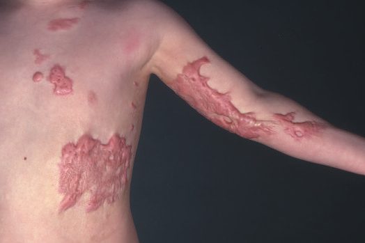
- Hypertrophic scars are raised scars that remain within the confines of the original wound and frequently regress spontaneously; they are more common in areas of increased tension, wounds crossing tension lines, deep dermal burns, and wounds left to heal by secondary intention for more than 3 weeks [1].
- Keloid scars, by contrast, extend beyond the boundaries of the original wound, do not spontaneously regress, are difficult to treat, occur more often after relatively minor trauma in darker-skinned individuals, and are more prevalent above the clavicles, on the trunk, upper extremities and face, developing in 15–20% of Black, Asian and Hispanic patients [1][2].
- Histologically, hypertrophic scars contain well-organised type III collagen with fibroblast/vessel/collagen "islands," while keloids contain disorganised type I and III collagen bundles forming thicker, acellular nodule-like structures in the deep dermis [2].
- Wound dehiscence presents as leakage of large amounts of pink "salmon-coloured" fluid from the wound and, if untreated, can progress to evisceration [3].
Etiology
- Numerous local and systemic factors influence wound healing.
- Local factors include skin tension, hypoxia and ischaemia, vascular insufficiency, lymphoedema, contamination, infection, presence of foreign bodies, and radiotherapy; systemic factors include advancing age, obesity, malnutrition, smoking, diseases such as diabetes mellitus and connective tissue disorders, immunocompromise, and medications such as steroids, immunosuppressants and chemotherapy [1].
- Impediments to wound healing include bacterial contamination above 10^5 organisms/cm² (which decreases oxygen content and causes collagen lysis and prolonged inflammation), devitalised tissue and foreign bodies (which retard granulation), cytotoxic drugs such as 5-fluorouracil, methotrexate and calcineurin inhibitors (which impair healing in the first 14 days after injury), diabetes (which impedes early inflammation via poor leukocyte chemotaxis from hyperglycaemia), serum albumin below 3.0 g/dL, corticosteroids (which inhibit macrophages, neutrophils and fibroblast collagen synthesis, reducing wound tensile strength), and wound ischaemia from fibrosis, pressure, poor arterial inflow, poor venous outflow, smoking, radiation, oedema or vasculitis [3].
- Vitamin A (25,000 IU daily) can counteract the inhibitory effects of steroids on wound healing [3].
- Genetic disorders of abnormal wound healing include osteogenesis imperfecta (type I collagen defect), Ehlers-Danlos syndrome (multiple collagen disorders, more than half due to defects in the α-chains of type V collagen), Marfan syndrome (FBN-1 gene mutation affecting fibrillin), epidermolysis bullosa, scurvy (vitamin C deficiency) and pyoderma gangrenosum [3][4].
- Keloid formation shows strong evidence of genetic susceptibility, including familial heritability, twin concordance and high prevalence in certain ethnic populations, with proposed molecular pathways including apoptosis, cytokine-cytokine receptor interaction, MAPK signalling and TGF-β/VEGF overexpression [2].
Systemic and local impediments in Schwartz's detail
- Age over 70 delayed epithelialisation of superficial defects by 1.9 days in volunteers, with normal collagen (DNA and hydroxyproline) but less non-collagenous protein, so age delays rather than prevents healing and the elderly's dehiscence and hernia rates largely reflect comorbidity and drugs [5].
- Hypoxia impairs fibroplasia despite initially stimulating it, raising inspired oxygen briefly during and after surgery increases collagen deposition and cuts infection, the subcutaneous capillary bed is exquisitely sensitive to volume, temperature and sympathetic tone from pain, mild normovolaemic anaemia is harmless but a haematocrit 15% below normal interferes with healing [5].
- Glucocorticoids in large or chronic doses inhibit inflammation (angiogenesis, neutrophil and macrophage migration, fibroblast proliferation) and lysosomal enzyme release in proportion to anti-inflammatory potency, matter less if started after days 3–4 yet always impair epithelialisation and contraction and raise infection, and topical or systemic vitamin A reverses their effect; antimetabolite chemotherapy inhibits early proliferation, DNA and protein synthesis, fibrin deposition and contraction, worst when given preoperatively, so a 2-week delay after injury is advised, and extravasation necroses tissue [5].
- Uncontrolled diabetes reduces inflammation, angiogenesis and collagen synthesis and adds macro- and microvascular hypoperfusion with defective granulocytes, capillary ingrowth and fibroblasts; in clean experimental human wounds type 1 diabetes reduced collagen accumulation regardless of glycaemic control whereas type 2 did not; insulin given early restores collagen and granulation in animals, and the diabetic wound is growth-factor deficient and highly proteolytic; uraemia lowers collagen synthesis and breaking strength (hard to separate from malnutrition) and calciphylaxis produces exquisitely painful wounds; uncomplicated obesity (over 60% of Americans overweight or obese) weakens wounds through proinflammatory adipokines and infiltrating preadipocytes, with complication estimates of 30% dehiscence, 17% surgical site infection, 30% incisional hernia, 19% seroma, 13% haematoma and 10% fat necrosis and a tenfold rise in leak, collection and infection with increased subcutaneous fat [5].
- Nutrition: 0–4% protein or 50% energy diets impair collagen, breaking strength and infection resistance in rats, malnourished patients deposit less hydroxyproline into implanted PTFE tubes, even brief preoperative deficits impair fibroplasia and brief enteral or parenteral repletion reverses it; arginine is the most active amino acid, 30 g arginine aspartate (17 g free arginine) or 30 g arginine HCl (24.8 g) daily for 14 days raised collagen deposition in young and elderly volunteers without affecting DNA synthesis or epithelialisation, and arginine with β-hydroxy-β-methylbutyrate and glutamine enhanced collagen in the elderly; vitamin C (RDA 60 mg, up to 2 g daily in severe injury or burns, no benefit from excess) is required for proline and lysine hydroxylation and its lack raises and worsens infection; vitamin A increases macrophage influx and collagen and EGF receptors, reverses steroid, diabetic, tumour, cyclophosphamide and radiation impairment, and 25,000–100,000 IU daily is advocated after severe injury; zinc is part of over 150 enzymes, deficiency lowers fibroblast proliferation, collagen, strength and epithelialisation, and supplementation helps only the deficient [5].
Heritable connective tissue disease in Schwartz's account
Ehlers-Danlos comprises ten disorders, over half with defective type V collagen α-chains giving thin friable skin with prominent veins, bruising, atrophic scars, recurrent hernias and hypermobile joints, plus GI bleeding, hiatal hernia, diverticula and rectal prolapse, fragile small vessels and large-vessel aneurysm, varicosity, fistula or rupture (type IV, the type III collagen defect, ruptures arteries, bowel and uterus; type VI lacks lysyl hydroxylase; type IX has a lysyl oxidase/copper defect with bladder diverticula and occipital horns; a recessive tenascin-X-deficient form exists); it should be considered in any child with recurrent hernias and coagulopathy, hernias resemble adult ones with a thin transversalis and dilated internal ring so mesh lowers recurrence, and dermal wounds are closed in two layers under tension with sutures left twice as long and reinforced by tape [5]. Marfan's (FBN1 fibrillin mutation, now understood to dysregulate TGF-β signalling especially in the aortic wall) gives tall stature, arachnodactyly, lax ligaments, myopia, scoliosis, pectus excavatum, ascending aortic aneurysm and hernias with hyperextensible skin but normal healing; osteogenesis imperfecta (type I collagen mutation; four subtypes from mild blue-sclera type I to prenatally lethal type II) brings brittle bones, osteopenia, hernias, thin bruisable skin but normal scarring; epidermolysis bullosa (simplex, junctional, dystrophic (recessive COL7A1 type VII collagen) and Kindler's) blisters with minimal trauma, compromises nutrition through oral erosions and oesophageal obstruction needing dilatation or gastrostomy, and requires meticulous incisions and non-adhesive bulky dressings; acrodermatitis enteropathica (recessive SLC39A4 on 8q24.3, failed intestinal zinc uptake) impairs granulation because zinc is a cofactor for DNA polymerase and reverse transcriptase, causes periorificial and acral pustular dermatitis, is diagnosed by low blood zinc and cured by 100–400 mg oral zinc sulphate daily [5].
Diagnosis
- Assessment of a wound relies on clinical history and examination (duration, cause, contamination, and local/systemic risk factors listed above) together with staging by phase of healing [1].
- A widely used wound classification, introduced in 1964 by the US National Research Council and later adapted by the US Centers for Disease Control and Prevention, describes the degree of bacterial contamination of surgical wounds at the time of surgery as Class I (clean), Class II (clean-contaminated), Class III (contaminated) or Class IV (dirty) [1].
- Diabetic foot ulcers typically occur at points of pressure related to neuropathy (e.g. at the Charcot joint, classically the second metatarsophalangeal joint), because loss of protective sensation allows repeated unnoticed trauma [3].
Reading an ulcer: the five edges
- When a wound fails to heal, the edge is the single most informative physical sign, and it is examined systematically rather than glanced at.
- After recording site, size and shape, examine the base, edge, depth, discharge and surrounding tissues, then the state of the local tissues and local lymph glands, before completing the general examination [6].
- For an irregular ulcer, draw it in the notes with dimensions, or lay a sterile transparent sheet over it and trace the outline with a felt-tipped pen [6].
The base carries its own clues: solid brown or grey dead tissue indicates full-thickness skin death; a slough resembling yellow-grey wash-leather is syphilitic; bluish unhealthy granulation tissue is tuberculous; and ischaemic ulcers contain poor granulation tissue with tendons and other structures lying bare [6]. The redness of the granulation tissue reflects its underlying vascularity and so indicates the ulcer's ability to heal, and healing epidermis is seen as a pale layer extending in over the granulation from the edge [6].
There are five types of edge, and each maps to a mechanism [6]:
| Edge | Mechanism | Classic lesion |
|---|---|---|
| Flat, gently sloping | Shallow, superficial ulcer; new skin growing in is pale pink and almost transparent | Venous ulcer (though many other types share it) |
| Square-cut or punched-out | Rapid full-thickness death and loss of skin with little attempt at repair | Trophic ulcer from a neurological deficit; classically tertiary syphilis, but today mostly diabetic neuropathy and peripheral arterial ischaemia |
| Undermined | Infection affecting subcutaneous tissue more than skin | Pressure necrosis of the buttock (subcutaneous fat is more susceptible to pressure than skin); classically tuberculous |
| Rolled | Slow growth of tissue in the edge | Basal cell carcinoma, "typical, and almost diagnostic", pale pink or white, with clumps of cells visible through a paper-thin squamous covering and telangiectases in the pearly edge |
| Everted | Tissue growing so rapidly it spills out to overlap normal skin | Squamous cell carcinoma, in skin, bowel, bladder and respiratory tract |
Table reformats the five ulcer edges [6]. Record the depth in millimetres and anatomically, by naming the structures the ulcer has reached, and describe the discharge as serous, sanguineous, serosanguinous or purulent [6]. A scab of coagulated discharge may hide all of these features and may have to be removed before the ulcer can be examined properly [6].
Surgical site infection: where the organisms come from
- Most surgical site infections are preventable, and most come from the patient rather than the theatre.
- SSI accounts for up to 20% of all healthcare-acquired infections and 72% of deaths from them, with an incidence of at least 0.5% to 9.5% across all surgical procedures in the EU and USA; the majority are caused by microorganisms from the patient's own flora introduced intraoperatively, with postoperative infection from external sources less common [7].
- The consequences are quality of life, morbidity including further operations, extended hospital and intensive care stay, higher mortality and increased cost [7].
Incidence varies by operation, and the laparoscopic-versus-open gap is the striking pattern [7]:
| Operation | Approximate mean SSI incidence |
|---|---|
| Hip or knee replacement | <1% |
| Spinal surgery (laminectomy) | 1% |
| Laparoscopic cholecystectomy | 1.5% (open biliary surgery 4%) |
| Caesarean section | 2% |
| Coronary artery bypass grafting | 3% |
| Colorectal surgery, laparoscopic | 7% (open 11%) |
Table reformats the reported incidence by operation type [7].
The likely organism is predictable from the operation, which is what makes prophylaxis rational. The commonest single organism overall is Staphylococcus aureus [7]:
| Operation type | Likely organisms |
|---|---|
| Grafts, implants or prostheses | S. aureus, coagulase-negative Staphylococcus |
| Cardiac, vascular, neurosurgery, breast | S. aureus, coagulase-negative Staphylococcus |
| Orthopaedic and trauma | S. aureus, coagulase-negative Staphylococcus, Gram-negative bacilli |
| Diabetic foot sepsis | Staphylococcus spp. including MRSA, streptococci, Gram-negative bacilli including Pseudomonas, anaerobes |
| Appendicectomy, colorectal, biliary tract | Gram-negative bacilli, anaerobes |
| Upper GI (gastroduodenal) | Gram-negative bacilli, streptococci, oropharyngeal anaerobes |
| Head and neck involving oropharyngeal mucosa | S. aureus, streptococci, oropharyngeal anaerobes |
| Obstetric and gynaecological | Gram-negative bacilli, enterococci, group B Streptococcus, anaerobes |
| Urology | Gram-negative bacilli |
Table reformats the expected organisms by operation [7]. Beyond the patient's own flora, sources are indirect contact (hands of healthcare workers, other patients, visitors, contaminated surfaces), direct inoculation (failure of aseptic technique, contaminated instruments or dressings, colonisation of indwelling drains, catheters and lines, a contaminated field such as bowel spillage at laparotomy), airborne (skin and clothing of staff, patients and visitors; theatre or ward air flow) and haematogenous (intravenous and intra-arterial lines, sepsis elsewhere, contaminated infusions) [7].
Two structural points from the same source are worth carrying: cells are labile (good regenerative capacity, such as surface epithelium), stable (slow regeneration, such as hepatocytes) or permanent (none, such as nerve and striated muscle), and tissue architecture itself limits repair, since complex arrangements such as renal glomeruli cannot be reconstructed once destroyed [7]. Myofibroblast-mediated contraction can reduce a tissue defect by up to 80%, which is beneficial in an open wound and harmful in a burn, where it produces contracture [7]. Zinc is the cofactor for the collagenases that mediate replacement of type III by type I collagen, which is why zinc deficiency delays healing [7].
Contamination, colonisation, infection and the chronic wound in Schwartz's terms
- Contamination is bacteria without multiplication, colonisation multiplication without host response, and infection a host response to deposition and multiplication, cellulitis, abnormal discharge, delayed healing, changed pain, abnormal granulation, bridging, abnormal colour and odour, so cultures alone neither define infection nor necessarily identify the responsible organism, and antibiotics are reserved for infection [5].
- A wound is chronic when it fails to heal after 4 weeks of treatment (most definitions use 3 months), with skin ulcers in traumatised or vascularly compromised tissue the largest group; chronicity is perpetuated by repeated trauma, poor perfusion and excess inflammation, by failed growth factor synthesis or their destruction in a proteolytic environment lacking antiprotease control, and by fibroblasts with reduced proliferative potential from senescence or lost receptors; any long-standing wound may transform (Marjolin's ulcer) into squamous or basal cell carcinoma, signalled by overturned edges and confirmed by edge biopsy [5].
- Wound infection runs at 5–10% nationally, unchanged for decades, rises markedly above 10⁵ organisms per gram (lower with foreign material), arises from endogenous skin, mucosal or hollow-organ flora, Staphylococcus, coagulase-negative species, enterococci and E. coli in order, tracks the class I–IV contamination of the operation, usually declares itself at 7–10 days though rarely years later and increasingly as an outpatient, and is classified as superficial incisional (three-quarters), deep incisional or organ/space; early infection shows as waxy oedema around the suture line, low-grade fever, unexplained leukocytosis or undue pain, a few staples are removed and a cotton-tipped applicator opens the subcutaneous layer, pus mandates opening the whole pocket with aerobic and anaerobic cultures, and systemic antibiotics are needed only for the immunosuppressed, tissue penetration, systemic toxicity or prostheses; deep infections beside the fascia present with fever and leukocytosis, pus between fascial sutures signals intra-abdominal extension and dehiscence may follow [5].
- Chronic granulomatous disease, a deficiency of NADPH oxidase diagnosed by the nitroblue tetrazolium test, causes recurrent pneumonia, lymphadenitis, hepatic abscess and osteomyelitis (S. aureus, Aspergillus, Klebsiella, Serratia, Candida), granulomatous obstruction of gastric antrum and urinary tract and slow healing, so preoperative lung function is checked, sutures are removed late and abscess drains left long [5].
Chronic ulcer types in Schwartz's description
- Ischaemic arterial ulcers are extremely painful, distal (interdigital clefts), shallow with smooth margins and a pale base amid dry hairless scaling skin with absent pulses and low ankle-brachial index, and do not heal without revascularisation; bed-bound ischaemic patients need restrictive stockings removed, repositioning and surveillance [5].
- Venous ulcers follow ambulatory venous hypertension from deep, superficial or perforator reflux or outflow obstruction: dermal capillaries dilate and leak fibrinogen into pericapillary fibrin cuffs that block oxygen exchange and, with α2-macroglobulin, trap growth factors, neutrophils plug capillaries, extravasated haemoglobin causes pruritus and pigmentation which with fat loss is lipodermatosclerosis; the typically painless ulcer sits over incompetent perforators, most often Cockett's above the medial malleolus, is shallow with irregular margins and fails to re-epithelialise despite good granulation; compression is the cornerstone (rigid zinc-oxide-impregnated inelastic bandage or four-layer graduated bandaging) with hydrocolloids for moisture, sprayed allogeneic keratinocytes and fibroblasts plus four-layer bandaging speeding healing over compression alone, correction of venous hypertension, and frequent recurrence from non-compliance [5].
- Diabetic ulcers affect 10–25% of diabetics and underlie 50,000–60,000 US amputations a year; 60–70% are neuropathic, 15–20% ischaemic and 15–20% mixed, sensory loss permitting unrecognised injury and motor neuropathy (Charcot's foot) collapsing interphalangeal and metatarsophalangeal joints; treatment is glycaemic control, eradication of infection with antibiotics reaching bone if osteomyelitis is present, wide debridement, offloading with orthotic shoes or casts, topical PDGF or GM-CSF (limited but significant success), engineered skin allografts and foot care [5].
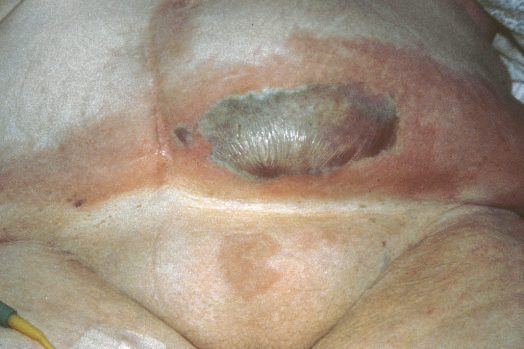
- Pressure ulcers occur in 2.7–9% of acute and 2.4–23% of long-term care patients where soft tissue is compressed between bone and surface with capillary collapse, worsened by friction, shear, moisture, immobility, altered mentation and nutrition, staged I non-blanching erythema of intact skin, II partial-thickness loss, III full-thickness to but not through fascia and IV involving muscle and bone, and treated by a multidisciplinary team with surgical (or enzymatic or hydrotherapy) debridement, moist non-desiccating dressings, pressure relief, nutritional and circulatory correction and flap rotation, though recurrence is extremely high [5].
Scoring and Severity
The National Nosocomial Infections Surveillance (NNIS) score is used to predict surgical site infection risk, stratifying wounds on an index from 0 (lowest risk) to 3 (highest risk), with one point allocated for each of several defined risk factors present [1]. A pressure sore has its own four-stage scheme in which the stage dictates the treatment [8]:
| Stage | Description | Treatment |
|---|---|---|
| I | Erythema and pain, no skin loss (epidermis) | Keep pressure off |
| II | Partial skin loss with yellow debris (into the dermis) | Local treatment, keep pressure off |
| III | Full-thickness skin loss (subcutaneous fat exposed) | Sharp debridement; likely to need a myocutaneous flap |
| IV | Involves bony cortex, muscle, adipose tissue, tendon | Myocutaneous flaps, for example a gluteal flap |
- Table reformats the pressure sore staging [8].
- The transition that changes management is between II and III: at stage III the wound stops being a dressing problem and becomes a reconstructive one.
- The CDC surgical wound classification (Class I–IV, described above under Diagnosis) is itself a severity/contamination grading system used to predict infection risk and guide antibiotic prophylaxis decisions [1].
Strength trajectories and the operative classification
Maximal wound strength is reached at about 6 weeks and a fully healed wound achieves only 75–80% of normal tissue; delayed healing (malnutrition, infection, severe trauma) lags but ultimately reaches normal strength once the cause is corrected, whereas impaired healing (diabetes, chronic steroids, irradiated tissue) never does, demanding care in incision placement, suture choice and aftercare; closed wounds heal mostly by epithelialisation, open wounds and chronic ulcers by contraction, connective tissue deposition and lesser epithelialisation [5]. Surgical wounds are clean (class I), clean-contaminated (II), contaminated (III) or dirty (IV), and appropriate prophylaxis reduces infection in classes II–IV to a third of untreated series [5].
Treatment and Management
- Essentials for promoting wound healing include maintaining a moist environment (avoiding desiccation), optimising oxygen delivery (fluids, smoking cessation, pain control, arterial revascularisation, supplemental oxygen, aiming for transcutaneous oxygen measurement >25 mmHg), avoiding oedema through elevation, and removing necrotic tissue [3].
- Suture removal timing differs by site: approximately 1 week for the face and 2 weeks for other areas [3].
- Delayed primary closure is used to reduce the risk of wound infection when there is concern about contamination [3].
- Wound dehiscence is managed with placement of retention sutures [3].
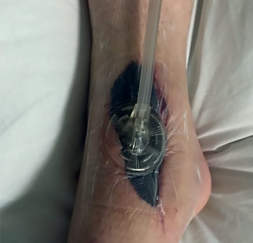
- Prevention of adverse (hypertrophic/keloid) scarring focuses on tension relief, hydration/occlusion and taping/pressure garments, applied as soon as the wound is closed and tolerated.
- Postsurgical taping for 3 months, moisturising lotions, and silicone sheets/gels can reduce scar thickness, discomfort and itching, and avoidance of sun exposure with SPF 50 sunscreen for a year postoperatively reduces scar hyperpigmentation [2].
- Established hypertrophic scars are treated with pressure therapy (6 weeks to 3 months post-injury), continued silicone therapy, and intralesional corticosteroid injections (triamcinolone acetonide 10–40 mg/mL every 2–4 weeks into the papillary dermis) for ongoing hypertrophy, with 50–100% of patients responding and up to 50% recurring; adverse effects include skin atrophy, hypopigmentation and telangiectasia [2].
- Keloids are treated first-line with intralesional corticosteroids combined with silicone dressings and pressure therapy.
- Intralesional 5-fluorouracil, bleomycin and verapamil are used as adjuncts, and refractory cases after 12 months may be considered for surgical excision combined with adjuvant therapy, since excision alone carries a 50–100% recurrence rate, whereas immediate postoperative electron-beam irradiation or brachytherapy reduces recurrence by 50–95% [2].
The UK surgical site infection bundle is defined by NG125, and a striking number of its recommendations are prohibitions rather than interventions. Reading it as "what not to do" is the fastest way to learn it.
| Phase | Do | Do not |
|---|---|---|
| Preoperative | Shower or bath with soap the day before or the day of surgery (1.2.1). Consider nasal mupirocin with a chlorhexidine body wash before procedures where S. aureus is a likely cause (1.2.2). If hair must be removed, use electric clippers with a single-use head on the day of surgery (1.2.5) | Do not use hair removal routinely (1.2.4). Do not use razors, which increase SSI risk (1.2.5). Do not use mechanical bowel preparation routinely (1.2.9) |
| Antibiotic prophylaxis | Give before clean surgery involving a prosthesis or implant, clean-contaminated surgery, and contaminated surgery (1.2.12). Consider a single intravenous dose on starting anaesthesia, but give earlier when a tourniquet is used (1.2.15). Repeat the dose when the operation is longer than the antibiotic's half-life (1.2.16). Give antibiotic treatment in addition to prophylaxis for surgery on a dirty or infected wound (1.2.17) | Do not use prophylaxis routinely for clean non-prosthetic uncomplicated surgery (1.2.13) |
| Intraoperative | Prepare the skin immediately before incision with an antiseptic (1.3.7). If an incise drape is required, use an iodophor-impregnated one unless iodine-allergic (1.3.4). Consider two pairs of gloves where glove perforation risk is high and contamination would be serious (1.3.6). Consider triclosan-coated sutures, especially in paediatric surgery (1.3.20). Consider gentamicin-collagen implants in cardiac surgery (1.3.19) | Do not use non-iodophor-impregnated incise drapes routinely, as they may increase SSI risk (1.3.3). Do not use diathermy for the surgical incision (1.3.11). Do not use wound irrigation (1.3.16) or intracavity lavage (1.3.17). Do not give insulin routinely to patients without diabetes to reduce SSI risk (1.3.15). Apply an antiseptic or antibiotic to the wound before closure only as part of a clinical research trial (1.3.18) |
| Postoperative | Aseptic non-touch technique for changing or removing dressings (1.4.1). Sterile saline for cleansing up to 48 hours; tap water after 48 hours if the wound has separated or been opened to drain pus (1.4.2; 1.4.4). Patients may shower safely 48 hours after surgery (1.4.3) | Do not use topical antimicrobials on wounds healing by primary intention (1.4.5). Do not use Eusol and gauze, moist cotton gauze or mercuric antiseptic solutions for wounds healing by secondary intention (1.4.6), nor Eusol and gauze, dextranomer or enzymatic treatments for debridement of an established SSI (1.4.10) |
Table reformats the NG125 recommendations [9].
- The antiseptic choice is a four-step cascade, not a preference.
- First choice is an alcohol-based solution of chlorhexidine, unless contraindicated or the surgical site is next to a mucous membrane; next to a mucous membrane use aqueous chlorhexidine; if chlorhexidine is contraindicated use alcohol-based povidone-iodine; and if both an alcohol-based solution and chlorhexidine are unsuitable, use aqueous povidone-iodine [9].
- Two safety points sit alongside it: be aware of the risk of severe chemical injuries from chlorhexidine, both alcohol-based and aqueous, in preterm babies, and where diathermy is to be used, dry the antiseptic by evaporation and avoid pooling of alcohol-based preparations [9].
NG125 also defines the two healing terms precisely. Healing by primary intention occurs when a wound has been sutured after an operation and heals to leave a minimal, cosmetically acceptable scar. Healing by secondary intention occurs when a wound is deliberately left open at the end of an operation because of excessive bacterial contamination, particularly by anaerobes, or a risk of devitalised tissue. It may be sutured within a few days (delayed primary closure), much later when clean and granulating (secondary closure), or left to heal naturally without suturing [9].
Where wound healing is likely to be a problem, NG125 asks for structure rather than a product: use a structured approach to care including preoperative assessments to identify people with potential wound healing problems, and ask a tissue viability nurse for advice on dressings for wounds healing by secondary intention [9].
- Pressure ulcers have their own NICE guideline, and its opening premise is deliberately absolute: be aware that all patients are potentially at risk of developing a pressure ulcer [10].
- Risk assessment is carried out and documented for adults admitted to secondary care or to care homes providing NHS care, and for adults receiving NHS care elsewhere, including in primary care, community care and emergency departments, if they have a risk factor such as significantly limited mobility, significant loss of sensation, a previous or current pressure ulcer, nutritional deficiency, inability to reposition themselves, or significant cognitive impairment [10].
- A validated scale, for example the Braden scale, the Waterlow score or the Norton risk-assessment scale, is used to support clinical judgement rather than replace it, and risk is reassessed whenever clinical status changes, for example after surgery, on worsening of an underlying condition, or with a change in mobility [10].
Two of the recommendations turn on how erythema behaves and how it looks on darker skin. The skin assessment checks integrity in areas of pressure, colour changes or discoloration, and variations in heat, firmness and moisture, and CG179 notes explicitly that non-blanchable erythema may present as colour changes or discolouration, particularly in darker skin tones or types [10]. Use finger palpation or diascopy to determine whether erythema or discolouration is blanchable, and where it is non-blanching, start preventative action and consider repeating the skin assessment at least every 2 hours until it resolves [10].
- Repositioning frequency is graded by risk level, and the two numbers are easy to transpose.
- Encourage adults at risk to change position at least every 6 hours, and adults at high risk at least every 4 hours, helping those who cannot reposition themselves and documenting the frequency required [10]. Do not offer skin massage or rubbing to prevent a pressure ulcer [10].
- High-specification foam mattresses are used for adults admitted to secondary care and for those assessed as high risk in primary and community care, with a high-specification foam theatre mattress or equivalent considered for all adults undergoing surgery [10].
- Nutrition and fluids are given for deficiency, never for the ulcer itself.
- Do not offer nutritional supplements to prevent a pressure ulcer in adults whose nutritional intake is adequate, nor subcutaneous or intravenous fluids where hydration is adequate; the same two prohibitions are then repeated for treatment [10].
- What is offered is a nutritional assessment by a dietitian or equivalent for every adult with a pressure ulcer, and supplements only where a deficiency exists [10].
- On measurement and grading, one instruction is a "do not".
- Document the surface area of all pressure ulcers, using a validated technique such as transparency tracing or a photograph, and document an estimate of depth and the presence of undermining, but do not routinely measure the volume [10].
- Categorise each ulcer with a validated classification tool such as the International NPUAP-EPUAP Pressure Ulcer Classification System, repeating and documenting it at each assessment [10].
Treatment is largely a list of things not to do, which is what makes the guideline memorable.
| Intervention | CG179 position |
|---|---|
| Standard-specification foam mattress | Do not use for adults with a pressure ulcer; use high-specification foam, escalating to a dynamic support surface if that is insufficient (1.4.9; 1.4.10) |
| Negative pressure wound therapy | Do not routinely offer, unless needed to reduce the number of dressing changes, for example in a heavily exuding wound (1.4.13) |
| Electrotherapy and hyperbaric oxygen | Do not offer (1.4.14) |
| Larval (maggot) therapy and enzymatic debridement | Do not routinely offer; consider larval therapy only if debridement is needed and sharp debridement is contraindicated, or where there is associated vascular insufficiency (1.4.17) |
| Debridement | Offer where assessment identifies the need: autolytic debridement with an appropriate dressing, considering sharp debridement if autolytic would take longer and prolong healing (1.4.15; 1.4.16) |
| Topical antiseptics or antimicrobials | Do not routinely use (1.4.22) |
| Gauze dressings | Do not offer; consider a dressing promoting a warm, moist environment for category 2, 3 and 4 ulcers (1.4.24; 1.4.25) |
Table reformats the CG179 treatment recommendations [10].
Systemic antibiotics have three named indications and two explicit exclusions. Offer them after skin assessment if there is clinical evidence of systemic sepsis, spreading cellulitis, or underlying osteomyelitis, discussing the choice with the local hospital microbiology department so it is effective against local strains [10]. Do not offer systemic antibiotics specifically to heal a pressure ulcer, and do not offer them on the basis of positive wound cultures alone without clinical evidence of infection [10].
Acute wound care and dressings in Schwartz's detail
- After history, examination (depth, configuration, non-viable tissue, foreign bodies; location, dimensions, drainage, base, necrosis, pain, edges, aetiology, circulation) and tetanus prophylaxis, the wound is anaesthetised with 0.5–1% lidocaine or 0.25–0.5% bupivacaine with 1:100,000–1:200,000 adrenaline (never adrenaline in fingers, toes, ears, nose or penis) irrigated with plain saline under high pressure (iodine, povidone-iodine, hydrogen peroxide and organic antibacterials injure neutrophils and macrophages and are not used), haematomas evacuated, marginal flaps resected or revascularised, hair clipped, surrounding skin prepared with povidone-iodine or chlorhexidine, and macerated or bevelled edges freshened; W- or Z-plasty is seldom used acutely but edges crossing the vermilion, eyebrow or hairline are aligned first; the smallest adequate suture is chosen, non-absorbable or slowly absorbable monofilament for fascia, braided absorbable for subcutaneous tissue avoiding fat, multilayer abdominal closure at some infection cost, flaps or delayed split-thickness grafts (porcine xenograft or cadaveric allograft in contaminated wounds) for tissue loss, and skin closed with staples or monofilament removed at 4–5 days on the face and 7–10 days elsewhere before tracts epithelialise, or buried absorbable dermal sutures with tapes, or octyl-cyanoacrylate glue for simple linear wounds even when contaminated, especially in children [5].
- Antibiotics are given only for infection (erythema, cellulitis, swelling, pus), single-agent for a suspected single organism and broad-spectrum for enteric contamination or immune impairment, topical delivery being of questionable efficacy [5].
- Dressings should keep the wound moist, absorb excess exudate, conform, control pain and odour, be non-allergenic, gas-permeable, safe, atraumatic to remove, cheap and convenient; occlusion reduces inflammation and necrosis and aids collagen synthesis and epithelial migration but is contraindicated in infected or highly exudative wounds; primary dressings sit on the wound and secondary dressings protect, absorb, compress and occlude; types are absorbent, non-adherent (paraffin, petroleum or water-soluble jelly needing a secondary layer), occlusive or semi-occlusive films (waterproof, microbe-impermeable, vapour- and oxygen-permeable, for clean minimally exudative wounds), hydrophilic/hydrophobic composites, hydrocolloids (leaving a washable yellow-brown gel) and hydrogels (high water content, high evaporation, useful in burns), alginates from brown algae (calcium exchanging to soluble sodium alginate, gelling and absorbing heavily; for skin loss, medium-exudate surgical wounds and full-thickness chronic wounds), absorbable haemostats (collagen, gelatin, oxidised cellulose) and medicated dressings (benzoyl peroxide, zinc oxide, neomycin, bacitracin-zinc) raising epithelialisation 28%; choice follows drainage, semi-occlusive for none, semi-occlusive or absorbent non-adherent under 1–2 mL/day, non-adherent plus absorbent plus occlusive at 3–5 mL/day, with a highly absorbent secondary layer above 5 mL/day; oedematous wounds are compressed, ischaemic ones are not; negative-pressure foam systems remove exudate and aid closure of diabetic and stage III–IV pressure ulcers, acute and traumatic wounds, flaps, grafts and dehisced incisions pending more randomised evidence [5].
Advanced modalities for chronic wounds in Schwartz's account
- Cellular and tissue-based products supplement but never replace the basics, blood flow, infection control, debridement, dressing, offloading and compression; dermoinductive products (Apligraf, Theraskin, Dermagraft) supply cells and factors, Apligraf achieving 56% healing at 65 days against 38% at 90 days with saline in its pivotal trial, while dermoconductive scaffolds such as Integra (bovine type I collagen, shark chondroitin-6-sulphate, silicone) prepare a neodermis for grafting and cover small areas of exposed bone or tendon, sometimes needing repeat application; a systematic review of 15 randomised trials found convincing though low-quality support for living cell-based substitutes; amniotic membrane-derived stem cell products (Epifix, Grafix) supply VEGF, PDGF, EGF and TGF but are extremely expensive and applied weekly [5].
- Cultured epithelial autografts expand keratinocytes from a postage-stamp biopsy over weeks under temporary allograft or xenograft but are fragile, infection-prone, contract and regenerate dermis poorly; cryopreserved allogeneic keratinocytes are available off the shelf, lack Langerhans cells and MHC antigens so persist until superseded, but lack dermal strength and risk disease transmission; fibroblast-seeded meshes make living dermis cheaply; bilayered equivalents are indicated with compression for venous ulcers and for neuropathic diabetic ulcers [5].
- Growth factor therapy delivers about 1000 times physiological concentrations; autologous platelet-rich plasma showed no value in a meta-analysis and only recombinant PDGF-BB gel is FDA-approved, for diabetic foot ulcers, where it raises complete healing and shortens time; gene delivery by viral vector, plasmid, electroporation or microseeding of IL-8, PDGF, IGF-1, KGF or laminin-5 has had modest, situation-specific effects and is unlikely to be a universal solution, so mesenchymal, marrow, cord, adipose or epidermal stem cells producing VEGF, PDGF, bFGF and MMP-9 are the newest vector [5].
- Hyperbaric oxygen, near-100% oxygen at a minimum 1.4 atmospheres absolute in mono- or multiplace chambers, has 14 UHMS/FDA-accepted indications including clostridial myonecrosis, crush injury, radiation soft-tissue and bone necrosis, necrotising infection, Wagner grade III or worse diabetic ulcers, refractory osteomyelitis and thermal burns; reviews to 2001 showed better graft survival, osteoradionecrosis, gas gangrene amputation rates, necrotising fasciitis mortality and diabetic wound size, and 18 of 29 studies from 2001–2016 (62%) had at least one positive outcome, with diabetic foot trials showing more healing at 1 year and less proximal amputation, though heterogeneity and poor randomisation limit the evidence [5].
- Over 60% of chronic wounds carry a biofilm ("an aggregate of bacteria tolerant to treatment and the host defense") formed by reversible adhesion, quorum-sensing-dependent permanent adhesion and maturation, and secretion of extracellular polymeric substance; staphylococci, streptococci and Pseudomonas attach in minutes, form microcolonies in 2–4 hours, resist disinfectants and antibiotics by 6–12 hours, mature in 2–4 days and recover from debridement within 24 hours, shedding to seed new colonies while phenotypic and horizontally transferred genetic plurality evade phagocytosis, degranulation and reactive oxygen species and quiescent cells escape antimicrobials; the host's macrophage and neutrophil proteases and radicals damage tissue and the exudate feeds the film, so treatment is repeated (suggested weekly) debridement, surfactant cleansers and topical silver, iodine or honey in combination, aiming at removal, eradication and prevention of recurrence [5].
Surgeries
- Surgical scar revision may be considered for permanent linear hypertrophic scars present after 1 year, using simple resection and primary closure, sometimes combined with adjacent tissue undermining, subcutaneous sutures, Z-plasty and postsurgical taping/silicone therapy [2].
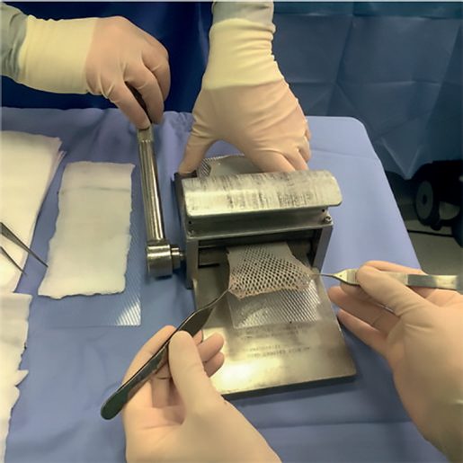
- Correction of a scar contracture (abnormal shortening of an immature scar causing functional impairment, particularly across a joint) generally requires surgery with Z-plasty, skin graft, or flap to release tension and restore function [2].
- Refractory keloids after prolonged medical therapy may be excised surgically in combination with adjuvant radiotherapy or other adjuncts, as excision alone has a high recurrence rate [2].
- Delayed primary closure, leaving a contaminated wound open initially, then surgically approximating the edges after debridement once the wound bed is clean, is itself a surgical strategy for high-risk wounds [1].
Antimicrobial prophylaxis and the surgical site in Schwartz's account
- Prophylaxis works only with adequate tissue levels at incision, a single intravenous dose within 60 minutes (vancomycin or fluoroquinolone 60–120 minutes) before incision, redosed at one to two half-lives (ampicillin/sulbactam and cefoxitin 2-hourly, cefazolin and cefuroxime 4-hourly, clindamycin 6-hourly, vancomycin 12-hourly) for operations over 3 hours, major blood loss or burns, continued up to 24 hours for long cases, prostheses or unexpected contamination, and tailored to the operation, expected contaminants and institutional resistance; cefazolin is 1 g under 80 kg and 2 g above, vancomycin (for MRSA-prevalent units, carriers or β-lactam allergy) 15 mg/kg to 1.5 g over 90 minutes to avoid hypotension at induction; colorectal surgery adds oral neomycin with erythromycin or metronidazole the day before to cefoxitin, cefotetan, cefazolin-metronidazole or ampicillin/sulbactam, biliary and oesophagogastric surgery are covered only in high-risk patients (age over 70, acute cholecystitis, obstructive jaundice, duct stones; obesity, obstruction, achlorhydria, bleeding, malignancy, perforation, immunosuppression), head and neck mucosal incisions take clindamycin or cefazolin-metronidazole, the whole dose precedes tourniquet inflation in orthopaedics, patients with valves or vascular or orthopaedic prostheses receive prophylaxis before bacteraemic procedures (penicillins for dental work, second-generation cephalosporin for urological instrumentation, anaerobic plus cephalosporin cover for GI surgery), and nasal screening and decolonisation of S. aureus carriers precede cardiac, orthopaedic and neurosurgical implant operations [5].
- Hunt showed the subcutaneous capillary bed vasoconstricts with hypovolaemia, hypothermia and stress, so euvolaemia, core temperature above 36–36.5°C and pain control each and together reduce infection, an FiO₂ of 0.8 through and just after surgery helped in most but not all trials, and moderate glycaemic control (120–180 mg/dL) from before surgery onward equals or beats tight control (80–100 mg/dL), which adds complications [5].
- Necrotising fasciitis, the most dangerous deep infection, is septic thrombosis of the vessels between skin and deep layers with haemorrhagic bullae and necrosis, fascial involvement wider than the skin suggests, a toxic febrile tachycardic hypovolaemic patient and mixed flora: Gram stain and cultures are taken, high-dose penicillin (20–40 million units a day) started for Clostridium perfringens with broad-spectrum cover, resuscitation completed before induction, all necrotic skin and fascia excised through multiple longitudinal incisions when skin is viable, re-inspection every 12–24 hours for further debridement, and homograft or xenograft cover until autografting [5].
Complications
- Hypertrophic and keloid scarring represent excessive, disorganised collagen deposition following aberrant wound healing; keloids in particular are frequently refractory to both medical and surgical intervention and can recur even after excision (50–100% recurrence with excision alone) [1][2].
- Wound dehiscence (leakage of pink "salmon-coloured" fluid) can progress to evisceration if untreated; the leading risk factor is deep wound infection, along with poor nutrition, COPD, diabetes mellitus and chronic coughing [3].
- Chronic non-healing wounds are a major complication of impaired healing: an estimated 3–4 million patients per year in the US are at risk of diabetic ulcers, up to 2 million per year develop chronic venous leg ulcers, and 2–3 million per year develop pressure ulcers from immobility, with these numbers rising alongside an ageing population and increasing prevalence of diabetes and smoking [2].
- Scar contractures across joints or involving the eyelid or mouth (causing ectropion) are a functional complication of excessive wound contraction [2].
Excess healing and adhesions in Schwartz's account
- Excess healing probably demands more operations than wound failure, mutilating scars and burn contractures, frozen tendon repairs, GI strictures, cirrhosis and pulmonary fibrosis, peritoneal adhesions.
- Hypertrophic scars stay within the wound, rarely rise over 4 mm, appear within 4 weeks (risk rising if epithelialisation exceeds 21 days), favour tension lines and flexor surfaces at right angles to joints and creases, and often regress; keloids extend beyond the wound, appear 3 months to years after even trivial insults (surgery, burns, acne, chickenpox, zoster, folliculitis, tattoos, vaccination, piercing) at earlobe, deltoid, presternal and upper back skin, spare eyelids, genitalia, palms, soles and joints, rarely regress and recur (often worse) after surgery, are 15 times commoner in darker-pigmented African, Hispanic and Asian populations with autosomal dominant, incompletely penetrant inheritance and equal sex incidence [5].
- Both show thickened epidermis without rete ridges; hypertrophic scar collagen is flatter, random and wavy, keloid collagen lies in haphazard loose sheets of thick fibres without myofibroblasts, keloid fibroblasts make collagen 20 times faster than normal and 3 times faster than hypertrophic scar fibroblasts, fibronectin synthesis persists for years, TGF-β is raised in hypertrophic scars and both respond to lower TGF-β concentrations, hypertrophic scars over-express IGF-1 (lowering collagenase mRNA), keloids over-express TGF-β1/β2, VEGF, PAI-1, PDGF receptors and anti-apoptotic genes, keratinocytes express HLA-2 and ICAM-1, keloids carry IgG, IgA, IgM and anti-nuclear antibodies with IgE correlation, hypertrophic scars more T cells and Langerhans cells, mast cells rise in both, and mononuclear-derived fibrocytes stimulate fibroblasts and collagen [5].
- Excision alone recurs in 45–100%, so surgery debulks or is second line and is combined with intralesional steroid, silicone, radiation or pressure, including the advancing dermal edge, following tension lines and closing without tension; silicone worn 24 hours a day for about 3 months (mechanism probably hydration) suits children; intralesional steroids (first line for keloids, second line for hypertrophic scars) every 2–3 weeks soften and flatten but cannot erase keloids or narrow wide scars and cause atrophy, hypopigmentation, telangiectasia, necrosis and ulceration; radiation alone recurs 10–100% but 1500–2000 rad after excision helps adults with resistant scars at the cost of pigmentation, pruritus, pain and possible malignancy; pressure garments at 24–30 mmHg worn 23–24 hours a day for a year or more, started early (scars over 6–12 months respond poorly), mature collagen and flatten burn scars; topical retinoids respond in 50–100%; intralesional IFN-γ fails alone through resistance, imiquimod after excision induces IFN-γ, and intralesional 5-fluorouracil alone or with steroid, bleomycin or mitomycin C treat steroid-resistant older scars [5].
- Adhesions are found at autopsy in 67% after surgery and 28% after intra-abdominal infection, cause 65–75% of small bowel obstructions (especially ileal, commoner after lower abdominal surgery, 11% within a year and 30% by 10 years after rectal, left or total colectomy), secondary infertility and pain, and account for 2% of surgical admissions and 3% of laparotomies; peritoneal injury from surgery, heat, ischaemia, inflammation or foreign body strips mesothelium, fibrin bridges opposed serosa and is normally lysed by plasmin from tPA and uPA, but tPA falls early and PAI-1 rises later under TNF-α, IL-1 and IL-6 so permanent collagenous adhesions form within a week; prevention rests on gentle handling, avoiding desiccation, ischaemia and excess cautery, laser and retraction, laparoscopy, and the three FDA-approved barriers, Interceed (oxidised regenerated cellulose, pelvic surgery), Seprafilm (hyaluronate–carboxymethylcellulose under the incision) and Adept (4% icodextrin, mainly pelvic), never placed over an anastomosis because of leak risk [5].
Prognosis
- Wound tensile strength never returns to that of unwounded skin; it reaches a maximum of approximately 80% of the original strength, at around 12 weeks (Bailey & Love) or over 6–12 months (Sabiston/Schwartz's ABSITE) post-injury [1][4].
- Hypertrophic scars often regress spontaneously once the inciting stimuli (tension, growth factor stimulation) are removed, whereas keloids are genetically predisposed, do not regress spontaneously, and are frequently refractory to treatment [2].
- Chronic wounds, by definition, fail to achieve anatomic and functional integrity within an expected 3-month timeframe and carry substantial morbidity, reduced quality of life and healthcare burden [2].
References
- Bailey & Love's Short Practice of Surgery, 28th ed., Ch. 3 Wound healing and tissue repair
- Sabiston Textbook of Surgery, 22nd ed., Ch. 23 Wound Healing
- The ABSITE Review, 2022, Ch. 14 Wound Healing
- Schwartz's Principles of Surgery: ABSITE and Board Review, Ch. 9 Wound Healing
- Schwartz's Principles of Surgery, 11th ed., Ch. 9, Wound Healing, Table 9-1, Fig. 9-1
- Browse's Introduction to the Symptoms and Signs of Surgical Disease, 6th ed., Ch. 1 History and examination
- Oxford Handbook of Clinical Surgery, 5th ed., Ch. 3 Surgical pathology
- The ABSITE Review, 2022, Ch. 18 Plastics, Skin, and Soft Tissues
- NICE Guideline NG125: Surgical site infections: prevention and treatment. National Institute for Health and Care Excellence, London, UK, 2019, updated 2020., 1.2.1 to 1.4.11; 1.3.8; 1.3.9, Table 1; 1.3.10; 1.4.8; 1.4.11; Terms used in this guideline www.nice.org.uk
- NICE Clinical Guideline CG179: Pressure ulcers: prevention and management. National Institute for Health and Care Excellence, London, UK, 2014., 1.1.1; 1.1.2; 1.1.3; 1.1.4; 1.1.5; 1.1.6; 1.1.7; 1.1.8; 1.1.9; 1.1.10; 1.1.11; 1.1.12; 1.1.13; 1.1.14; 1.4.1; 1.4.2; 1.4.3; 1.4.4; 1.4.5; 1.4.7; 1.4.8; 1.4.9 to 1.4.25; 1.4.18; 1.4.19; 1.4.20; 1.4.21 www.nice.org.uk