



Surgical Site Infection
Summary
- Surgical site infection (SSI) is infection of the tissues, organs, or spaces exposed by surgeons during an invasive procedure, arising from invasion of organisms into tissues following a breakdown of local and systemic host defences [1][2].
- SSIs are classified into incisional (superficial, limited to skin/subcutaneous tissue; or deep, involving deeper musculofascial layers) and organ/space infections, and their development depends on the degree of microbial contamination of the wound, the duration of the procedure, and host factors such as diabetes, malnutrition, obesity and immunosuppression [2].
- The majority of wound infections arise from endogenous sources within the patient, although exogenous infection from the ward or theatre environment also occurs [1].
Definition
- The infection of a wound is defined as the invasion of organisms into tissues following a breakdown of local and systemic host defences, leading to cellulitis, lymphangitis, abscess formation or bacteraemia.
- Most surgical wound infection is superficial surgical site infection (SSSI), with the other categories being deep SSI (infection in the deeper musculofascial layers) and organ/space infection (e.g. an abdominal abscess after an anastomotic leak) [1].
- By definition, an incisional SSI has occurred if a surgical wound drains purulent material or if the surgeon judges it to be infected and opens it [2].
- A major SSI is a wound that either discharges significant quantities of pus spontaneously or needs a secondary procedure to drain it, with systemic signs such as tachycardia, pyrexia and raised white cell count.
- Minor wound infections discharge pus or infected serous fluid without excessive discomfort, systemic signs or delayed discharge [1].
Pathophysiology
- Pathogens resist host defences by releasing toxins that favour their spread, enhanced in anaerobic or necrotic wound tissue; for example, Clostridium perfringens (gas gangrene) releases hyaluronidase, lecithinase and haemolysin to spread through tissues [1].
- The human body harbours approximately 10^14 organisms, released into tissues before, during or after surgery.
- Contamination is most severe when a hollow viscus perforates [1].
- Natural protective mechanisms against infection are chemical (low gastric pH), humoral (antibodies, complement, opsonins) and cellular (phagocytes, macrophages, polymorphonuclear cells, killer lymphocytes), all of which may be compromised by surgery and its treatment [1].
- The chance of developing SSI depends on organism pathogenicity and inoculum size balanced against host response.
- Devitalised tissue, dead space, haematoma and foreign material (sutures, drains) all increase infection risk [1].
- There is up to a 4-hour interval (the "decisive period") before bacterial growth becomes established enough to cause infection after a tissue breach.
- Prophylactic antibiotic strategies become ineffective after this window, so tissue antibiotic levels should exceed the minimum inhibitory concentration (MIC90) for expected pathogens during this period [1].
- When enteral feeding is suspended perioperatively, bacteria (particularly aerobic Gram-negative bacilli) can colonise the normally sterile upper GI tract and translocate to mesenteric nodes, releasing endotoxin (lipopolysaccharide) that triggers a cytokine-mediated systemic inflammatory response (SIRS) and multiple organ dysfunction syndrome (MODS) even with negative blood cultures [1].
- At least 10^5 bacteria are generally needed to cause a wound infection, though fewer organisms suffice in the presence of a foreign body [3].
Host defences and the microbial inoculum in Schwartz's account
- Koch's four postulates (organism present in every case and absent from health; isolated in pure culture; reproducing disease in a healthy animal; re-isolated from it), McBurney's 1889 report of early appendicectomy as the first intra-abdominal "source control", Treves draining Edward VII's periappendiceal abscess in 1902, Fleming's 1928 penicillin and Meleney's and Altemeier's demonstration that aerobes and anaerobes synergise in soft tissue and intra-abdominal infection frame Schwartz's account, with Osler's 1904 observation that the patient "appears to die from the body's response to infection rather than from it" [4].
- Barriers are physical, chemical (sebaceous secretions, shedding) and microbial, colonisation resistance by commensals: skin carries Staphylococcus, Streptococcus, Corynebacterium and Propionibacterium with enterococci, E. coli and Candida below the umbilicus; the urogenital, biliary, pancreatic and distal respiratory tracts are sterile unless disease, stones, foreign body, catheter or aspiration intervene; the acid, low-motility stomach holds only 10²–10³ CFU/mL (more with acid suppression), the terminal ileum 10⁵–10⁸, and the colon 10¹¹–10¹² CFU/g with anaerobes (Bacteroides fragilis, distasonis and thetaiotaomicron, Bifidobacterium, Clostridium, Eubacterium, Fusobacterium, Lactobacillus, Peptostreptococcus) outnumbering aerobes 100:1, the flora that resists Salmonella, Shigella and Vibrio yet supplies the inoculum after perforation, of which only a few species predominate in established infection [4].
- Within a sterile cavity, lactoferrin and transferrin sequester iron, polymerising fibrinogen traps microbes, the diaphragmatic pump expels peritoneal particles through stomata into thoracic lymphatics, and omentum and ileus wall off infection, all favouring abscess formation; resident macrophages sense pathogen- and danger-associated molecular patterns through Toll-like receptors and secrete TNF-α, IL-1β, IL-6, IL-8 and IFN-γ, countered by TNF-binding protein, IL-1 receptor antagonist, IL-4 and IL-10; opsonisation by C1q, C3bi and Fc, the C5b-9 membrane attack complex, phagocytic killing, complement fragments C3a, C4a and C5a raising permeability, and neutrophil chemotaxis to C5a, N-formyl-methionine peptides and IL-8 bring diapedesis within minutes, peaking over hours to days; outcome depends on inoculum size, proliferation versus containment, virulence and host potency [4].
- Outcomes are eradication, containment as pus (furuncle, abscess), locoregional spread (cellulitis, lymphangitis, aggressive soft tissue infection, metastatic abscess) or systemic bacteraemia or fungaemia [4].
Clinical features
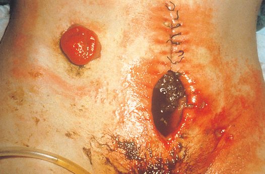
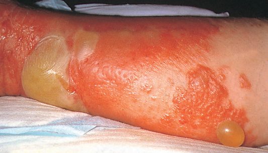
- Presentations include: abscess (calor, rubor, dolor, tumor, and functio laesa, per Celsus), usually forming 7–10 days after surgery, with as many as 75% of SSIs presenting after the patient has left hospital [1][3]; cellulitis and lymphangitis, non-suppurative, poorly localised spreading infection with systemic signs (chills, fever, rigors) but often negative blood cultures [1]; gas gangrene, severe local wound pain, crepitus, thin brown sweet-smelling exudate, oedema and spreading gangrene [1]; necrotising fasciitis/necrotising soft tissue infection, pain out of proportion to physical findings, mental status changes, WBC >20, thin grey foul-smelling ("dishwater") drainage, skin blistering/necrosis (overlying skin may look normal early, then progresses from pale red to purple with bullae), induration, oedema, crepitus or soft-tissue gas on X-ray, and can progress to sepsis [2][3].
- Surgical infections presenting within 48 hours of a procedure suggest bowel injury with leak or invasive soft-tissue infection with Clostridium perfringens or beta-haemolytic streptococcus, which produce exotoxins [3].
- Fournier's gangrene is a severe perineal/scrotal infection from mixed organisms in diabetic or immunocompromised patients [3].
Etiology
- Common causative organisms: Staphylococcus aureus (coagulase-positive) is the most common organism overall in SSIs; Staphylococcus epidermidis (coagulase-negative) forms biofilms on prosthetic surfaces limiting antibiotic effectiveness; Escherichia coli is the most common Gram-negative rod in surgical wound infections; Bacteroides fragilis is the most common anaerobe, acting in synergy with aerobic Gram-negative bacilli [1][3]. β-haemolytic Streptococcus (Streptococcus pyogenes, Lancefield group A) spreads via streptolysin, streptokinase and streptodornase; Streptococcus faecalis (enterococcus) often acts synergistically with other organisms [1].
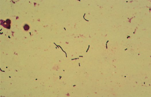
- Clostridium perfringens causes gas gangrene; Clostridium tetani causes tetanus; Clostridium difficile causes pseudomembranous colitis following disruption of normal gut flora by antibiotic therapy [1].
- Necrotising fasciitis is usually caused by beta-haemolytic group A streptococcus or MRSA.
- C. perfringens myonecrosis follows a decreased tissue oxidation-reduction potential in necrotic tissue and releases alpha toxin [3].
- Risk factors are grouped as patient factors (older age, immunosuppression, obesity, diabetes mellitus, chronic inflammatory process, malnutrition, smoking, renal failure, peripheral vascular disease, anaemia, radiation, chronic skin disease, chronic Staphylococcus carriage, recent operation), local factors (open vs laparoscopic surgery, poor skin preparation, contamination of instruments, inadequate antibiotic prophylaxis, prolonged procedure, local tissue necrosis, blood transfusion, hypoxia, hypothermia), and microbial factors (prolonged hospitalisation with nosocomial organisms, toxin secretion, resistance to clearance) [2].
- Additional risk factors cited include long operations, haematoma or seroma formation, advanced age, chronic disease (COPD, renal failure, liver failure, diabetes mellitus), malnutrition, and immunosuppressive drugs [3].
Wound classes, rates and risk factors in Schwartz's figures
- SSI risk rests on contamination at operation, duration and host factors, age, immunosuppression, obesity, diabetes, chronic inflammation, malnutrition, smoking, renal failure, peripheral vascular disease, anaemia, radiation, skin disease, staphylococcal carriage, recent surgery; open rather than laparoscopic access, poor preparation, contaminated instruments, inadequate prophylaxis, long procedures, necrosis, transfusion, hypoxia and hypothermia; and prolonged hospitalisation, toxins and capsules [4].
- Class I clean wounds (hernia, breast biopsy) infect at 1–2%, class I D adds a prosthesis such as mesh or valve; class II clean-contaminated (cholecystectomy, non-colonic GI surgery) at 2.1–9.5%, but colorectal surgery, classically class II, runs 4–14% (9–25% in recent series, two-thirds presenting after discharge, higher with rectal dissection, and cut from 9.8% to 4.0% by one multidisciplinary programme); class III contaminated (early open wounds, major sterile breaks such as open cardiac massage, gross spillage, incision through inflamed non-purulent tissue) 3.4–13.2%; class IV dirty (delayed traumatic wounds with necrosis, overt pus, perforated viscus) 3.1–12.8%; class I infections are skin flora, class II colonic surgery skin or colonic flora or both; classes I–II are closed primarily while class III–IV skin closure infects in about 25–50%, so they are packed open or selectively closed late, except perforated or gangrenous appendicitis in healthy patients, closed primarily under aerobic–anaerobic antibiotics with 3–4% SSI [4].
- Resistance is innate (Gram-negatives lack penicillin-binding proteins) or acquired by mutation, plasmid or transposon through target modification, permeability change, efflux or inactivation; community MRSA producing Panton-Valentine leukocidin now causes a rising share of SSIs because it evades standard prophylaxis; ESBL Klebsiella and E. coli carry plasmid-mediated inducible β-lactamases that appear cephalosporin-sensitive in vitro yet fail rapidly, so a carbapenem is chosen; vancomycin-resistant E. faecium carries transposon-borne vanA, with the feared transfer to S. aureus in co-infected hosts [4].
Diagnosis
- Surgical wounds are classified by presumed bacterial load at surgery: Class I (clean), no infection present, only skin flora, no hollow viscus entered, infection rate 1–2%; Class II (clean-contaminated), a hollow viscus (respiratory, alimentary, genitourinary tract) is entered under controlled conditions without significant spillage, infection rate roughly 2–9.5% for non-colonic GI surgery and 4–14% for colorectal surgery; Class III (contaminated), open accidental wounds, major breaks in sterile technique, gross spillage of viscus contents, or incision through inflamed non-purulent tissue, infection rate 3.4–13.2%; Class IV (dirty), traumatic wounds with delayed treatment and necrotic tissue, wounds created in the presence of overt infection/pus, or access to a perforated viscus with high contamination, infection rate 3.1–12.8% [2].
- Similar figures are given elsewhere: clean (hernia) 2%, clean-contaminated (elective colon resection, prepped bowel) 3–5%, contaminated (e.g. gunshot wound to colon with repair) 5–10%, gross contamination (abscess) 30% [3].
- Bailey & Love gives comparable rates with and without prophylaxis: clean 1–2% either way; clean-contaminated 3% with prophylaxis vs 6–9% without; contaminated 6% vs 13–20%; dirty 7% vs 40% [1].
- Diagnosis of an established SSI is clinical: purulent drainage, or the surgeon's judgement that the wound is infected, prompting opening of the wound; culture of pus or wound fluid should ideally be taken before antibiotics are started, with the larger the pus volume sent, the more likely accurate organism identification [1][2].
- Necrotising soft-tissue infection is a clinical diagnosis suspected with sepsis, skin changes or crepitus, and pain out of proportion to examination findings.
- Imaging (X-ray for soft-tissue gas) can support but should not delay surgical exploration [2].
The surveillance definitions, and why they matter more than the clinical impression
Surgical site infection is a defined event with a date window, not a clinical impression, and the definition differs by depth. A superficial incisional SSI has a date of event within 30 days of the operative procedure (day 1 is the date of operation), involves only skin and subcutaneous tissue, and requires at least one of: purulent drainage from the superficial incision; organisms identified from an aseptically obtained specimen by culture or non-culture-based testing performed for clinical diagnosis or treatment; or a superficial incision deliberately opened by a surgeon where testing is not performed, together with at least one of localised pain or tenderness, localised swelling, erythema or heat, or a diagnosis of superficial incisional SSI by a physician [5].
A deep incisional SSI has a date of event within 30 or 90 days depending on the procedure, involves deep soft tissues such as fascial and muscle layers, and requires purulent drainage from the deep incision or a deep incision that is deliberately opened or aspirated or spontaneously dehisces, with organisms identified from the deep soft tissues, and at least one of fever above 38°C, localised pain or tenderness, or an abscess or other evidence of infection on gross anatomical, histopathological or imaging examination [5]. The third category, organ/space SSI, is defined anatomically deeper than the fascial and muscle layers [5].
- The reason to know these definitions precisely is administrative as well as clinical.
- SSI data have emerged as the leading publicly reported surgical outcome, and SSIs are tied to payment determinations [5].
- International prevention guidelines were published in 2016 and updated in 2018, and national guidance followed in 2019, yet SSIs remain prevalent despite these concerted global efforts [5].
- The whole mechanism reduces to one sentence, which is worth quoting because it organises everything else. Acquisition of an SSI is dependent on bacterial exposure via an operative incision and the host response to control that bacterial contamination [5].
- Risk factors therefore split into patient-related and procedure-related, and the emphasis in the patient list is that many of them are modifiable: obesity, immunosuppression, hyperglycaemia, tobacco use, malnutrition and staphylococcal colonisation [5].
- Obesity increases risk through decreased delivery of oxygen, nutrients and antibiotics. Hyperglycaemia impairs the innate immune response and so compromises wound healing, and patients with intraoperative and postoperative hyperglycaemia are more likely to develop SSI than those with normoglycaemia, in both diabetics and non-diabetics [5].
- Where the disease pathology allows, temporarily holding immunomodulating medication in the perioperative period may optimise infectious outcomes [5].
The contamination classification is defined by what was encountered, not by what was expected [5]:
| Class | Criteria |
|---|---|
| Clean | Atraumatic; no inflammation or infection encountered; no break in sterile technique; no entry into the gastrointestinal, genitourinary or respiratory tracts |
| Clean-contaminated | Controlled entry into the gastrointestinal, genitourinary or respiratory tracts without unusual contamination |
| Contaminated | Open, fresh, accidental wounds; major breaks in sterile technique; acute, non-purulent inflammation encountered |
| Dirty-infected | Old, traumatic wounds with retained devitalised tissue; existing clinical infection or perforated viscera |
Table reformats the operative wound contamination stratification [5]. The distinction between contaminated and dirty-infected turns on whether inflammation is non-purulent (contaminated) or whether clinical infection or perforation is already present (dirty).
- On bowel preparation, the surgical literature and the UK guidance diverge, and it is worth knowing that they do.
- Sabiston records that recent large studies have shown value in bowel preparation, that many surgical society guidelines recommend it when colorectal intervention is planned, and that combining mechanical with enteral antibiotic bowel preparation results in fewer SSIs than no preparation at all [5].
- The UK recommendation, set out below, is not to use mechanical bowel preparation routinely for the purpose of reducing SSI, which is a narrower claim about mechanical preparation alone.
Sepsis definitions and nosocomial diagnoses in Schwartz's account
- SIRS, core temperature over 38.3°C or under 36°C, heart rate over 90, tachypnoea, altered mentation, positive balance over 20 mL/kg in 24 hours, non-diabetic hyperglycaemia, white count over 12,000 or under 4,000, over 10% bands, CRP or procalcitonin over 2 SD, hypotension (systolic under 90, MAP under 70 or a fall over 40), hypoxaemia, oliguria, creatinine rise, coagulopathy, ileus, thrombocytopenia, hyperbilirubinaemia, hyperlactataemia and poor capillary refill, signals physiological stress but is neither specific nor life-threatening in itself; Sepsis-3 (2016) defines sepsis as life-threatening organ dysfunction from a dysregulated host response, quantified as a SOFA rise of 2 or more (PaO₂/FiO₂, platelets, bilirubin, MAP or pressor dose, GCS, creatinine or urine output; a 2-point rise carries about 10% in-hospital mortality), screens with qSOFA (two of altered mentation, systolic ≤100 mmHg, respiratory rate over 22), retires "severe sepsis", and defines septic shock as vasopressor dependence to keep MAP ≥65 with lactate over 2 mmol/L after adequate volume, with mortality over 40%; sepsis is the commonest cause of death in non-coronary ICUs and the 11th overall in the United States (10.3 per 100,000 in 2010), 1.1 million cases a year costing $24 billion, a third surgical, with mortality now under 30% [4].
- Gram stain colours (blue positive, red negative), morphology, grouping and spores classify bacteria; surgical Gram-positives are S. aureus, S. epidermidis, S. pyogenes and enterococci (low virulence but nosocomial UTI and bacteraemia in the immunocompromised), Gram-negatives the Enterobacteriaceae (E. coli, Klebsiella, Serratia, Enterobacter, Citrobacter, Acinetobacter), Pseudomonas and Stenotrophomonas; anaerobes lack catalase; acid-fast bacilli (M. tuberculosis (one in four European deaths in the 17th–18th centuries) avium-intracellulare, leprae, Nocardia) grow over weeks unless DNA-detected; fungi are stained with potassium hydroxide, India ink, methenamine silver or Giemsa and grown at 25°C and 37°C, Candida in polymicrobial and fungaemic infection, Mucor, Rhizopus and Absidia in aggressive soft tissue infection, Aspergillus, Blastomyces, Coccidioides and Cryptococcus in the immunocompromised, treated with amphotericin (broad, cheap, nephrotoxic; liposomal form dearer), azoles (fluconazole narrow; itraconazole no CSF penetration and negative inotropy; posaconazole oral with zygomycete activity; voriconazole broad with visual disturbance and a diluent that accumulates in renal failure) or echinocandins (broad, IV only, poor CNS penetration); viruses are PCR-detected and mostly matter in transplant recipients [4].
- Postoperative UTI is diagnosed by pyuria, bacteriuria or leukocyte esterase with over 10⁴ CFU/mL when symptomatic or 10⁵ when not, treated 3–5 days with a single urine-concentrated agent and prevented by early catheter removal; ventilator pneumonia needs two of purulent sputum, leukocytosis and fever plus new radiographic change, bronchoalveolar lavage for Gram stain and quantitative culture, and early weaning; of several million intravascular catheters a year about 25% colonise and 5% cause bacteraemia (risks: duration, emergency or non-sterile insertion, hyperalimentation, multiple lumens), full barrier precautions with chlorhexidine reduce infection, peripherally inserted lines infect as often as subclavian or jugular ones, paired peripheral and catheter cultures growing the same organism raise suspicion, exit-site pus, unexplained severe sepsis or Gram-negative or fungal bacteraemia mandates removal, S. epidermidis infections clear in 50–60% with 14–21 days of antibiotics when no other access exists, antibiotic-bonded catheters, chlorhexidine sponges and ethanol or antimicrobial locks reduce colonisation, and systemic prophylaxis for lines is useless and contraindicated [4].
Classification and severity
There is no dedicated SSI severity score. Severity is expressed through the CDC/NRC wound classification system (Classes I–IV) and associated expected infection rates detailed under Diagnosis above, and through the major/minor SSI distinction (systemic signs, need for secondary drainage procedure, and delayed discharge define a major SSI) [1][2].
Treatment and Management
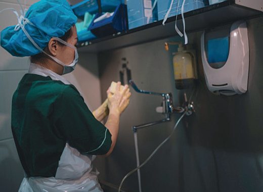
- Prevention: a short preoperative hospital stay lowers risk of acquiring resistant organisms; hand hygiene (with ≥70% alcohol gel, though this does not kill C. difficile spores) is essential; preoperative skin shaving should be performed immediately before surgery (not the night before) as SSI rates after clean surgery may double with night-before shaving due to enhanced bacterial colonisation of minor skin injury, clippers are preferred over razors [1][3].
- Skin preparation uses chlorhexidine or povidone-iodine; alcohol-based skin preparation achieves >95% reduction in bacterial count [1].
- Theatre discipline (minimising staff numbers/movement, laminar flow ventilation, gentle tissue handling, avoiding dead space/haematoma) reduces infection.
- There is high-level evidence that perioperative avoidance of hypothermia and use of supplemental oxygen during recovery significantly reduce SSI rates [1].
- The CDC's 2017 SSI prevention guidelines recommend maintaining perioperative blood glucose <200 mg/dL (11.1 mmol/L), and support increased FiO2 during and after surgery in patients with normal pulmonary function under general anaesthesia, although evidence on hyperoxia's benefit is mixed [2].
- Similarly, keeping glucose 80–120 mg/dL, keeping the patient warm (OR at 70°F, forced-air warming), and chlorhexidine skin preparation with iodine-impregnated drapes are cited as SSI-prevention measures [3].
- Prophylactic antibiotics are indicated for clean-contaminated and contaminated operations and whenever a prosthesis is implanted, but have limited value in non-prosthetic clean surgery where baseline infection rates are already low; they should be given intravenously at induction of anaesthesia so that maximal tissue levels are present at incision, repeated at 4-hourly intervals in long operations or with excessive blood loss, and not continued after surgery (no evidence of benefit, and continuation promotes resistance) [1].
- Prophylactic antibiotics should be given within 1 hour of incision and stopped within 24 hours of the end of the operation (48 hours for cardiac surgery) [3].
- Patients with valvular heart disease or an implanted vascular/orthopaedic prosthesis should receive prophylactic antibiotics for dental, urological or open visceral surgery to prevent bacteraemic seeding [1].
- Treatment of an established SSI: if under tension or clearly suppurating, sutures/clips should be removed with curettage to allow drainage; heavily contaminated wounds are best managed by delayed primary or secondary closure, or left to heal by secondary intention; abscess cavities need drainage (percutaneous/ultrasound-guided where possible) and, if left open to drain freely, do not require additional antibiotics, antibiotics are used if the cavity is closed after drainage [1].
- Empirical antibiotic choice should be based on the likely spectrum of organisms and local policy, then refined by culture and sensitivity, typically available in 2–3 days.
- A narrow-spectrum agent treats a known sensitive organism (e.g. vancomycin/teicoplanin for MRSA), while broad-spectrum combinations (e.g. teicoplanin or meropenem) are used when multiple synergistic gut organisms are suspected, such as after perforated or ischaemic bowel surgery [1].
- Topical antiseptics should be used only briefly on heavily contaminated wounds as they impair epithelial ingrowth and wound healing [1].
- Necrotising soft-tissue infection requires immediate surgical exploration with radical debridement of affected tissue, antibiotics alone are not sufficient treatment [2][3].
- High-dose penicillin is used for confirmed group A streptococcal or clostridial necrotising infection, with broader-spectrum cover if polymicrobial infection is suspected [3].
- Gas gangrene requires large-dose intravenous penicillin and aggressive debridement [1].
The evidence-supported prevention bundle
- Surgical site infection affects between 0.5% and 3% of patients having a surgical procedure and can increase hospital length of stay by 7 to 11 days [6].
- A 2023 review identified six prevention strategies supported by the literature: (1) avoidance of razors for hair removal, using clippers instead; (2) decolonisation with intranasal antistaphylococcal agents and antistaphylococcal skin cleansers for high-risk procedures; (3) chlorhexidine gluconate with alcohol-based skin preparation; (4) maintenance of normothermia with active warming to keep core temperature above 36°C; (5) perioperative glycaemic control, keeping glucose below 150 mg/dL; and (6) negative-pressure wound therapy [6].
- Guidelines also support an appropriate preoperative antibiotic dose, timing and choice based on the procedure [6].
- Adherence is the weak link rather than knowledge, and electronic medical record systems providing automated decision support and reminders have been shown to improve compliance with several of these steps [6].
NG125 is the UK surgical site infection guideline, and the fastest way to hold it is as a list of things it tells you not to do.
| Phase | Do | Do not |
|---|---|---|
| Preoperative | Shower or bath with soap the day before or the day of surgery (1.2.1). Consider nasal mupirocin with a chlorhexidine body wash where S. aureus is a likely cause, determined locally by procedure type, patient risk factors, the increased risk of side effects in preterm infants, and the potential impact of infection (1.2.2). If hair must be removed, use electric clippers with a single-use head on the day of surgery (1.2.5) | Do not use hair removal routinely (1.2.4); do not use razors, which increase SSI risk (1.2.5); do not use mechanical bowel preparation routinely (1.2.9) |
| Prophylaxis | Give before clean surgery involving a prosthesis or implant, clean-contaminated surgery and contaminated surgery (1.2.12). Consider a single intravenous dose on starting anaesthesia, but earlier where a tourniquet is used (1.2.15). Repeat the dose when the operation is longer than the antibiotic's half-life (1.2.16). Give treatment in addition to prophylaxis for a dirty or infected wound (1.2.17). Inform the patient beforehand if prophylaxis will be needed, and afterwards if antibiotics were given during the operation (1.2.18) | Do not use prophylaxis routinely for clean non-prosthetic uncomplicated surgery (1.2.13) |
| Theatre discipline | Wash hands before the first operation with an aqueous antiseptic surgical solution and a single-use brush or pick for the nails; before subsequent operations use either an alcoholic hand rub or an antiseptic surgical solution, washing again with antiseptic solution if hands are soiled (1.3.1, 1.3.2). Remove hand jewellery, artificial nails and nail polish (1.2.10, 1.2.11). Sterile gowns throughout; consider two pairs of gloves where glove perforation risk is high and contamination would be serious (1.3.5, 1.3.6) | Keep movements in and out of the operating area to a minimum (1.2.8) |
| Intraoperative | Prepare the skin immediately before incision (1.3.7). If an incise drape is required, use an iodophor-impregnated one unless iodine-allergic (1.3.4). Consider triclosan-coated sutures, especially in paediatric surgery (1.3.20). Consider gentamicin-collagen implants in cardiac surgery (1.3.19). Cover the incision with an appropriate interactive dressing at the end of the operation (1.3.22) | Do not use non-iodophor-impregnated incise drapes routinely, as they may increase SSI risk (1.3.3). Do not use diathermy for the surgical incision (1.3.11). Do not use wound irrigation (1.3.16) or intracavity lavage (1.3.17). Do not give insulin routinely to people without diabetes to reduce SSI risk (1.3.15). Apply an antiseptic or antibiotic to the wound before closure only as part of a clinical research trial (1.3.18) |
Table reformats the NG125 recommendations [7].
- The antiseptic choice is a four-step cascade, not a preference.
- First choice is an alcohol-based solution of chlorhexidine, unless contraindicated or the surgical site is next to a mucous membrane; next to a mucous membrane, aqueous chlorhexidine; if chlorhexidine is contraindicated, alcohol-based povidone-iodine; and if both an alcohol-based solution and chlorhexidine are unsuitable, aqueous povidone-iodine [7].
- Two safety clauses travel with it: be aware of the risk of severe chemical injuries from chlorhexidine, both alcohol-based and aqueous, in preterm babies, and where diathermy will be used, dry the antiseptic by evaporation and avoid pooling of alcohol-based preparations [7].
- Two intraoperative recommendations are about the patient's physiology rather than about asepsis, and they connect this guideline to the hypothermia one. Maintain patient temperature in line with the NICE guideline on hypothermia in adults having surgery, and maintain optimal oxygenation, giving sufficient oxygen during major surgery and in the recovery period to ensure a haemoglobin saturation of more than 95% [7].
- Adequate perfusion should likewise be maintained [7].
- The hypothermia guideline supplies the operative numbers: warm from induction with a forced-air device if anaesthesia will exceed 30 minutes, keep the ambient theatre temperature at least 21°C while the patient is exposed, and warm intravenous fluids of 500 ml or more and all blood products to 37°C [8].
Where wound healing is likely to be a problem, NG125 asks for a system rather than a product. Use a structured approach to care to improve overall management of surgical wounds, including preoperative assessments to identify people with potential wound healing problems, supported by enhanced education of healthcare workers, patients and carers and by sharing of clinical expertise [7].
Antimicrobial principles and durations in Schwartz's detail
- CDC prevention: full-body soap or antiseptic wash the night before, hair removed in theatre with clippers not razors, alcohol-based skin antisepsis, no proven benefit from antibiotic irrigation or soaking prostheses, antibiotic given so bactericidal tissue levels precede incision (within 30 minutes) and redosed by half-life in long operations, no more than 24 hours of routine prophylaxis and ideally a single dose (postoperative doses add only cost and resistance), glucose under 200 mg/dL (11.1 mmol/L), normothermia and tissue oxygenation, hyperoxia showed early promise in colorectal surgery, a small effect in meta-analysis and CDC 2017 support for increased FiO₂ during and after general anaesthesia in normal lungs [4].
- Schwartz's prophylaxis table: cefazolin or cefuroxime for cardiovascular surgery (vancomycin or clindamycin if allergic); cefazolin for gastroduodenal and unobstructed small bowel; cefazolin, cefoxitin, cefotetan, ceftriaxone or ampicillin-sulbactam for open or high-risk laparoscopic biliary surgery and nothing for low-risk laparoscopic cholecystectomy; cefoxitin, cefotetan or cefazolin-metronidazole for uncomplicated appendicectomy; cefazolin or ceftriaxone plus metronidazole, ertapenem, cefoxitin, cefotetan or ampicillin-sulbactam for colorectal and obstructed small bowel surgery; cefazolin or cefuroxime plus metronidazole or ampicillin-sulbactam for clean-contaminated head and neck; cefazolin for neurosurgery, orthopaedics (or ceftriaxone), breast and hernia; allergic alternatives pair clindamycin or vancomycin with an aminoglycoside, aztreonam or fluoroquinolone, or metronidazole with an aminoglycoside or fluoroquinolone [4].
- Empiric therapy, for high-risk disease (ruptured appendix), intraoperative contamination or septic critical illness with a suspected site, starts broad within the first hour because delay raises mortality, uses unit-specific sensitivities, is de-escalated at 48–72 hours on cultures, and stops within 3–5 days if no infection is proven, since prolonged culture-negative empiric therapy in the critically ill increases mortality; monomicrobial nosocomial infections are treated 3–5 days for UTI, 7–8 for pneumonia and 7–14 for bacteraemia (longer courses breed superinfection; procalcitonin trends allow earlier cessation), osteomyelitis, endocarditis and irremovable prosthetic infection 6–12 weeks, agents chosen by MIC against a 10⁵ CFU/mL inoculum as the least toxic and cheapest effective drug, with IV-to-oral switch after 1–2 weeks only in improving patients using high-bioavailability drugs such as fluoroquinolones [4].
- Polymicrobial infection is treated by source control with broad aerobic–anaerobic cover; cultures matter less because a few species predominate, so worsening prompts a search for failed source control rather than an antibiotic change; the 2016 Surgical Infection Society guideline limits antibiotics to 24 hours after traumatic bowel perforation repaired within 12 hours, gastroduodenal perforation repaired within 24 hours, non-perforated ischaemic bowel and gangrenous appendicitis or cholecystitis, and to 4 days for more extensive peritonitis such as perforated appendicitis, with a normal white count, no bands and temperature under 38°C giving near-complete assurance of eradication and failure to improve by 5–7 days demanding re-evaluation [4].
- Penicillin allergy is reported in 0.7–10% but cross-reactivity is about 1% for carbapenems, 5–7% for cephalosporins and near zero for monobactams; true allergy (urticaria, bronchospasm) must be distinguished from dyspepsia, anaphylaxis bars the class except to save life, intradermal testing pathways cut vancomycin use to 16% in "allergic" surgical patients, and desensitisation by escalating doses is possible; antibiotic misuse costs over $20 billion a year in the United States and drives toxicity, C. difficile and resistance, so the responsible surgeon confines prophylaxis to the operation, does not drift into empiric therapy, fixes duration at prescription, stops when evidence is lacking and never treats drains or tubes prophylactically [4].
- Incisional SSI is treated by incision and drainage alone, antibiotics being added only for significant cellulitis or SIRS, the wound healing by secondary intention with negative-pressure dressings for large complex wounds (inspected if sepsis is unexplained, since suction hides odour and drainage), topical antimicrobials unproven, and cultures taken in hospitalised or long-term-care patients for resistant organisms; US hospitals must survey SSIs for 30 days and, since 2012, report them to CMS, the National Healthcare Safety Network deriving procedure-specific risk indices from 847 hospitals and nearly a million patients, while NSQIP, IHI, NCQA, SIS and CDC drive quality [4].
Sepsis management in Schwartz's summary of the 2016 Surviving Sepsis guidelines
Resuscitate immediately for hypotension or raised lactate with at least 30 mL/kg of crystalloid in the first 3 hours (a 3-hour delay worsens outcome), then guide fluid by dynamic monitoring and response (heart rate, pressure, urine output) rather than the old CVP 8–12 target, aiming for MAP ≥65 mmHg, urine ≥0.5 mL/kg/h, mixed venous saturation over 65% and lactate normalisation; culture before antibiotics without delaying them, start broad intravenous therapy within the first hour, reassess daily, de-escalate, stop at 7–10 days for most infections or sooner by procalcitonin, image promptly and achieve source control as soon as possible after initial resuscitation, removing suspect vascular devices; use crystalloid with albumin as an adjunct for large volumes and never hydroxyethyl starch or gelatin; noradrenaline is first-line, vasopressin is added to raise MAP or spare noradrenaline, adrenaline is an alternative with more splanchnic vasoconstriction, dopamine only for bradycardic low-arrhythmia-risk patients (no "renal-dose" role), phenylephrine not recommended, arterial lines for anyone on pressors, dobutamine for persistent hypoperfusion, and hydrocortisone under 300 mg/day (about 200 mg) for shock refractory to fluid and pressors; transfuse below 7 g/dL unless ischaemia or haemorrhage, do not correct INR with plasma absent bleeding, give platelets below 10,000 prophylactically, below 20,000 with bleeding risk and below 50,000 with bleeding or procedures; ventilate lung injury at 6 mL/kg with plateau under 30 cmH₂O, PEEP, conservative fluids, prone positioning when PaO₂/FiO₂ is under 150 in preference to high-frequency oscillation, weaning protocols and no routine pulmonary artery catheter; minimise sedation, target glucose ≤180 mg/dL, give stress-ulcer and DVT prophylaxis, and discuss realistic goals with families [4].
Surgeries
Surgical management of SSI centres on source control rather than a defined "operation": incision and drainage (with curettage of the cavity) for abscesses; removal of sutures/clips with wound de-roofing for infected surgical incisions; delayed primary or secondary closure for heavily contaminated wounds; and radical surgical debridement (incision with direct visualisation of deep soft tissue, fascia and muscle, and resection of affected tissue) for necrotising soft-tissue infection, which may require repeated debridement [1][2]. Fulminant Clostridium difficile (pseudomembranous) colitis with perforation requires emergency total abdominal colectomy with ileostomy [3].
Complications
- Local complications include chronic abscess, antibioma (partly sterilised abscess), and sinus/fistula formation, particularly with Mycobacterium or Actinomyces infection [1].
- Uncontrolled spreading infection can progress to bacteraemia and a systemic inflammatory response.
- Gas gangrene can cause early systemic circulatory collapse and organ failure if not promptly treated [1].
- Necrotising fasciitis and other necrotising soft-tissue infections are associated with sepsis and septic shock, often without an obvious cause identified initially [2].
- Gut colonisation and bacterial translocation can precipitate SIRS and multiple organ dysfunction syndrome (MODS) [1].
- Antibiotic overuse or inappropriate switching promotes antibiotic resistance and risks complications such as C. difficile enteritis [1].
Intra-abdominal, organ-specific and necrotising infection in Schwartz's account
- Primary peritonitis, haematogenous or direct inoculation in ascites or peritoneal dialysis, monomicrobial (Gram-positives on dialysis; otherwise E. coli, Klebsiella, S. pneumoniae), diagnosed by diffuse tenderness without a surgical source and over 250 neutrophils/mL on paracentesis, is treated 14–21 days with device removal for recurrence and rarely needs surgery; secondary peritonitis from perforation or organ infection needs source control plus aerobic–anaerobic cover chosen before laparotomy because colonic perforation is the most morbid possibility, oral step-down when ileus resolves, and carries 5–6% mortality with control against over 40% without, response rates having stayed at 70–90% for decades; failures become abscess, anastomotic leak or tertiary peritonitis, persistent infection in the immunosuppressed with E. faecalis and faecium, S. epidermidis, Candida and Pseudomonas and mortality over 50%; abscesses are CT-drained percutaneously, surgery being reserved for multiple collections, hazardous proximity to vital structures or an ongoing leak, with 3–5 days of antibiotics and drains kept until the cavity collapses, output falls below 10–20 mL/day and no source persists [4].
- Hepatic abscesses (15 per 100,000 admissions; 80% pyogenic, the rest parasitic or fungal; once pylephlebitic from neglected appendicitis or diverticulitis, now often after biliary manipulation and cryptogenic in nearly half; E. coli, Klebsiella, enterococci, Pseudomonas, Bacteroides, anaerobic streptococci, Fusobacterium, Candida) are sampled and treated 4–6 weeks when small and multiple or drained percutaneously when large, recurrent hepatic or splenic abscesses needing unroofing and marsupialisation or splenectomy [4].
- Infected pancreatic necrosis follows 10–15% of severe necrotising pancreatitis; Bradley and Allen pioneered repeated debridement; prophylactic antibiotics are not indicated, nasojejunal feeding beyond the ligament of Treitz reduces infection, suspicion arises when SIRS fails to settle or sepsis returns at 2–3 weeks, CT-guided aspiration with positive Gram stain or culture or pancreatic gas mandates intervention, intervention within the first 2 weeks raises mortality so delay to 4 weeks is sought, endoscopic necrosectomy carries 5% mortality and 30% complications with a median of four sessions, laparoscopic series 6% mortality in 65 patients, and the randomised step-up of percutaneous or transgastric drainage then video-assisted retroperitoneal debridement if no improvement at 72 hours cut the composite of death and complications from 69% to 40% with fewer hernias, less new diabetes and less enzyme supplementation than open necrosectomy [4].
- Aggressive soft tissue infections kill 80–100% if missed and 16–24% even when treated; the old eponyms (Meleney's synergistic gangrene, Fournier's, gas gangrene) give way to classification by layer and pathogen; compromised fascial blood supply in the elderly, diabetic, immunosuppressed and vascular patient is the common thread though streptococcal cases strike the healthy; limbs, perineum, trunk and torso are affected in that order; "dishwater pus" from a small break or sinus, bronze hue, brawny induration, blebs, crepitus and pain out of proportion mandate immediate incision, direct inspection of fascia and muscle and radical resection without radiology, which delays and confuses; tissue fluid is Gram-stained, vancomycin plus a carbapenem and 16–20 million units of aqueous penicillin G daily (for clostridia) are given, about half are polymicrobial with the rest S. pyogenes, Pseudomonas or C. perfringens, scheduled returns to theatre continue until progression stops, and hyperbaric oxygen has weak support only for gas-forming organisms [4].
Blood-borne exposure and biological agents in Schwartz's account
- Six surgeons had seroconverted to HIV occupationally by 2011 with none since 1999, against 60 nurses and 19 non-surgeon physicians among about 200 workers; percutaneous exposure carries 0.3% risk and mucous membrane 0.09%, higher with hollow needles, large volumes, intravascular injection and high viral load; universal precautions (barriers, immediate washing, careful sharps handling) and three-drug post-exposure prophylaxis started within hours, begun while testing when the source is unknown but high-risk, not for deceased sources or sharps-bin needles, reduce annual risk from 1 in 200,000 to 1 in 10,000,000 in a Glasgow model [4].
- Hepatitis B kills about 30% of chronic carriers through cirrhosis or hepatocellular cancer, vaccination cut US cases from about 250,000 a year in the 1980s to 3350 in 2010 and is mandatory for surgeons; hepatitis C becomes chronic in 75–80% (three-quarters developing liver disease), transmits by needlestick in about 1.8%, has no vaccine, immunoglobulin or post-exposure prophylaxis, and is now treated with direct-acting antivirals such as sofosbuvir rather than ribavirin and interferon [4].
- Biological weapons, chosen for inhalational spread: anthrax (1979 Sverdlovsk release; 1–6 days' incubation, then respiratory distress, chest pain, widened mediastinum and effusions; ciprofloxacin or doxycycline prophylaxis, amoxicillin if sensitive, ciprofloxacin–clindamycin–rifampicin treatment, clindamycin blocking toxin), plague (Y. pestis, catapulted corpses at Caffa; pneumonic with bloody sputum if aerosolised, bubonic if flea-borne; bipolar safety-pin Gram-negative rod on bubo aspirate; doxycycline prophylaxis, streptomycin, aminoglycoside, doxycycline, fluoroquinolone or chloramphenicol treatment), smallpox (viable in scabs 13 years; 10–12 days' incubation, centripetal rash, 30% fatality, vaccine effective up to 4 days post-exposure, cidofovir promising) and tularaemia (tick reservoir, intramacrophage growth, pneumonia with adenopathy in 85%, agglutination diagnosis, aminoglycoside or doxycycline or ciprofloxacin) [4].
Prognosis
- Since the introduction of prophylactic antibiotics, SSI in contaminated, immunosuppressed or prosthetic-surgery patients has become the exception rather than the rule, and faecal peritonitis is no longer inevitably fatal, incisions made in the presence of such contamination can heal primarily without infection in over 90% of patients with appropriate antibiotic therapy [1].
- Two-thirds of SSIs after colorectal surgery may present after hospital discharge, underscoring the importance of follow-up [2].
- Sepsis mortality overall has fallen to under 30% with improvements in care delivery and evidence-based treatment strategies such as the Surviving Sepsis Campaign guidelines [2].
- Severe forms of tetanus requiring ventilation are associated with high mortality [1].
References
- Bailey & Love's Short Practice of Surgery, 28th ed., Ch. 5 Surgical infection
- Schwartz's Principles of Surgery: ABSITE and Board Review, Ch. 6, citing Schwartz 11th ed., p. 169
- The ABSITE Review, 2022, Ch. 5 Infection
- Schwartz's Principles of Surgery, 11th ed., Ch. 6, Surgical Infections
- Sabiston Textbook of Surgery, 22nd ed., Ch. 25 Surgical Site Infections, Table 25.1
- Sabiston Textbook of Surgery, 22nd ed., Ch. 9 Safety in the Surgical Environment
- NICE Guideline NG125: Surgical site infections: prevention and treatment. National Institute for Health and Care Excellence, London, UK, 2019, updated 2020., 1.2.1 to 1.3.22; 1.3.8; 1.3.9, Table 1; 1.3.10; 1.3.12; 1.3.13; 1.3.14; 1.4.11 www.nice.org.uk
- NICE Clinical Guideline CG65: Hypothermia: prevention and management in adults having surgery. National Institute for Health and Care Excellence, London, UK, 2008, updated 2016., 1.3.4; 1.3.6; 1.3.7 www.nice.org.uk