



Fluid and Electrolyte Management
Summary
- Fluid and electrolyte management aims to keep the surgical patient neither fluid-depleted nor fluid-overloaded by replacing what is lost, since success in this area can be the difference between an uncomplicated postoperative course and admission to intensive care [1].
- Roughly two-thirds of body weight is water, two-thirds of which is intracellular and one-third extracellular (of which two-thirds is interstitial and one-third intravascular) [2].
- Sodium determines the intracellular/extracellular osmotic gradient and proteins determine plasma/interstitial oncotic pressure, so disorders of fluid balance and of individual electrolytes (sodium, potassium, calcium, magnesium, phosphate) and acid-base status are closely interrelated in the perioperative period [2][3].
Definition
- Total body water is approximately 50–60% of total body weight in adults (60% in young men, 50% in young women), around 80% in newborns falling to about 65% by one year, and should be adjusted down 10–20% in obese individuals and up 10% in malnourished individuals [3].
- Fluid balance is defined against approximate average daily losses: water loss of about 2500 mL/day (insensible loss from skin, respiratory tract, GI tract, and urine), sodium loss of 1–2 mmol/kg/day, and potassium loss of 0.7–1 mmol/kg/day, each increased by pyrexia, diarrhoea, vomiting or high-output fistulae [1].
- Acid-base balance is defined by maintenance of arterial pH between 7.35 and 7.45 through respiratory (PaCO2) and metabolic (bicarbonate) regulation [1].
Pathophysiology
- Water shifts from areas of low solute concentration to areas of high solute concentration to achieve osmotic equilibration.
- Serum osmolality is calculated as (2 × Na) + (glucose/18) + (BUN/2.8), normally 280–295 [2].
- Third-space fluid is interstitial fluid sequestered from the functional extracellular compartment [2].
- Volume overload is most commonly iatrogenic, with weight gain as its first sign [2].
- Isotonic crystalloids remain largely extracellular acutely, whereas colloids (including blood) produce more lasting intravascular volume expansion because crystalloid rapidly redistributes into the interstitium; when crystalloid is used to replace blood loss, 3–4 times the lost volume must be given because only one-quarter to one-third remains intravascular [1].
- Acid-base regulation follows CO2 + H2O ⇌ H2CO3 ⇌ H+ + HCO3⁻, with respiratory compensation occurring within minutes and renal (bicarbonate) compensation over hours to days [1][2].
- Nasogastric suction causes loss of hydrogen and chloride ions from gastric secretions, producing hypochloraemic, hypokalaemic metabolic alkalosis.
- The kidney's compensatory sodium/hydrogen and potassium/hydrogen exchange produces a paradoxical aciduria [2].
Compartments, exchange and volume control in Schwartz's figures
- Sodium is confined to the extracellular fluid, so sodium-containing fluids expand the interstitium about three times as much as the plasma; for univalent ions 1 mEq equals 1 mmol, 1 mmol of sodium chloride contributes 2 mOsm, calculated osmolality is 2 × sodium + glucose/18 + BUN/2.8, and both compartments sit at 290–310 mOsm with water redistributing until effective osmotic pressures equalise [5].
- A healthy adult takes about 2000 mL of water a day (75% by mouth, the rest from food) and loses 800–1200 mL in urine, 250 mL in stool and 600 mL insensibly (75% skin, 25% lungs, more with fever, hypermetabolism and hyperventilation); the kidneys must pass 500–800 mL a day to clear metabolic products, dietary salt is 3–5 g with renal sodium excretion adjustable from 1 to 5000 mEq a day, sweat is hypotonic and GI losses isotonic to slightly hypotonic (gastric 1000–2000 mL with sodium 60–90 and chloride 100–130; small bowel 2000–3000 mL with sodium 120–140 and bicarbonate 30–40; pancreas 600–800 mL with bicarbonate 95–115; bile 300–800 mL) [5].
- Osmoreceptors drive thirst and hypothalamic vasopressin release; aortic arch and carotid sinus baroreceptors act neurally and through renin–angiotensin, aldosterone, atrial natriuretic peptide and renal prostaglandins [5].
- Only 2% of body potassium (4.5 mEq/L × 14 L, about 63 mEq) is extracellular against 98% intracellular, with intake 50–100 mEq and renal excretion 10–700 mEq a day, so small shifts from acidosis, hyperosmolality, stress, injury or catabolism move serum values sharply; serum calcium is 40% protein-bound, 10% complexed and 50% ionised, the ionised fraction governing neuromuscular stability and rising with acidosis; magnesium (2000 mEq total, half in bone, a third of the extracellular fraction albumin-bound, intake 20 mEq a day, renal loss under 1 mEq a day in deficiency) is a poor serum indicator in hypoalbuminaemia and should be replaced to the upper normal limit [5].
Clinical features
- Assessment of volume status combines history, examination, fluid balance charts, and blood results.
- The "dry" (hypovolaemic) patient feels thirsty, has a dry mouth, low JVP, dry mucous membranes, reduced skin turgor, falling blood pressure, rising pulse, low urine output (<1 mL/kg/h), and a body weight several kilograms below baseline.
- Blood tests show high sodium, potassium, creatinine and disproportionately raised urea [1].
- The "overfilled" patient does not feel thirsty, has a raised JVP, dependent oedema, pulmonary oedema on auscultation, a high central venous pressure that plateaus with fluid challenges, and may have low serum sodium [1].
- Electrolyte-specific clinical features include: hyperkalaemia, peaked T waves progressing to flattened P waves, prolonged PR interval, widened QRS, sine-wave pattern and ventricular fibrillation, with gastrointestinal (nausea, vomiting, colic) and neuromuscular (weakness to ascending paralysis) symptoms [2][3]; hypokalaemia, fatigue, weakness, muscle cramps/twitches, flattened T waves [2]; hyponatraemia, headache, nausea, vomiting, seizures [2]; hypernatraemia, restlessness, irritability, seizures [2]; hypocalcaemia, perioral tingling and numbness (first symptom), hyperreflexia, Chvostek's sign, Trousseau's sign (carpopedal spasm on blood pressure cuff inflation), and prolonged QT interval [2]; hypomagnesaemia, irritability, confusion, hyperreflexia, seizures, resembling hypocalcaemia [2]; hypophosphataemia (classically in refeeding syndrome), failure to wean from ventilator, muscle weakness, confusion [2].
Etiology
- Hyperkalaemia often occurs with renal failure [2].
- Hypokalaemia usually results from over-diuresis or diarrhoea [2].
- Hyponatraemia is usually due to fluid overload with dilute urine, but can also occur from isotonic GI fluid loss compensated by water retention.
- Postoperative causes include excess intraoperative saline infusion, antipsychotic medication, excess oral water intake, and transient decreases (or, less often, syndrome of inappropriate secretion) of antidiuretic hormone.
- Pseudohyponatraemia occurs with hyperglycaemia or hyperlipidaemia [2][3].
- Hypernatraemia is usually due to poor fluid intake with concentrated urine [2].
- Diabetes insipidus (low ADH) causes hypernatraemia with dilute polyuria and can follow alcohol use or head injury.
- SIADH (high ADH) causes hyponatraemia with concentrated, low-volume urine and can also follow head injury [2].
- Hypercalcaemia is most commonly caused by hyperparathyroidism (most common benign and overall cause) or malignancy (breast cancer most common malignant cause), with undiagnosed hyperparathyroidism plus a stressor such as surgery being the most common cause of hypercalcaemic crisis [2].
- Hypocalcaemia commonly follows parathyroidectomy or previous thyroidectomy (injury to parathyroid glands) [2].
- Hypomagnesaemia typically occurs with massive diuresis, chronic total parenteral nutrition without magnesium replacement, or alcohol abuse [2].
- Metabolic acidosis with an elevated anion gap arises from exogenous acid ingestion (ethylene glycol, salicylate, methanol) or endogenous acid production (ketoacidosis, lactic acidosis, renal insufficiency), summarised by the mnemonic MUDPILES (methanol, uraemia, diabetic ketoacidosis, paraldehydes, isoniazid, lactic acidosis, ethylene glycol, salicylates) [2][3].
- Metabolic acidosis with a normal anion gap arises from loss of bicarbonate via diarrhoea, enteric/pancreatic/biliary fistulae, ureterosigmoidostomy, or renal tubular acidosis, or from rapid infusion of bicarbonate-deficient (high-chloride) fluid [2][3].
- Metabolic alkalosis is usually a contraction alkalosis from fluid loss such as nasogastric suction or over-diuresis with loop diuretics [2].
Absorption of irrigation fluid: post-TURP syndrome
A surgical cause of acute dilutional hyponatraemia that is specific to the operating theatre is the systemic absorption of irrigation fluid during transurethral resection of the prostate. Post-TURP syndrome is a hyponatraemia secondary to irrigation with water, and it can precipitate seizures through cerebral oedema. Treatment is careful correction of sodium with diuresis [6].
Causes of concentration and composition disorders in Schwartz's account
- Extracellular deficit is the commonest surgical fluid disorder: acute deficits show cardiovascular and CNS signs, chronic deficits add tissue signs (turgor, sunken eyes), BUN rises with haemoconcentration, urine osmolality exceeds serum and urine sodium is under 20 mEq/L, serum sodium may be high, normal or low, and causes are GI losses (nasogastric suction, vomiting, diarrhoea, fistula) and sequestration in injured soft tissue, burns, peritonitis, obstruction or prolonged surgery; excess is iatrogenic or from renal, cardiac or hepatic failure, tolerated by the fit but tipping the elderly and cardiac into failure [5].
- Hyponatraemia is dilutional (excess water, postoperative ADH secretion (self-limiting as hyponatraemia and expansion suppress ADH) antipsychotics, tricyclics, ACE inhibitors, the elderly most susceptible) or depletional (low-sodium diet or enteral feeds, GI or renal loss with diuretics or renal disease), or apparent from hyperglycaemia or mannitol (sodium falls 1.6 mEq/L per 100 mg/dL of glucose above normal) or pseudohyponatraemia from lipids and protein; depletion gives urine sodium under 20 mEq/L, renal wasting over 20, dilution a hypervolaemic circulation, and normovolaemia prompts a search for SIADH [5].
- Hypernatraemia is hypervolaemic from sodium-containing fluids, excess bicarbonate or mineralocorticoid excess (urine sodium over 20 mEq/L, osmolality over 300), normo- or hypovolaemic from renal loss (diabetes insipidus, diuretics, renal disease; urine sodium under 20, osmolality under 300–400) or non-renal loss (diarrhoea, fever, tracheostomy hyperventilation, thyrotoxicosis, hypertonic peritoneal dialysate; urine sodium under 15, osmolality over 400), symptomatic only with impaired thirst or restricted access and rarely below 160 mEq/L, when cellular dehydration puts traction on cerebral vessels and can cause subarachnoid haemorrhage alongside restlessness, seizures, coma and dry sticky mucous membranes [5].
- Hyperkalaemia follows intake (supplements, transfused red cell lysis), release (haemolysis, rhabdomyolysis, crush, GI bleeding, acidosis, hyperglycaemia or mannitol) or impaired excretion (potassium-sparing diuretics, ACE inhibitors, NSAIDs, renal failure); hypokalaemia, far commoner, follows poor intake, renal loss (hyperaldosteronism, diuretics, magnesium-wasting amphotericin, aminoglycosides, cisplatin and ifosfamide, refractory until magnesium is replaced), GI loss and alkalosis or insulin shifts, potassium falling 0.3 mEq/L per 0.1 rise in pH [5].
- Hypercalcaemia is primary hyperparathyroidism in outpatients and malignancy (bone metastases or PTH-related protein) in hospital; hypocalcaemia follows pancreatitis (fatty-acid chelation), necrotising fasciitis, renal failure, pancreatic and small bowel fistulas, hypoparathyroidism, toxic shock, magnesium disorders, tumour lysis and rhabdomyolysis (phosphate precipitation), citrate in massive transfusion, osteoblastic breast and prostate metastases and the hungry bone and post-adenomectomy states, rarely low intake alone; hyperphosphataemia is mostly renal, plus hypoparathyroidism, hyperthyroidism, cell destruction and excess intake; hypophosphataemia is chronic from malabsorption, binders or malnutrition and acute from intracellular shift with respiratory alkalosis, insulin, refeeding or hungry bone; hypermagnesaemia needs renal failure with magnesium antacids or laxatives, TPN, massive trauma, burns or acidosis, and hypomagnesaemia (common in the critically ill) follows starvation, alcoholism, unsupplemented fluids or TPN, diuretics, amphotericin, aldosteronism, diarrhoea, malabsorption and pancreatitis, producing hypocalcaemia and persistent hypokalaemia [5].
Diagnosis
- Normal reference ranges: potassium 3.5–5.0 mEq/L, sodium 135–145 mEq/L, calcium 8.5–10.0 mg/dL (ionized 1.0–1.5), magnesium 2.0–2.7 mg/dL, phosphate 2.5–4.5 mg/dL, arterial pH 7.35–7.45, PaCO2 4.7–6.0 kPa, bicarbonate 22–26 mmol/L, base excess −2 to +2 mEq/L [1][2].
- The anion gap = [Na] − [Cl + HCO3], normally <12 mmol/L (or 8–16 mmol/L per Oxford Handbook), and must be corrected for hypoalbuminaemia (corrected AG = actual AG + [2.5 × (4.5 − albumin)]) [1][3].
- The Flenley acid-base nomogram is a diagnostic aid: assess pH first (acidaemia <7.35, alkalaemia >7.45), then check whether PaCO2 and bicarbonate changes are concordant (primary respiratory acidosis if pH<7.35 with PaCO2>6.0 kPa; primary respiratory alkalosis if pH>7.45 with PaCO2<4.7 kPa; primary metabolic acidosis if pH<7.35 with HCO3<22 mmol/L; primary metabolic alkalosis if pH>7.45 with HCO3>26 mmol/L), with discordant changes indicating compensation [1].
- Calculated serum osmolality = 2×Na + glucose/18 + BUN/2.8, normal 280–295 mOsm.
- Each 180 mg/dL rise in glucose raises osmolality by approximately 10 mOsm [2][3].
- Corrected serum calcium: add/subtract 0.8 mg/dL of calcium for every 1 g/dL decrease/increase in albumin from a normal of 4 g/dL [2][3].
- The free water deficit for correcting hypernatraemia is calculated from total body water (estimated as 50% of lean body mass in men, 40% in women) multiplied by (serum sodium − 140)/140 [3].
- Potassium changes with pH: potassium falls by approximately 0.3 mEq/L for every 0.1 rise in pH above normal in alkalosis [3].
- Fractional excretion of sodium (FeNa) is the best test for azotaemia.
- Prerenal failure shows FeNa <1%, urine sodium <20, BUN/creatinine ratio >20, and urine osmolality >500 mOsm [2].
Clinical and ECG signatures in Schwartz's tables
- Hyponatraemia produces headache, confusion, altered reflexes, seizures, coma and raised intracranial pressure with hypertension and bradycardia, weakness and cramps, anorexia, nausea, watery diarrhoea, lacrimation, salivation and oliguria; hypernatraemia restlessness, lethargy, ataxia, tonic spasms, delirium, seizures, tachycardia, hypotension, syncope, a red swollen tongue and fever [5].
- Hyperkalaemia gives nausea, colic, diarrhoea, weakness ascending to paralysis and respiratory failure, and an ECG of peaked T waves, widened QRS, flattened P, prolonged PR, sine wave and ventricular fibrillation; hypokalaemia gives ileus, constipation, weakness, diminished reflexes, paralysis and arrest with U waves, T flattening, ST changes and digitalis arrhythmias, symptoms masked in volume depletion and unmasked by dilution [5].
- Total calcium is corrected down 0.8 mg/dL per 1 g/dL fall in albumin; hypercalcaemia (total over 8.5–10.5 mg/dL, ionised over 4.2–4.8) causes neurological impairment, weakness, bone pain, renal dysfunction, GI upset, hypertension, arrhythmia, worse digitalis toxicity, a short QT, long PR and QRS, high QRS voltage, flat wide T waves and AV block; hypocalcaemia is symptomatic once ionised calcium falls below 2.5 mg/dL (or with alkalosis at normal total calcium) with perioral and acral paraesthesia, cramps, carpopedal spasm, stridor, tetany, seizures, hyperreflexia, Chvostek's and Trousseau's signs, reduced contractility and heart failure, and a long QT, T inversion, heart block and ventricular fibrillation [5].
- Hypermagnesaemia mimics hyperkalaemia on ECG (long PR, wide QRS, tall T) with nausea, weakness, hyporeflexia, hypotension and arrest; hypomagnesaemia mimics hypocalcaemia (hyperreflexia, tremor, tetany, Chvostek and Trousseau, delirium, seizures) with long QT and PR, ST depression, P flattening, torsades de pointes and arrhythmias; hypophosphataemia shows cardiac dysfunction and weakness only when severe and prolonged hyperphosphataemia deposits calcium-phosphate in soft tissue [5].
- Acid–base: respiratory compensation is prompt, renal compensation begins after about 6 hours and continues for days, so respiratory disorders are acute before and chronic after it; predicted PCO₂ is 1.5 × HCO₃⁻ + 8 in metabolic acidosis and 0.7 × HCO₃⁻ + 21 in alkalosis, and pH changes 0.008 per mmHg in acute and 0.003 in chronic respiratory acidosis, 0.008 and 0.017 in acute and chronic alkalosis, a pH beyond prediction means a mixed disorder; the anion gap (Na − Cl − HCO₃⁻) is normally under 12 mmol/L and is corrected upward by 2.5 × (4.5 − albumin), raised with ethylene glycol, salicylate, methanol, ketones, lactate and renal organic acids and normal with HCl or ammonium, diarrhoea, fistulas, ureterosigmoidostomy, renal tubular acidosis (low urinary ammonium; proximal reabsorptive or distal secretory) and acetazolamide [5].
- Metabolic alkalosis needs both bicarbonate generation and impaired excretion, chloride-losing (urine chloride over 20: mineralocorticoid excess, profound potassium depletion), chloride-sparing (under 20: gastric loss, diuretics), alkali excess (acetate in TPN, citrate, antacids, bicarbonate, milk-alkali) with low GFR or hypercarbic or hypokalaemic bicarbonate reabsorption; obstructed-pylorus vomiting (infantile pyloric stenosis, duodenal ulcer) loses only chloride- and acid-rich gastric fluid, giving hypochloraemic hypokalaemic alkalosis with paradoxical aciduria as aldosterone-driven sodium retention wastes potassium and hydrogen; respiratory acidosis follows narcotics, CNS injury, secretions, atelectasis, mucus plugs, pneumonia, effusion, incisional pain, distension, compartment syndrome and ascites, and acute hypocapnia drives potassium and phosphate into cells and calcium onto albumin [5].
Thresholds and severity
There is no dedicated severity score for fluid and electrolyte disturbance. Severity is expressed through reference ranges and thresholds for specific electrolytes and acid-base parameters (e.g. hypercalcaemia symptomatic usually above calcium 13; hyperkalaemia ECG progression from peaked T waves to sine-wave arrhythmia) as detailed under Diagnosis and Clinical features above [2].
Treatment and Management
- Maintenance IV fluid can be calculated by the "4-2-1" rule: 4 mL/kg/h for the first 10 kg, 2 mL/kg/h for the second 10 kg, and 1 mL/kg/h for each kilogram thereafter [2].
- For major adult GI surgery, lactated Ringer's (Hartmann's) solution is used intraoperatively and for the first 24 hours, then changed to D5 half-normal saline with 20 mEq K⁺ thereafter, since 5% dextrose stimulates insulin release and limits protein catabolism [2].
- Urine output should be maintained at a minimum of 0.5 mL/kg/h and is the best indicator of adequate volume replacement, but should not itself be "topped up" if it reflects normal postoperative diuresis [2][7].
- Fluid resuscitation is tailored to the source of loss: sweat and gastric losses (e.g. gastric outlet obstruction) are replaced with normal saline; pancreatic, biliary or small-bowel losses with lactated Ringer's (may need added bicarbonate); large-bowel losses (e.g. diarrhoea) with lactated Ringer's (may need added potassium); GI losses are generally replaced volume-for-volume [2].
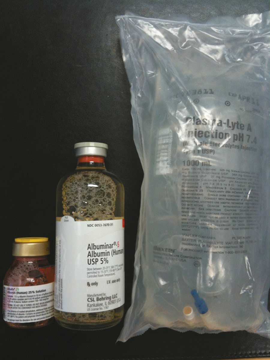
- Common crystalloids: Hartmann's solution (130 mmol/L Na, 103 mmol/L Cl, 28 mmol/L lactate, 4 mmol/L K, 1.5 mmol/L Ca, iso-osmolar/isotonic); 0.9% normal saline (154 mmol/L Na, 154 mmol/L Cl; large volumes can cause hyperchloraemic acidosis); 5% glucose (no electrolytes, becomes hypotonic as glucose is metabolised, should not be used for volume replacement as it causes hyponatraemia) [1][3].
- Colloids (Gelofusine, albumin) produce more lasting volume expansion than crystalloid but albumin resuscitation can cause pulmonary oedema via increased intravascular oncotic pressure, and hydroxyethyl starch is associated with postoperative bleeding in cardiac/neurosurgical patients [1][3].
- Hypertonic (7.5%) saline is used in closed head injury to increase cerebral perfusion and reduce intracranial pressure/brain oedema, but carries a bleeding risk as an arteriolar vasodilator and should not be used for initial resuscitation of hypovolaemic shock generally [3].
- Hyperkalaemia management: calcium gluconate first (membrane stabilisation, does not lower potassium), then measures that shift potassium intracellularly (sodium bicarbonate, insulin with dextrose, nebulised albuterol) followed by potassium-removing measures (Kayexalate, loop diuretics, dialysis if refractory) [2][3].
- Hyponatraemia: water restriction is first-line for fluid-overload hyponatraemia (then diuresis); correction should proceed no faster than 1 mEq/L/h to avoid central pontine myelinolysis; isotonic-loss hyponatraemia is treated with isotonic fluids [2].
- SIADH is managed with fluid restriction and slow diuresis first-line, with conivaptan/tolvaptan (vasopressin V2-receptor antagonists) if refractory.
- Diabetes insipidus is managed with free water first-line and DDAVP if refractory [2].
- Hypercalcaemia is treated with normal saline (200–300 mL/h) followed by furosemide once euvolaemic, avoiding lactated Ringer's (contains calcium) and thiazide diuretics (retain calcium).
- Malignancy-related hypercalcaemia may need calcitonin, bisphosphonates, glucocorticoids or dialysis [2].
- Hypocalcaemia and hypomagnesaemia often need magnesium repletion before calcium will correct [2].
- Respiratory acidosis is treated by increasing minute ventilation; respiratory alkalosis by lowering minute ventilation.
- Metabolic acidosis treatment targets the underlying cause, keeping pH above 7.20 with bicarbonate if needed (correction of acidosis can precipitate hypokalaemia).
- Metabolic alkalosis from nasogastric losses is treated with normal saline to correct the chloride deficit [2].
- Insensible fluid losses average 10 mL/kg/day (75% skin, 25% respiratory), increased by fever, burns, large open wounds, and mechanical ventilation [2].
- Neonatal maintenance fluids require 10% glucose with appropriate electrolytes per local NICU protocol, with watch kept for hyperglycaemia and hypernatraemia (which increases intraventricular haemorrhage risk in preterm infants), and nasogastric/stoma losses over 15 mL/kg/day replaced millilitre-for-millilitre with 0.9% NaCl plus 0.15% KCl [8].
- CG174 states the adult maintenance prescription as three numbers per kilogram per day, and they are the numbers UK practice is audited against.
- If patients need intravenous fluids for routine maintenance alone, restrict the initial prescription to 25 to 30 ml/kg/day of water, approximately 1 mmol/kg/day of potassium, sodium and chloride, and approximately 50 to 100 g/day of glucose to limit starvation ketosis [9].
- NICE adds that this quantity will not address nutritional needs, that weight-based potassium prescriptions should be rounded to the nearest commonly available fluids, and that potassium should not be added to intravenous fluid bags, as this is dangerous [9].
Two adjustments follow. For patients who are obese, adjust to ideal body weight, use lower-range volumes per kilogram (patients rarely need more than a total of 3 litres per day), and seek expert help if BMI is above 40 kg/m² [9]. And consider prescribing less, for example 20 to 25 ml/kg/day, for patients who are older or frail [9].
Resuscitation has a defined bolus and a defined list of triggers, and the paediatric figures differ, which is the commonest transposition error.
| Adults (CG174) | Children and young people (NG29) | Term neonates (NG29) | |
|---|---|---|---|
| Fluid | Crystalloid, sodium 130 to 154 mmol/l | Glucose-free crystalloid, sodium 131 to 154 mmol/l | Glucose-free crystalloid, sodium 131 to 154 mmol/l |
| Bolus | 500 ml over less than 15 minutes | 10 ml/kg over less than 10 minutes | 10 to 20 ml/kg over less than 10 minutes |
| Escalation | Seek expert help for complex imbalance or significant comorbidity | Seek expert advice if 40 to 60 ml/kg or more is needed | As for children |
Table compares the adult and paediatric resuscitation prescriptions [9][10]. Do not use tetrastarch in children and young people [10].
The indicators that a patient may need urgent fluid resuscitation are a six-item list, and one of them is a manoeuvre rather than a number [9]:
- systolic blood pressure less than 100 mmHg
- heart rate more than 90 beats per minute
- capillary refill time more than 2 seconds, or peripheries cold to touch
- respiratory rate more than 20 breaths per minute
- National Early Warning Score (NEWS) of 5 or more
- passive leg raising suggests fluid responsiveness
CG174 describes the manoeuvre precisely: with the patient semi-recumbent, tilt the entire bed through 45°, or lie the patient flat and passively raise the legs to more than 45°. Haemodynamic improvement at 30 to 90 seconds indicates that volume replacement may be required; deterioration, in particular breathlessness, indicates the patient may be fluid overloaded [9].
- Replacement and redistribution are a third prescription, separate from resuscitation and maintenance.
- Adjust the prescription, adding to or subtracting from maintenance needs, to account for existing fluid or electrolyte deficits or excesses, ongoing losses, or abnormal distribution [9]. Seek expert help for a complex redistribution issue or imbalance, or significant comorbidity: gross oedema, severe sepsis, hyponatraemia or hypernatraemia, renal, liver or cardiac impairment, postoperative fluid retention and redistribution, and malnutrition with refeeding issues [9].
- In children, use 0.9% sodium chloride containing potassium to replace ongoing losses [10].
- Around an operation, two NG180 recommendations change what is given and when.
- Tell people having surgery, including dental surgery, that they may drink clear fluids until 2 hours before their operation, that doing so can help reduce headaches, nausea and vomiting afterwards, and that clear fluids means water, fruit juice without pulp, coffee or tea without milk, and ice lollies [11]. Consider carbohydrate drinks before surgery for people having abdominal major or complex surgery [11].
- Intraoperatively, consider using intravenous crystalloid for maintenance, following the CG174 recommendations on resuscitation and routine maintenance [11].
- And on glucose, NG180 is explicit that tight control is not the target: do not use glucose-lowering medicines to achieve tight blood glucose control of 4 to 6 mmol/litre for people having surgery who have type 2 diabetes or do not have diabetes [11].
Fluids, correction rates and protocols in Schwartz's detail
- Lactated Ringer's (sodium 130, chloride 109, potassium 4, lactate 28, calcium 3; 273 mOsm) is slightly hypotonic, its lactate (more stable than bicarbonate in storage) converted by the liver even in haemorrhagic shock; 0.9% saline (154/154; 308 mOsm) is mildly hypertonic, loads the kidney with chloride and can cause hyperchloraemic acidosis, yet is ideal for deficits with hyponatraemia, hypochloraemia and metabolic alkalosis; Plasma-Lyte (sodium 140, chloride 98, potassium 5, bicarbonate equivalents 27, magnesium 3; 295 mOsm) most closely resembles plasma, buffers acidosis and has become one of the most popular operative fluids, with care in renal impairment for its potassium; 0.45% saline (with 5% dextrose, 407 mOsm) supplies free water for insensible loss and enough sodium for renal adjustment, dextrose (200 kcal/L) being added to anything under 0.45% to prevent haemolysis; 3% saline is 513/513 (1026 mOsm) [5]. 7.5% saline (2565 mOsm) raises cerebral perfusion and lowers intracranial pressure but vasodilates arterioles and showed no mortality benefit across 11 studies in severe brain injury; colloids (5% albumin 70,000 Da; 25% albumin 1500 mOsm; dextran 40 and 70; hetastarch 450,000; Hextend 670,000; Gelofusine 30,000) expand plasma more efficiently until shock increases capillary permeability and they worsen oedema, showed no 30-day mortality gain in a large ICU trial, no randomised support over crystalloid in a Cochrane review, cost far more and (for hydroxyethyl starch) increase renal replacement therapy, so their use is limited [5].
- Hypernatraemia: restore volume with normal saline first, then replace the water deficit, (serum sodium/140 − 1) × total body water, estimated as 50% of lean mass in men and 40% in women, with 5% dextrose, dextrose-quarter-saline or enteral water at no more than 1 mEq/h and 12 mEq/day when acute and 0.7 mEq/h when chronic to avoid cerebral oedema and herniation, using half- or quarter-saline or even Ringer's rather than dextrose alone when a volume deficit coexists; hypernatraemia is rarer than hyponatraemia but carries a worse prognosis and independently predicts ICU death [5].
- Hyponatraemia: water restriction and, if severe, sodium; symptoms rarely appear above 120 mEq/L with normal kidneys, neurological symptoms take 3% saline at no more than 1 mEq/L/h until 130 or symptoms improve, asymptomatic patients no more than 0.5 mEq/L/h and 12 mEq/day, slower when chronic, because rapid correction causes pontine myelinolysis (seizures, weakness, paresis, akinesia, unresponsiveness, permanent damage or death; serial MRI to confirm) [5].
- Symptomatic hyperkalaemia: stop all sources, remove potassium with Kayexalate (15–30 g orally in 50–100 mL of 20% sorbitol, or 50 g rectally in 200 mL) or dialysis, shift it with an ampoule of D50 plus 5–10 units of insulin (glucose alone may not release enough insulin in the acutely ill), an ampoule of bicarbonate and nebulised salbutamol 10–20 mg, and counter ECG changes at once with 5–10 mL of 10% calcium chloride or gluconate (cautiously with digitalis), remembering Kayexalate and bicarbonate load sodium and volume and all measures last 1–4 hours [5].
- Hypokalaemia: oral repletion when mild, intravenous no faster than 10 mEq/h unmonitored, up to 40 mEq/h with continuous ECG and more for imminent arrest, cautiously in oliguria; Schwartz's protocol gives 40 mEq KCl enterally or 20 mEq IV 2-hourly × 2 for asymptomatic levels under 4.0 and 20 mEq IV hourly × 4 when symptomatic, rechecking 2 hours after infusion [5].
- Calcium: hypercalcaemia is treated when symptomatic, usually above 12 mg/dL, 15 mg/dL being the critical level, by repleting volume then forcing saline diuresis; acute symptomatic hypocalcaemia takes 10% calcium gluconate to a serum level of 7–9 mg/dL after correcting magnesium, potassium and pH (refractory until magnesium is fixed), protocol doses being calcium carbonate 1250 mg/5 mL 6-hourly enterally or calcium gluconate 2 g IV over an hour for ionised calcium under 4.0 mg/dL, and routine calcium with massive transfusion is no longer recommended [5].
- Phosphate: binders (sucralfate, aluminium antacids, calcium acetate when hypocalcaemic) or dialysis for excess; Neutra-Phos 2 packets 6-hourly or potassium or sodium phosphate 0.15 mmol/kg IV over 6 hours for levels 1.0–2.5 mg/dL and 0.25 mmol/kg for under 1.0, rechecked 4 hours after infusion [5].
- Magnesium: excess is managed by stopping sources, correcting volume and acidosis, 5–10 mL of calcium chloride for acute symptoms and haemodialysis if persistent; deficiency by 0.5 mEq/kg magnesium sulphate in 250 mL saline over 24 hours for 3 days at 1.0–1.8 mEq/L and 1 mEq/kg then 0.5 mEq/kg daily under 1.0, with 1–2 g over 15 minutes for severe or symptomatic deficits and over 2 minutes under ECG monitoring for torsades, calcium gluconate countering rapid-rise toxicity; protocols are suspended in renal failure or a creatinine clearance under 30 mL/min and use lean body weight [5].
- Lactic acidosis is treated by restoring perfusion, not bicarbonate, which has not improved outcome in lactic or ketoacidosis, generates CO₂ that diffuses into cells while bicarbonate stays outside (worsening intracellular acidosis) and compounds hypercarbia in ARDS; respiratory acidosis is treated at its cause with non-invasive bilevel pressure or intubation as needed [5].
Perioperative fluid strategy and ERAS in Schwartz's account
- Maintenance for a fasting healthy patient is 100 mL/kg/day for the first 10 kg, 50 for the next 10 and 20 thereafter (a 60 kg woman needs 2300 mL), or replacement of urinary, stool and insensible loss with hypotonic saline, either way about 100 mL/h of 5% dextrose–0.45% saline with potassium if renal function is normal, and never a "routine" order [5].
- Pre-existing deficits, obvious GI losses, poor intake, and third-space or non-functional losses in obstruction, peritoneal or bowel inflammation, ascites, crush, burns and necrotising fasciitis, are diagnosed clinically (tachycardia and orthostasis with oliguria and haemoconcentration when acute) and corrected before operation with a 1–2 L isotonic bolus then infusion, guided by vital signs, urine output of 0.5–1 mL/kg/h and base deficit, with ICU monitoring of central venous pressure or output for non-responders, the elderly and renal impairment, and symptomatic electrolyte abnormalities corrected to relief before surgery [5].
- Anaesthesia removes compensatory mechanisms, so uncorrected deficits declare as hypotension; beyond blood loss, open abdominal surgery, large wounds, complex fractures and burns lose functional extracellular fluid as bowel wall, peritoneal and wound oedema (parasitic loss, sequestration, third-space oedema), saline was withheld intraoperatively until the 1960s as an "inappropriate challenge to salt intolerance", and replacement typically needs 500–1000 mL/h of balanced salt solution, albumin adding nothing to cardiac function or extravascular lung water in major vascular surgery [5].
- Postoperatively an isotonic solution is given first, adequacy judged by vital signs, urine output and in complex cases base deficit or lactate, with straight leg raise, point-of-care ultrasound and arterial respiratory variation as adjuncts; after 24–48 hours fluids change to dextrose–half-saline with potassium if renal function allows, electrolytes rarely need checking in an uncomplicated first few days, diuresis may need potassium replacement, and all measured losses are replaced by composition [5].
- Kehlet's ERAS pathways cut stay, cost and complications; the 2011 European Society of Anaesthesiology guidance allows clear fluids to 2 hours before surgery, carbohydrate–electrolyte drinks improve hydration and metabolic response, intra- and postoperative sodium and fluid are restricted because overload prolongs ileus and disturbs coagulation, goal-directed therapy reduces morbidity and stay independently of other ERAS elements, and early enteral intake with prompt cessation of intravenous fluids targets euvolaemia [5].
- Postoperative volume excess is common from overestimated third-space or unmeasurable GI losses, weight gain is its earliest sign, a patient without nutritional support should lose 0.11–0.23 kg a day to catabolism, and peripheral oedema may coexist with a depleted circulating plasma volume [5].
Procedural interventions
Fluid and electrolyte management is a supportive discipline rather than an operative one. Dialysis is the procedural intervention the source texts reference, for refractory hyperkalaemia and severe acid-base or electrolyte derangement [2].
Complications
- Uncorrected severe electrolyte derangement produces cardiac arrhythmia (hyperkalaemia progressing to sine-wave pattern and ventricular fibrillation; severe hypocalcaemia causing decreased cardiac contractility and heart failure) [2][3].
- Overly rapid correction of hyponatraemia risks central pontine myelinolysis [2].
- Excessive normal saline administration can cause hyperchloraemic metabolic acidosis [1][2].
- Albumin used for resuscitation can precipitate pulmonary oedema [3].
- Correction of acidosis can lead to hypokalaemia [2].
- Contrast-induced and myoglobin-associated (rhabdomyolysis) acute renal failure are recognised complications requiring fluid-based prevention/treatment strategies (prehydration, bicarbonate and N-acetylcysteine for contrast; hydration and urinary alkalinisation for myoglobinuria) [2].
- Tumour lysis syndrome, precipitated by treatment of leukaemias/lymphomas, produces hyperphosphataemia, hyperkalaemia, hyperuricaemia and hypocalcaemia, and can cause acute renal failure [2].
- Chronic renal failure produces hyperkalaemia, hypermagnesaemia, hyperphosphataemia, elevated urea/creatinine, hyponatraemia, hypocalcaemia (from reduced active vitamin D and calcium-binding protein) and anaemia (from reduced erythropoietin) [2].
Special populations in Schwartz's account
- SIADH follows head injury and neurosurgery, morphine, NSAIDs, oxytocin, pulmonary and endocrine disease (hypothyroidism, glucocorticoid deficiency) and malignancy (small cell lung cancer most often; pancreatic carcinoma, thymoma, Hodgkin's), presents as euvolaemic hyponatraemia with high urine sodium and osmolality, and is treated by water restriction, frusemide for free water loss, isotonic or hypertonic saline if restriction fails (isotonic saline can worsen it when urine sodium exceeds infused sodium unless a loop diuretic prevents concentration) and demeclocycline or lithium in chronic cases; central diabetes insipidus follows pituitary surgery, closed head injury and anoxic encephalopathy and nephrogenic DI hypokalaemia, contrast, aminoglycosides and amphotericin, diagnosed by a paradoxical rise in urine osmolality with water deprivation and treated with free water then vasopressin 5 units subcutaneously 6–8-hourly, monitoring for iatrogenic SIADH; cerebral salt wasting is a diagnosis of exclusion (natriuresis with a contracted extracellular volume) in which hyponatraemia is secondary, unlike SIADH [5].
- Refeeding syndrome after rapid feeding of the starved, alcoholic, anorexic or massively weight-reduced shifts metabolism from fat to carbohydrate, releases insulin and drives phosphate, magnesium, potassium and calcium into cells (with hyperglycaemia from blunted basal insulin), causing arrhythmia, confusion, respiratory failure and death; prevention corrects deficits, gives thiamine before feeding and escalates calories slowly over the first week [5].
- In acute renal failure prerenal azotaemia is corrected promptly, established tubular necrosis is managed by matching intake to urine plus insensible and GI losses, oliguria demands early hyperkalaemia treatment and dialysis, hyponatraemia from catabolism and free water, hypocalcaemia (verified as ionised), hypermagnesaemia, hyperphosphataemia and acidosis follow, and continuous renal replacement may improve renal recovery [5].
- In cancer, hyponatraemia is hypovolaemic from diuretics or cisplatin salt-wasting, cerebral salt wasting or SIADH (cervical cancer, lymphoma, leukaemia, chemotherapy); hypokalaemia follows radiation enteritis, chemotherapy diarrhoea or villous adenoma; tumour lysis causes hyperkalaemia; hypocalcaemia follows thyroid or parathyroid surgery, central neck dissection, hungry bone after secondary or tertiary hyperparathyroidism, osteoblastic prostate and breast metastases and hyperphosphataemia; hypomagnesaemia follows ifosfamide and cisplatin; hypophosphataemia follows hyperparathyroidism, oncogenic osteomalacia, myeloma and Bence Jones tubulopathy, chemotherapy and phosphate uptake by proliferating leukaemic cells; and malignancy is the commonest inpatient cause of hypercalcaemia, lytic metastases (breast, renal, myeloma), calcitriol production in Hodgkin's and non-Hodgkin's lymphoma, or humoral PTH-related protein, treated with saline expansion, then a loop diuretic, then bisphosphonates (etidronate, pamidronate; slow onset, 2-week effect), calcitonin (2–4 hours, limited by tachyphylaxis, mitigated by corticosteroids, which also work alone), gallium nitrate (long-acting, nephrotoxic), mithramycin (hepatic, renal and haematological toxicity, now confined to Paget's disease) and dialysis when volume cannot be tolerated [5].
- Tumour lysis, uric acid, potassium and phosphate released faster than the kidneys clear them, with hypocalcaemia and renal failure, typically in poorly differentiated lymphoma and leukaemia during chemo- or radiotherapy, is treated by volume expansion and correction of electrolytes, leaving asymptomatic hypocalcaemia untreated to avoid metastatic calcification, with dialysis as needed [5].
Outcomes
Outcome depends on the underlying cause rather than on fluid and electrolyte management as a topic: hyperkalaemia progresses to fatal arrhythmia if untreated, hypercalcaemic crisis is life-threatening, and tumour lysis syndrome risks acute renal failure [2].
References
- Oxford Handbook of Clinical Surgery, 5th ed., Ch. 2 Principles of surgery, Acid–base balance, Fig. 2.7
- The ABSITE Review, 2022, Ch. 9; verified by Schwartz's ABSITE Ch. 3 describing a positive Trousseau-type sign as hypocalcaemia
- Schwartz's Principles of Surgery: ABSITE and Board Review, Ch. 3 Fluid and Electrolyte Management of the Surgical Patient
- Sabiston Textbook of Surgery, 22nd ed., Ch. 33
- Schwartz's Principles of Surgery, 11th ed., Ch. 3, Fluid and Electrolyte Management of the Surgical Patient, Figs. 3-1 and 3-2
- The ABSITE Review, 2022, Ch. 39 Urology
- Oxford Handbook of Clinical Surgery, 5th ed., Fluid optimization
- Bailey & Love's Short Practice of Surgery, 28th ed., Ch. 18 Neonatal surgery
- NICE Clinical Guideline CG174: Intravenous fluid therapy in adults in hospital (2013, updated 2017), 1.2.1; 1.3.1; 1.4.1; 1.4.2; 1.4.3; 1.5.1; 1.5.2 www.nice.org.uk
- NICE Guideline NG29: Intravenous fluid therapy in children and young people in hospital. National Institute for Health and Care Excellence, London, UK, 2015, updated 2020., 1.3.1; 1.3.2; 1.3.3; 1.3.6; 1.5.3 www.nice.org.uk
- NICE Guideline NG180: Perioperative care in adults. National Institute for Health and Care Excellence, London, UK, 2020., 1.4.1; 1.4.2; 1.4.3; 1.4.4; 1.4.7 www.nice.org.uk