



Pediatric Appendicitis
Summary
- Appendicitis in children most commonly affects those aged 10–19 years, with an overall incidence of approximately 20 cases per 10,000 person-years, and by age 20 about 4% of children and adolescents will have undergone appendectomy [1].
- It is a leading surgical cause of the pediatric acute abdomen from age 5 upward [2], but diagnosis is often delayed in young children because of atypical presentation and diagnostic mimics, leading to disproportionately high perforation rates in infants and toddlers [1].
- Management follows the same principles as adult appendicitis, fluid resuscitation, antibiotics, and appendectomy (increasingly laparoscopic), with nonoperative antibiotic-only management emerging as an option for selected uncomplicated cases [1][3].
Definition
Acute appendicitis is inflammation of the vermiform appendix; in children it is discussed as a "special consideration" population within the general acute appendicitis literature because of differences in incidence, presentation, and diagnostic accuracy compared with adults [1].
Pathophysiology
- The pathophysiology of appendicitis is commonly believed to follow this sequence: (1) closed-loop obstruction caused by a fecalith or other nidus (calculus, neoplasm, or lymphoid swelling) leads to swelling of mucosal and submucosal lymphoid tissue at the appendiceal base; (2) intraluminal pressure increases as the mucosa secretes fluid against the fixed obstruction; (3) wall pressure exceeds capillary pressure, causing mucosal ischemia; and (4) luminal bacterial overgrowth and bacterial translocation across the appendiceal wall cause inflammation, edema, ischemia, and ultimately necrosis, with perforation if the appendix is not removed [1].
- However, some evidence suggests obstruction may be only one of several mechanisms, as some patients with a fecalith have a histologically normal appendix and most appendicitis patients show no fecalith on imaging [1].
- In children, lymphoid hyperplasia (enlarged/inflamed Peyer's patches) is the most common cause of luminal obstruction, and can follow a viral illness, in contrast to a fecalith, which is the most common cause in adults [4].
- Luminal obstruction is followed by distention of the appendix, venous congestion and thrombosis, ischemia, gangrene, necrosis, and finally rupture [4].
- The midpoint of the antimesenteric border is the area most likely to perforate [4].
Clinical features
- Pain classically begins as vague, colicky central abdominal pain (referred pain via the tenth thoracic visceral innervation) before localizing to the right iliac fossa as the parietal peritoneum becomes involved; this pattern is almost diagnostic but occurs in only about half of patients [5].
- Anorexia typically comes first, followed by abdominal (periumbilical) pain, then vomiting, with pain gradually migrating to the right lower quadrant as peritonitis develops [4].
- Diarrhoea may be a delayed symptom and, especially in children, can lead to a mistaken diagnosis of gastroenteritis [5].
- Atypical presentations are common in the very young (as well as in pregnant women and the very old) [5].
- Right-sided basal pneumonia can occasionally mimic appendicitis, especially in children [5].
- Palpable neck lymph glands and enlarged tonsils suggest mesenteric adenitis rather than appendicitis, though appendicitis itself often follows a viral infection [5].
- In early appendicitis there is a fever of 37.3–38.4°C, anorexia, a few episodes of vomiting, and central abdominal pain settling in the right iliac fossa.
- Persistent guarding in the right iliac fossa distinguishes it from self-resolving non-specific abdominal pain [3].
- Children often have higher fever and more vomiting and diarrhea than adults, and appendicitis is infrequent in infants but carries a higher propensity to rupture, largely because of delayed diagnosis [4].
- Among infants age 0–4, up to two-thirds present with perforation, disproportionately common because infants present later due to the difficulty of obtaining an accurate history [1].
Signs to elicit on examination
- Bailey & Love groups four named signs together as the signs to elicit in appendicitis: the pointing sign, Rovsing's sign, the psoas sign and the obturator sign [6].
- The pointing sign is elicited by asking the child to point to where the pain began and where it moved [6]. Rovsing's sign is pain felt in the right iliac fossa on deep palpation of the left iliac fossa [2][6].
- The psoas sign reflects an inflamed appendix lying on the psoas muscle, so that the patient lies with the right hip flexed for pain relief [6].
- The obturator test, described by Zachary Cope, causes pain in the hypogastrium when the flexed hip is internally rotated and the inflamed appendix is in contact with obturator internus [2][6].
- These signs are indicators of localised peritonitis rather than specific tests for appendicitis, and are largely of historical interest [7].
- Cutaneous hyperaesthesia may be demonstrable in the right iliac fossa but is rarely of diagnostic value [6].
- One temperature threshold is specific to children: a temperature greater than 38.5°C suggests a cause other than appendicitis, such as mesenteric adenitis [6].
Etiology
- Appendicitis in the pediatric population most commonly afflicts children aged 10 to 19 years, with an overall incidence of approximately 20 cases per 10,000 person-years; by age 20, approximately 4% of children and adolescents will have undergone an appendectomy [1].
- Among those younger than 20, infants aged 0–4 have the lowest incidence of appendicitis (2 cases per 10,000 person-years) [1].
- By age group, appendicitis becomes one of the leading causes of abdominal pain from ages 5–12 and remains a leading cause above age 12, whereas it is not among the top listed causes from birth to age 5 [2].
- Appendicitis is uncommon under age 4 [8].
Diagnosis
- The diagnosis in children is complicated by diseases of childhood that mimic appendicitis, including mesenteric adenitis (inflammation of mesenteric lymph nodes presenting with fever and RLQ pain), streptococcal pharyngitis, bacterial meningitis, ovarian cysts, ovarian torsion, urinary tract infection, pelvic inflammatory disease, and complications of a Meckel diverticulum [1].
- Non-specific abdominal pain (including mesenteric adenitis), Meckel's diverticulitis, and ovarian cyst/menstrual symptoms in peri-menarchal girls are noted as key differential diagnoses in children [8].
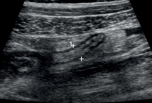
- For children with an equivocal history, examination, and labs, ultrasound is the preferred initial imaging study, as it avoids ionizing radiation and does not require contrast or sedation, although it is operator dependent.
- A meta-analysis of over 7000 patients found pooled sensitivity of 88% and specificity of 94% for sonographic diagnosis of appendicitis in children, though sensitivity fell as BMI increased (76% for BMI <25 vs. 37% for BMI >25 in one study) [1].
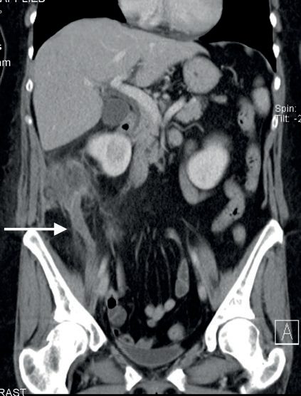
- When ultrasound is indeterminate, MRI is preferred as second-line cross-sectional imaging in children able to tolerate the study, given equivalent or better sensitivity/specificity to CT without ionizing radiation (one series reported 97% sensitivity and specificity, with median imaging time of 11 minutes).
- CT remains widely available and rapid but carries a small theoretical increase in lifetime cancer risk from radiation in childhood (estimated 0.18% for a 1-year-old and 0.11% for a 15-year-old after a CT scan; a large study found one excess leukemia and one excess brain tumor per 10,000 head CTs) [1].
- Efforts to standardize care, incorporating labs and ultrasound findings, have reduced CT utilization from 21% to 4% without changing the negative appendectomy rate [1].
- Investigations and scoring systems may help distinguish appendicitis from non-specific abdominal pain, but neither replaces regular clinical review [3].
- CT diagnostic criteria in general include an appendiceal diameter greater than 7 mm or wall thickness greater than 2 mm (a "bull's eye" appearance), fat stranding, and absence of contrast in the appendiceal lumen [4].
- Patients can have a normal WBC count despite appendicitis [4].
Schwartz's practice guideline for the uncertain child
Children under 5 often present perforated because coexisting viral syndromes and their inability to describe pain delay diagnosis; CT is reliable, ultrasound is very useful in experienced centres when the appendix is seen and excludes ovarian causes, and MRI offers high accuracy without radiation, yet diagnosis remains clinical: localised right lower quadrant tenderness with low-grade fever and leukocytosis in a boy prompts exploration, girls need ovarian or uterine pathology considered with ultrasound for cysts, torsion or tumour in menstruating girls, uncertain cases are observed, rehydrated and reassessed, and persistent pain with negative studies warrants diagnostic laparoscopy, removing even a normal-looking appendix unless another cause is definitively found and appendicectomy would add substantial morbidity [10]. Urinary infection brings less vomiting and dysuria, frequency and pressure; constipation rarely causes fever or abnormal blood tests; ovarian torsion is silent until abrupt severe pain whereas appendicitis builds gradually with nausea; and gastroenteritis produces persistent vomiting and diarrhoea before the pain [10].
Scoring and Severity
- The most widely used score is the Alvarado (MANTRELS) score, which totals 10 points: migratory right iliac fossa pain (1), anorexia (1) and nausea/vomiting (1) for symptoms; right iliac fossa tenderness (2), rebound tenderness (1) and elevated temperature (1) for signs; and leucocytosis (2) with a left shift (1) for laboratory values [1][6].
- A score of 7 or more is strongly predictive of acute appendicitis, and in patients with an equivocal score of 5 or 6, ultrasound or contrast-enhanced CT further reduces the negative appendicectomy rate [6].
- One prospective study reported that an Alvarado score of 7 or more in males, or 9 or more in females, was equivalent to CT imaging consistent with acute appendicitis, though such scores do not markedly improve diagnostic accuracy and have become increasingly marginalised as imaging has improved [1].
- Several other predictive models exist, including the pediatric appendicitis score, the appendicitis inflammatory response (AIR) score and the adult appendicitis score (AAS)
- The most recent consensus guidelines recommend the AIR or AAS score over the Alvarado score, which is useful for ruling out appendicitis but is not sufficiently specific [7].
Treatment and Management
- Treatment starts with intravenous fluids, analgesia, and broad-spectrum antibiotics.
- Early appendicitis in children is managed laparoscopically, though some mild cases may resolve with antibiotics alone [3].
- Early data suggest feasibility of nonoperative management (NOM) with intravenous antibiotics among children with acute appendicitis: in a prospective, nonrandomized cohort study of 102 children aged 7–17 years with suspected uncomplicated acute appendicitis offered a choice of NOM or appendectomy, the 1-year failure rate of NOM (rate of appendectomy) was 24%, with potential benefits of NOM including fewer disability days and lower healthcare costs at 1 year, despite a longer initial hospital stay [1].
- Laparoscopic (or open) appendectomy for uncomplicated acute appendicitis remains a safe procedure with very low complication rates, and NOM carries recurrence rates that can be as high as 35% overall (27% in the APPAC trial, exceeding a predefined acceptability threshold) [1].
- Patients with evidence of an appendicolith on imaging should not undergo NOM given its association with complicated appendicitis [1].
- Nonoperative situations where the CT scan shows a walled-off perforated appendix are managed with percutaneous drainage and interval appendectomy at a later date as long as symptoms are improving [4].
- NICE has published no guideline on appendicitis, in children or adults, and that absence is itself the first thing to know about the UK pathway.
- A search of the NICE guidance corpus returns no clinical guideline, technology appraisal or HealthTech guidance on the diagnosis or management of acute appendicitis.
- The condition appears only inside guidance on other subjects.
- UK practice is therefore set by surgical association guidance and local protocol, and the NICE documents that do bear on a child with suspected appendicitis are the ones about the unwell child and about contaminated surgery
- The traffic light system is the UK instrument for deciding how sick a febrile child is, and it applies before any diagnosis is made.
- Assess children with feverish illness for symptoms and signs predicting the risk of serious illness using the traffic light system, and direct management by the level of risk: any red feature means high risk.
- Any amber feature with no red means intermediate risk.
- Green features only means low risk [11].
- The circulation and hydration row is the one that matters most in a child with peritonitis [11]:
| Domain | Amber, intermediate risk | Red, high risk |
|---|---|---|
| Colour | Pallor reported by parent or carer | Pale, mottled, ashen or blue |
| Activity | Not responding normally to social cues, no smile, wakes only with prolonged stimulation, decreased activity | No response to social cues, appears ill to a healthcare professional, does not wake or if roused does not stay awake, weak, high-pitched or continuous cry |
| Respiratory | Nasal flaring; tachypnoea >50/min at 6 to 12 months or >40/min over 12 months; oxygen saturation ≤95% in air; crackles | Grunting; tachypnoea >60/min; moderate or severe chest indrawing |
| Circulation and hydration | Tachycardia >160/min under 12 months, >150/min at 12 to 24 months, >140/min at 2 to 5 years; capillary refill ≥3 seconds; dry mucous membranes; poor feeding in infants; reduced urine output | Reduced skin turgor |
| Other | Age 3 to 6 months with temperature ≥39°C; fever ≥5 days; rigors; swelling of a limb or joint; non-weight bearing or not using an extremity | Age under 3 months with temperature ≥38°C; non-blanching rash; bulging fontanelle; neck stiffness; status epilepticus; focal neurological signs or seizures |
Table reformats the NG143 traffic light system [11]. NICE adds that the table should be interpreted with the individual child's learning disability taken into account where one is present [11].
- On the operation itself, the applicable NICE guidance is the surgical site infection guideline, and appendicectomy sits in the categories that require prophylaxis.
- Antibiotic prophylaxis is given before clean-contaminated and contaminated surgery, and antibiotic treatment in addition to prophylaxis is given to patients having surgery on a dirty or infected wound, which is where a perforated appendix falls [12].
- For the paediatric patient specifically, NG125 singles out one closure material: consider using antimicrobial triclosan-coated sutures, especially for paediatric surgery, to reduce the risk of surgical site infection [12].
Perforation, abscess and the antibiotics-alone question in Schwartz's account
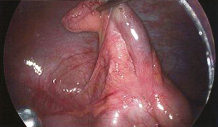
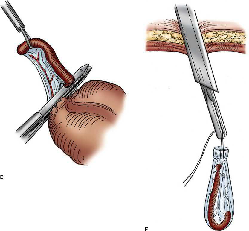
- Fluids correct the dehydration of fever and vomiting and a second-generation cephalosporin is started; laparoscopic appendicectomy through an umbilical and two lower abdominal incisions delivers the appendix through the umbilicus, an unruptured case drinks on waking and eats the next day, surgical site infection is the commonest complication, most children return to school in about a week and to full activity in 2–3 weeks [10].
- Perforation mimics gastroenteritis or presents as obstruction with a lower abdominal mass; after 4–5 days of symptoms CT with intravenous, oral and rectal contrast shows abscess, phlegmon or faecolith, generalised peritonitis, obstruction or toxicity mandates appendicectomy after resuscitation and antibiotics (laparoscopy giving a view of the pelvis and all quadrants, adhesions lysed, abscesses drained, drains seldom used, skin closed and any extraluminal faecolith retrieved), an abscess without peritonitis is drained percutaneously with antibiotics and the appendix removed 6–8 weeks later as an outpatient, phlegmon is handled similarly, children under 4–5 years localise poorly and more often need early surgery, and symptoms of no more than 4 days favour early appendicectomy before inflammation peaks [10].
- Trials show antibiotics alone treat nearly 80% of children effectively, but the failure rate is considered unacceptably high and responders cannot be predicted, so a US randomised trial of over 1500 children in eight states (NCT02800785) is comparing antibiotics with surgery [10].
Surgeries
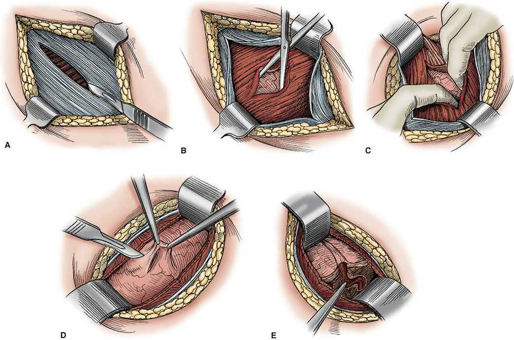
- Early appendicitis is managed laparoscopically in children [3].
- If, at operation for suspected appendicitis, no evidence of appendicitis is found, a thorough exploration of the peritoneum must be performed to identify an alternative cause [13].
- If presumed appendicitis instead reveals a ruptured ovarian cyst, thrombosed ovarian vein, or regional enteritis not involving the cecum, appendectomy should still be performed to prevent future diagnostic confusion [4].
Complications
- Perforation is disproportionately common in young children: up to two-thirds of infants aged 0–4 with appendicitis present with perforation, attributed to delayed presentation from the difficulty of obtaining an accurate history in this age group [1].
- Children and elderly patients have a higher propensity to rupture secondary to delayed diagnosis [4].
- A perforated patient is generally more ill and can show evidence of sepsis [4].
Appendix mass and appendix abscess
- Besides perforation, the local complications of appendicitis are a right iliac fossa appendix mass (appendicitis with densely adherent caecum, omentum and ileum), a right iliac fossa abscess (usually secondary to perforated retrocaecal appendicitis) and a pelvic abscess (usually secondary to perforated pelvic appendicitis) [8].
- CT is the best investigation for a suspected appendix mass or abscess [8].
- Where a mass is present and the patient's condition is satisfactory, the standard treatment is the conservative Ochsner–Sherren regime, on the premise that the inflammatory process is already localised and that operating on it is difficult and potentially dangerous, since the appendix may be impossible to find and a faecal fistula may form [6].
- The limits of the mass are marked on the abdominal wall with a skin pencil, temperature and pulse are recorded 4-hourly, a fluid balance record is kept, antibiotics are given and any abscess is drained radiologically [6][8].
- Conservative treatment is stopped (and early laparotomy indicated) for a rising pulse rate, increasing or spreading abdominal pain, or an increasing size of the mass [6].
- Clinical improvement is usually evident within 24–48 hours and about 90% of cases resolve without incident
- Failure of the mass to resolve should raise suspicion of a carcinoma or Crohn's disease [6].
- When symptoms settle without removing the appendix, delayed elective (interval) appendicectomy after 3 months is recommended because the recurrence rate is high [8].
References
- Maingot's Abdominal Operations, 13th ed., Ch. 41 Appendix and Small Bowel Diverticula, Table 41-1
- Sabiston Textbook of Surgery, 22nd ed., Ch. 85 The Acute Abdomen, Table 85.2
- Bailey & Love's Short Practice of Surgery, 28th ed., Ch. 17 Paediatric surgery
- The ABSITE Review, 2022, Ch. 35 Small Bowel
- Browse's Introduction to the Symptoms and Signs of Surgical Disease, 6th ed., Ch. 15 Acute lower abdominal pain
- Bailey & Love's Short Practice of Surgery, 28th ed., Ch. 76 The vermiform appendix, Summary box 76.4
- Sabiston Textbook of Surgery, 22nd ed., Ch. 94 The Appendix
- Oxford Handbook of Clinical Surgery, 5th ed., Ch. 26 Emergency surgery topics
- Bailey & Love's Short Practice of Surgery, 28th ed., Ch. 8
- Schwartz's Principles of Surgery, 11th ed., Ch. 39, Pediatric Surgery
- NICE Guideline NG143: Fever in under 5s: assessment and initial management. National Institute for Health and Care Excellence, London, UK, 2019, updated 2021., 1.2.3; 1.2.4; Table 2 www.nice.org.uk
- NICE Guideline NG125: Surgical site infections: prevention and treatment. National Institute for Health and Care Excellence, London, UK, 2019, updated 2020., 1.2.12; 1.2.17; 1.3.20 www.nice.org.uk
- Schwartz's Principles of Surgery: ABSITE and Board Review, Ch. 30 Appendix
- Maingot's Abdominal Operations, 13th ed., Ch. 10