



Laparoscopic Principles
Summary
- Laparoscopic (minimal access) surgery uses a rigid endoscope introduced through ports into a gas-distended peritoneal cavity to perform surgical therapeutic goals with minimal somatic and psychological trauma compared with open surgery [1].
- It relies on pneumoperitoneum, created most commonly with carbon dioxide, to provide working space and visualisation, and on either a closed (Veress needle) or open (Hasson) technique for safe abdominal access [2].
- Since the first video-laparoscopic cholecystectomy performed by Mühe in 1985 and popularised by Mouret in 1987, laparoscopy has spread to most general surgical procedures and is now a required competency in surgical residency training [2].
Definition
Minimal access surgery is "a product of modern technology and surgical innovation that aims to accomplish surgical therapeutic goals with minimal somatic and psychological trauma," offering reduced wound access trauma, shorter operating and hospital stays, and faster recuperation compared with conventional open techniques [1]. Laparoscopy specifically involves introducing a rigid endoscope through a port into the peritoneal cavity, distended by pneumoperitoneum, to visualise and manipulate intra-abdominal structures [1][2].
Origins of the component technologies in Schwartz's account
Kelling performed primitive laparoscopy in 1901 by placing a cystoscope in an insufflated abdomen lit by dangerous hot elements; Hopkins' rod lens in the late 1950s transmitted light through solid quartz without heat, quartz fibres opened fibre-optics and flexible endoscopy, and compact charge-coupled devices mounted on endoscopes drove the explosion of video surgery; the first endoscopic operation was Shinya and Wolfe's colonoscopic polypectomy, and Gauderer and Ponsky's percutaneous endoscopic gastrostomy of 1981 may have been the first NOTES procedure, with the German NOTES registry of 551 women showing conversion and complication rates for transvaginal cholecystectomy and appendicectomy comparable to laparoscopy [3]. Balloon angioplasty, stents, TIPS and aortic stent grafts extended the minimal-access philosophy through fluoroscopy, CT guides drainage, biopsy and radiofrequency ablation, and MRI-guided surgery is limited by slow image refresh, the need for non-metallic instruments and bulky magnets, though open magnets allow frameless stereotaxy [3].
Indications
The case for a minimal access approach rests on a consistent set of advantages: a smaller wound, with reduced wound infection, dehiscence, bleeding, herniation and nerve entrapment; less wound pain and improved mobility; reduced wound trauma and heat loss; and improved visualisation of the operative field [1].
Elective to emergency: minimal access methods were established first in elective surgery, but those advantages have driven increasing uptake in emergency operating, including perforated viscus repair such as omental patch repair of a perforated peptic ulcer, lavage of a localised perforation of diverticular disease, intrathoracic debridement of empyema, and surgery for pneumothorax and haemothorax [1].
Trauma: more recently some experienced surgeons have extended minimal access approaches to trauma, for initial assessment and treatment in haemodynamically stable patients [1].
Physiological effects
- Pneumoperitoneum is achieved by insufflating carbon dioxide into the peritoneal cavity.
- CO2 is preferred because it dissolves rapidly in blood, is nonflammable, and carries a low risk of gas embolism, with intra-abdominal pressure maintained at 10-15 mm Hg (normal range) via an insufflator [2][4].
- Cardiopulmonary dysfunction can occur once intra-abdominal pressure exceeds 20 mm Hg [4].
- Increased intra-abdominal pressure raises mean arterial pressure, pulmonary artery pressure, heart rate, systemic vascular resistance, central venous pressure, mean airway pressure, peak inspiratory pressure, and end-tidal CO2, while decreasing pH, venous return (from IVC compression), cardiac output, and renal blood flow secondary to decreased cardiac output.
- Hypovolaemia lowers the pressure threshold at which compromise occurs, and PEEP worsens these effects [4].
- CO2 also causes some decrease in myocardial contractility [4].
- In the hypovolaemic patient, excessive IVC pressure combined with reverse Trendelenburg positioning and loss of lower-extremity muscle tone can decrease venous return and cardiac output, an effect not seen in the normovolaemic patient [5].
- The most common arrhythmia during laparoscopy is bradycardia, caused by a vagovagal response to rapid stretch of the peritoneal membrane, occasionally with hypotension [5].
- Increased intra-abdominal pressure decreases renal blood flow, glomerular filtration rate, and urine output via direct pressure on the kidney and renal vein, secondarily increasing plasma renin release and sodium retention.
- Hormonally mediated changes (elevated ADH) can suppress urine output for up to an hour after the procedure ends, so intraoperative oliguria during an uncomplicated case should not by itself prompt extra fluid administration [5].
- Because insensible fluid losses through an open abdomen are eliminated with laparoscopy, intraoperative fluid needs are generally lower than for an equivalent open operation [5].
- Laparoscopy is associated with an increased risk of venous thromboembolism from pneumoperitoneum-induced IVC/iliac vein compression combined with prolonged operative time and reverse Trendelenburg positioning [2][6].
The pneumoperitoneum in Schwartz's detail
- Air pneumoperitoneum via a sphygmomanometer bulb was abandoned because nitrogen is poorly soluble and slowly absorbed; nitrous oxide is inert, rapidly absorbed, gives better analgesia under local anaesthesia, does not support combustion in controlled trials and lowers end-tidal CO₂ and minute ventilation, but its effects on tumour biology, port-site metastasis and pregnancy are unknown [3].
- CO₂ absorbed across the peritoneum forms carbonic acid; body buffers, chiefly bone, absorb up to 120 L before respiratory acidosis develops, after which the ventilator must clear it, but rates above 20 breaths/min exchange gas less efficiently and larger tidal volumes risk barotrauma and disturb the upper abdominal field, so pressure is reduced or the abdomen desufflated to let the anaesthetist catch up; severe acidosis causes arrhythmia, and hypercarbia raises heart rate, systemic resistance, blood pressure and myocardial oxygen demand [3].
- Bradycardia from vagovagal peritoneal stretch is the commonest arrhythmia, treated by desufflation, atropine and volume; caval compression in reverse Trendelenburg with hypovolaemia reduces venous return and cardiac output (not in the normovolaemic), promotes lower-limb thrombosis and pulmonary embolism when prophylaxis is omitted in long procedures though short operations may not warrant extensive prophylaxis; pressure transmitted across the paralysed diaphragm raises central venous and cardiac filling pressures and peak inspiratory pressure, cardiac output being maintained below 20 mmHg, and bleb rupture is rare except in oesophageal surgery [3].
- Renal blood flow, GFR and urine output fall from direct renal and renal vein compression, renin raises sodium retention and ADH raises free-water reabsorption for up to an hour afterwards, so intraoperative oliguria does not reflect volume and fluid should not chase urine output; insensible loss is eliminated, so fluid need only match limb pooling, third-space loss and (lesser) blood loss [3].
- Helium, neon and argon avoid metabolic effects but are poorly soluble and embolise; gas embolism is suspected with hypotension on insufflation, diagnosed by a "mill wheel" murmur on oesophageal stethoscope, and treated by left lateral head-down positioning to trap gas in the right ventricular apex and aspiration through a central line; abdominal lift devices through a 10–12 mm umbilical port avoid these effects but give inferior exposure, displace bowel into the field and cause more pain [3].
- Cortisol is often higher after laparoscopic than open surgery but stress hormones and cytokines normalise faster and immune suppression is less; a CO₂ pneumothorax during transhiatal dissection is managed by enlarging the pleural defect, placing an 18 F red rubber catheter with side holes across it and evacuating to a water seal at closure with pressure below 8 mmHg, keeping a formal chest tube after oesophagectomy because fluid siphons through the defect [3].
- Thoracoscopy needs no positive pressure within the bony thorax (which would reduce venous return, shift the mediastinum and demand sealed ports) but requires a double-lumen tube and ipsilateral lung collapse, allowing standard instruments through extended ports; extraperitoneal spaces (Retzius for hernia, retroperitoneum for nephrectomy and necrosectomy) are developed by balloon then held at about 10 mmHg, higher pressures forcing CO₂ into soft tissue to cause emphysema and acidosis, and they eliminate bowel injury, adhesion, port herniation and ileus [3].
- Anaesthesia favours short-acting agents, non-narcotic analgesia such as ketorolac where haemostasis allows, and liberal ondansetron and steroids because nausea, pain and urinary retention drive admission [3].
Patient selection
Preoperative assessment focuses on patient factors that affect tolerance of pneumoperitoneum and positioning: cardiopulmonary reserve, history of venous thromboembolism, prior abdominal surgery (adhesions), umbilical abnormalities, ascites, and range of motion for positioning [2].
Pregnancy, cancer, the elderly and cirrhosis in Schwartz's account
- In pregnancy the fundus reaches the umbilicus at 20 weeks, so most surgeons use Hasson entry, tilt the patient left to decompress the cava, apply compression devices for the thrombotic risk, avoid maternal respiratory acidosis because fetal pH tracks maternal pH linearly, accept 15 mmHg since mid-pregnancy contractions exceed it, shield the fetus from X-rays, operate in the second trimester, and may convert if fetal heart-rate decelerations reversibly follow insufflation; more than 100 laparoscopic cholecystectomies in pregnancy have been reported with uniformly good results [3].
- Cancer surgery follows open principles (R0 resection with an adequate lymphadenectomy, generally 10–15 nodes) with staging laparoscopy for liver assessment before pancreatic, gastric or hepatic resection and palliative bypass when curative resection is precluded; of the three major GI resections only oesophagectomy is routinely laparoscopic in many centres, distal pancreatectomy is common, laparoscopic-assisted gastrectomy is popular in Japan for early gastric cancer, and segmental colectomy is the commonest laparoscopic cancer operation, validated in a multicentre randomised trial [3].
- The elderly convert more often because of chronic disease and are harder to manage intraoperatively but gain most from early mobilisation; cirrhotic patients risk haemorrhage at trocar sites and in dilated venous fields, ascitic leak with bacterial peritonitis (so every port is closed watertight), and hepatic failure or hepatorenal syndrome, severity being judged by MELD or Child's class, portal hypertension being a relative contraindication until decompressed (paracentesis or TIPS with diuresis before repairing an incarcerated umbilical hernia), with reduced insufflation pressure and minimal sodium-sparing fluid [3].
- Savings accrue when hospital stay shortens (cholecystectomy, fundoplication, splenectomy, adrenalectomy) but not for already-ambulatory hernia repair or colectomy still needing 4–7 days [3].
Intraoperative confirmation
Correct intraperitoneal placement of the Veress needle is confirmed intraoperatively by the hanging-drop method, free flow of saline with no aspirate of blood/bowel content, and an opening pressure below 10 mmHg (Bailey & Love) or below 5-6 mmHg (Maingot's) [6][7].
Skills assessment
- There is no severity score for a technique, but there are validated instruments for the operator, and laparoscopy has the most heavily studied of them.
- The Objective Structured Assessment of Technical Skills (OSATS) is a seven-item global rating scale, each domain scored on its own 5-point scale and summed.
- Introduced in 1997, it has been the focus of over 440 studies, used whole or modified, and scored either by live direct observation or asynchronously from digital recordings [8].
- For laparoscopy specifically, the Global Operative Assessment of Laparoscopic Skills (GOALS) measures five domains, depth perception, bimanual dexterity, efficiency, tissue handling and autonomy, each on a 5-point scale, and has more than 100 referenced validation studies across a broad range of laparoscopic procedural skills, with demonstrated relevance in the operating room and not only the simulator [8].
- The robotic instrument, GEARS, was built by taking these same five GOALS domains and adding a sixth for robot control [8].
- Competence is certified separately from these rating scales.
- Assessment of laparoscopic skills is conducted through the Fundamentals of Laparoscopic Surgery (FLS) examination, a requirement for graduates of US residency programmes and implemented worldwide, complemented by dedicated simulation centres using high-fidelity models, box trainers and augmented or virtual reality simulators [2].
- Laparoscopy, initially performed only by attending surgeons, is now introduced early in residency [2].
Simulation mandates and the ethics of new procedures in Schwartz's account
Box trainers appeared in the 1990s, virtual reality platforms measure psychomotor skill objectively and improve operative performance in randomised trials, skills laboratories are mandatory for residency accreditation, FLS became prerequisite to the American Board of Surgery qualifying examination in 2010 and FES in 2015, a Fundamentals of Robotic Surgery examination is anticipated, and telementoring links remote surgeons by two-way audiovisual communication [3]. Schwartz sets out a drug-development-like path for innovation: identify an unsolved problem, search other disciplines for technology, test in the appropriate animal model until the procedure is reproducible, effective and free of serious side effects, secure the medical board, chief of staff and institutional review board, obtain brutally honest documented consent (with psychological evaluation where benefit is small and risk unknown), keep an experienced team together through the 10–50 cases until operative times converge (phase I), test efficacy non-randomised at several centres (phase II), then randomise against the old procedure (phase III), learners seeking expert assistance rather than presuming equal skill, and every surgeon asking whether the procedure is safe, whether they would undergo it, whether it equals or betters its predecessor and whether they can perform it as well as the experts [3].
Conduct of the procedure
Patient selection and contraindications: Patients with massive abdominal distension precluding safe insufflation, or unstable haemodynamics unable to tolerate pneumoperitoneum, should not undergo laparoscopy; patients for whom a possible laparotomy would pose prohibitive risk should not be offered any surgical approach, including laparoscopy, given the possibility of conversion [2].
Equipment: A functioning laparoscopic "stack" is required, comprising monitor, light source, insufflator, camera, diathermy, and irrigation/aspiration system [2][9]. Laparoscopes are available in 5 and 10 mm diameters with 0°, 30°, 45°, and 50° viewing angles [2].
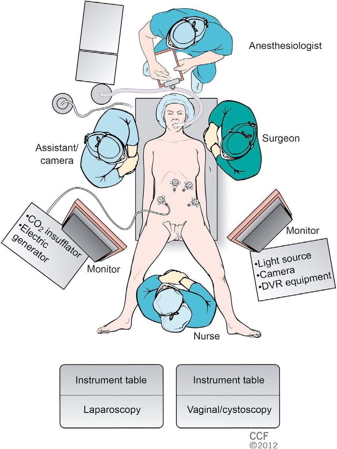
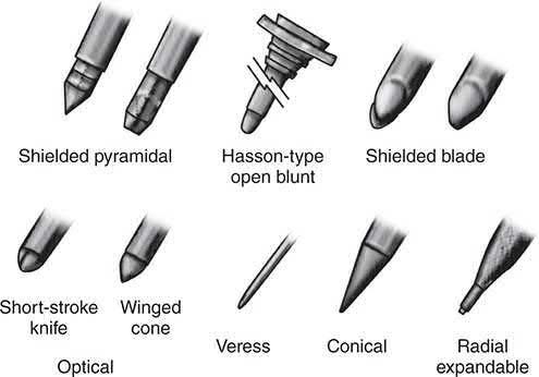
- Access techniques: There are two fundamental ways to access the abdomen laparoscopically: the open (Hasson's or modified Hasson's) technique and the closed technique (Veress needle and/or visual entry trocar); complication rates do not differ significantly between them, so the surgeon's preferred technique should be used, though blind trocar insertion without a Veress needle or visual control is avoided [7].
- In the Hasson (open) technique, a 1-2 cm skin incision is made at the umbilicus, subcutaneous tissue and fascia are divided under direct vision, the peritoneum is grasped and opened, and a blunt trocar is secured with fascial stay sutures before insufflation to 15 mmHg [2][6].
- In the Veress needle (closed) technique, a spring-loaded needle is inserted (commonly at the umbilicus or Palmer's point, 3 cm below the left costal margin in the mid-clavicular line).
- Correct placement is confirmed by the hanging-drop test, free saline flow without aspirate, and an opening pressure <10 mmHg, after which CO2 insufflation proceeds at low flow (1-4 L/min) to a maximum pressure of 15 mmHg [7].
- The Veress needle's most catastrophic complication is major vessel injury, so its trajectory must avoid the aorta and iliac vessels [6].
- Direct trocar insertion without prior pneumoperitoneum and optical trocar entry (a 0° scope inside a clear trocar visualising each abdominal wall layer during insertion) are alternative techniques used by some surgeons [6].
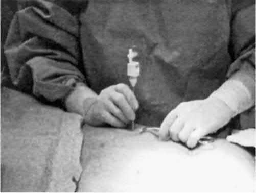
- Port placement: Secondary trocars should always be inserted under direct vision, perpendicular to the abdominal wall, using a two-handed or controlled technique to avoid sudden "plunging" [7].
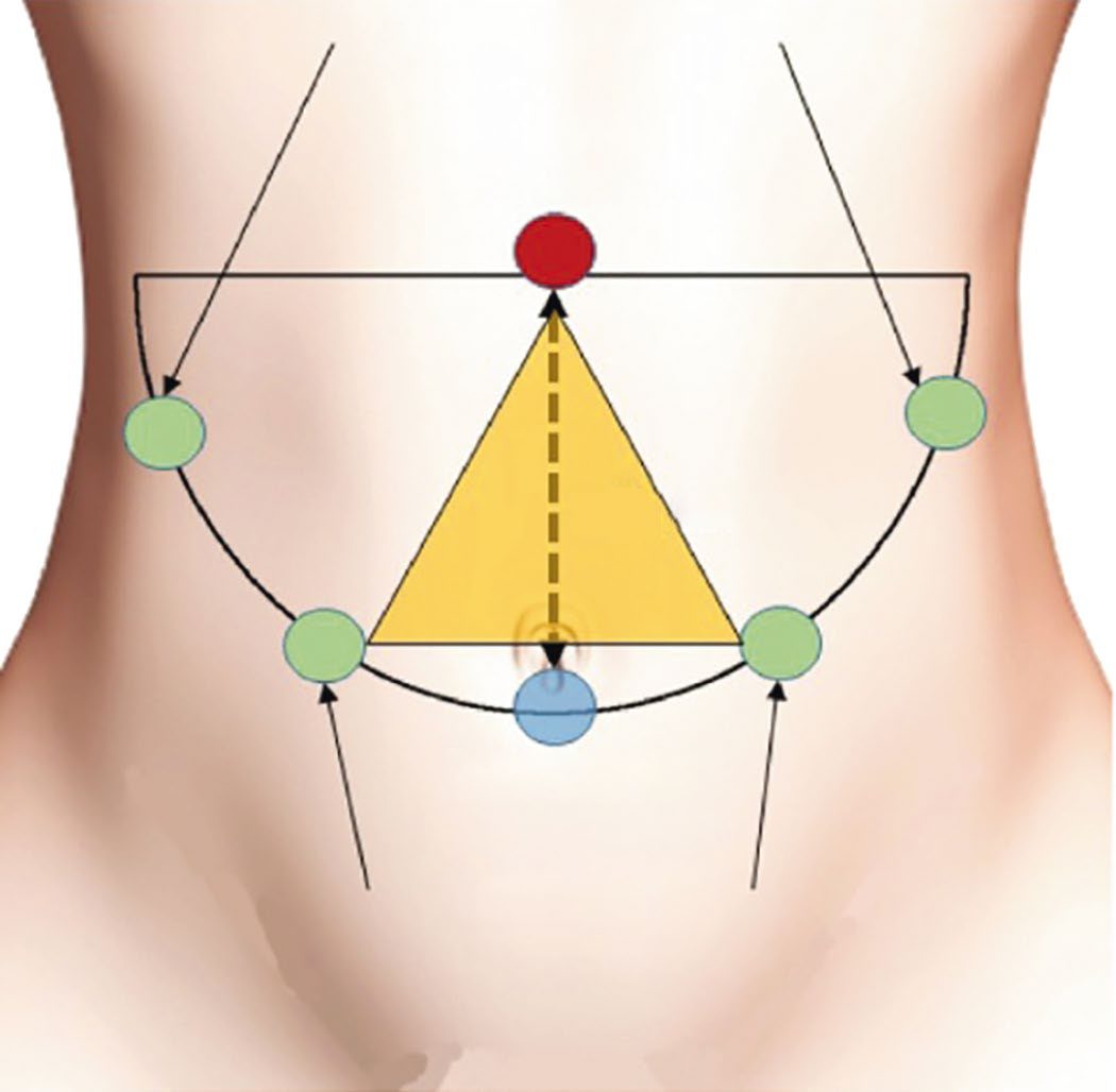
- Ports should be spaced to avoid the "crossing/sword-fighting" effect, ideally forming an equilateral triangle or "diamond of success" around the target, with an angle of 60-90 degrees between working instruments and the fulcrum roughly 15 cm from the target for standard 30 cm instruments [2][6].
- Smaller-diameter trocars reduce postoperative pain, port-site hernia, and improve cosmesis.
- All fascial defects from 10 mm or larger bladed trocars should be closed [6][7].
Positioning: Trendelenburg position (head down) improves access to lower abdominal/pelvic organs; reverse Trendelenburg improves access to the upper GI tract and biliary tree [2]. Sequential compression devices are used given the VTE risk from pneumoperitoneum and positioning [2].
- Dissection and tissue handling: Laparoscopic dissection is typically two-handed (an assisting grasper providing countertraction, an active dissecting tool that may be nonenergised or energised) and falls into blunt, sharp, or electrosurgical categories, the last carrying the highest injury risk if the electrode tip is not kept visible [2].
- A short-duration, high-voltage coagulation current causes rapid tissue heating and carries the highest thermal injury risk, whereas lower-voltage, higher-wattage cutting current causes less thermal injury.
- Bipolar electrosurgery confines current between two adjacent electrodes and provides the safest small-vessel coagulation [5].
- Additional energy devices include the harmonic scalpel (ultrasonic, disrupts protein hydrogen bonds to coagulate, cost-effective for medium vessels) and the argon beam coagulator (non-contact, smokeless, good for hepatic/splenic haemostasis) [4].
Laparoscopy in pregnancy: Laparoscopic surgery is considered safe at any stage of pregnancy; recommended precautions include obstetric involvement, left lateral decubitus positioning to avoid caval compression, sequential compression devices, entry remote from the gravid uterus (open Hasson technique preferred by many organisations), and maintaining CO2 pressures up to 15 mmHg [2][5].
There is no NICE guideline on laparoscopic surgery as a discipline. The UK position is assembled from procedure-specific appraisals, and the oldest of them still carries the governance rule. TA105 recommends laparoscopic, including laparoscopically assisted, resection as an alternative to open resection for people with colorectal cancer in whom both laparoscopic and open surgery are considered suitable [10]. It is the wording of the two accompanying recommendations that matters more than the first.
- On the surgeon, laparoscopic colorectal surgery should be performed only by surgeons who have completed appropriate training in the technique and who perform the procedure often enough to maintain competence
- The exact criteria are to be determined by the relevant national professional bodies, and cancer networks and constituent trusts must ensure local practice meets those criteria as part of their clinical governance arrangements [10].
- NICE thus delegates the volume threshold to the specialty associations but makes compliance a trust governance obligation.
On the decision, the choice between open and laparoscopic must be made after informed discussion between the patient and the surgeon, considering the suitability of the lesion for laparoscopic resection, the risks and benefits of the two procedures, and (stated explicitly) the experience of the surgeon in both procedures [10]. The surgeon's own experience is a factor the patient is entitled to weigh.
TA105 replaced TA17 and was last reviewed in December 2011, with nothing new found that affected the recommendations. Its content is now also carried within the NICE colorectal cancer guideline [10]. For gallstone disease, the corresponding UK position sits in CG188 [11].
- Laparoscopy in trauma occupies an awkward middle ground, and the reason is worth stating precisely.
- Traditionally, haemodynamically stable trauma patients were managed with serial abdominal examination and paracentesis.
- Rapid helical CT and bedside ultrasound have since displaced laparoscopy as the primary assessment tools [2].
- But laparoscopy surpasses both CT and ultrasound in detecting penetrating thoracoabdominal trauma and injuries to the diaphragm, hollow organs and the retroperitoneum, and studies report fewer undetected injuries, shorter stays and faster recovery, with limited cost data [2].
- The constraints are unchanged: it is not recommended in haemodynamic instability, preparation and positioning should mirror a trauma laparotomy so conversion can be immediate, open instruments must be available, and the abdomen must be assessed systematically to avoid missed injuries [2].
- A history of previous abdominopelvic surgery is assessed before the operation, not discovered during it.
- Adhesive disease can make laparoscopy in a previously operated abdomen either straightforward or impossible, so the preoperative history should establish which operation was performed, since that indicates where adhesions will lie, and whether it was emergent or complicated by bleeding [2].
- Prior incisions are inspected for hernia, which may need repair, and new incisions placed as far as possible from previous operative sites [2].
- During adhesiolysis, combine blunt and sharp dissection with traction and countertraction to define the boundaries of adhesions, use an angled-lens laparoscope, and use electrosurgical tools cautiously [2]. Extensive adhesiolysis remote from the field of interest should be avoided, because operating in a reoperative field increases the risk of bleeding and visceral injury.
- Visceral injuries usually follow excessive traction or tissue misidentification [2].
- Enhanced recovery is the other half of the benefit, and it predates laparoscopy's dominance.
- The "fast-track surgery" concept was introduced in 1997 by the Danish colorectal surgeon Henrik Kehlet for patients undergoing open sigmoid colectomy, demonstrating that reducing surgical stress and modifying conventional practice hastened recovery and shortened stay.
- European surgeons formed the Enhanced Recovery After Surgery study group in the early 2000s [2].
- The approach combines preoperative counselling, avoiding mechanical bowel preparation, minimising preoperative fasting, balanced fluid therapy, laparoscopy and short-acting anaesthetic agents, adequate analgesia, and early oral feeding and mobilisation [2].
- Combined with laparoscopy, enhanced recovery protocols deliver opioid-sparing analgesia and optimised fluid therapy, and a meta-analysis of randomised trials comparing laparoscopic with open colorectal surgery under such protocols showed reduced length of stay, complications and pain, attributed to a decreased surgical stress response [2].
- On cost, the honest position is that the up-front premium may be recovered rather than avoided.
- Laparoscopy carries high up-front costs from specialised instruments and longer operative times, but savings from shorter stays, fewer readmissions and fewer complications may balance them.
- A cost-effectiveness study in rectal cancer found no significant long-term societal cost difference between laparoscopic and open surgery despite the higher immediate costs, and laparoscopic colectomy has been associated with decreased healthcare costs and usage in both the immediate and long-term postoperative periods [2].
Browse's 6e is a text on the symptoms and signs of surgical disease and does not cover laparoscopic technique; it contributes nothing to this topic beyond the clinical assessment of the abdomen described elsewhere.
Set-up, access, ports and imaging in Schwartz's account
- The monitor sits across the table with the patient and operative field between surgeon and screen, at the feet for pelvic work, at 10 o'clock for cholecystectomy with the surgeon at 4 o'clock on the patient's left, with insufflator and monitoring visible; the surgeon stands between the legs on Allen stirrups or leg boards for the hiatus and left lobe, lateral decubitus with table flexion suits nephrectomy and adrenalectomy, 45° tilt suits splenectomy, and sequential compression devices are used whenever knees are bent or reverse Trendelenburg lasts more than minutes [3].
- Seldinger guidewire access serves vessels, PEG, transhepatic biliary and upper urinary tract entry; the Veress needle, held at its serrated collar with the wall lifted by towel clips, gives two distinct pops through fascia and peritoneum at the umbilicus where the wall is thin even in the obese, pressure and flow readings confirm placement, CO₂ is limited to 14–15 mmHg (under local anaesthesia N₂O is stopped at 2 L or 10 mmHg), the first trocar passes over a radially dilating sheath or as an optical viewing trocar with a vented stylet, shield or dilating tip and always away from the sacral promontory and great vessels, and the Hasson technique, Kocher clamps on fascia, Mayo scissors, a finger sweep for adherent bowel and stay sutures to the winged trocar, is preferred after previous surgery and in pregnancy, after which the telescope is passed through a secondary port to inspect the entry site [3].
- Ports are removed under vision; bleeding stops with 3–5 minutes of instrument pressure or Foley balloon tamponade, or a full-thickness wall suture; 5 mm sites need no closure and 10 mm sites off the midline, through dilating sheaths or above the transverse mesocolon usually need none, but dilated extraction sites and lower abdominal ports of 10 mm or more are closed, with crochet-type suture passers in the obese, to prevent incarcerated hernia [3].
- Working trocars sit at least 10 cm apart with the telescope between and behind them forming an equilateral triangle of 10–15 cm sides, the target at the apex of a second triangle to make a "baseball diamond" with the telescope at home plate and the target at second base, the table adjusted so the surgeon's elbows rest at the sides bent 90°; robotic primary ports are 12 mm for the stereo scope with 8 mm working ports; hand-assisted devices preserve pneumoperitoneum for solid-organ and colon surgery and ease the learning curve; and SILS through a 1–3 cm umbilical incision with separate low-profile or multilumen trocars crowds the ports, loses triangulation and forces crossed hands, needing articulating, curved or varied-length instruments and a deflectable-tip scope [3].
- Rigid telescopes are usually 30 cm and 2–12 mm (doubling rod diameter quadruples illumination, so 10 mm is needed in a bloody abdomen) with a 70° field viewed straight (0°) or obliquely (30°, 45°), light cables lose over 90% of a 300 W source, HD chips raise resolution from 480 to 1080 lines and need HD monitors (standard monitors manage about 700 lines), priorities are illumination, then resolution, then colour, flickering two-image 3-D systems tire the eye whereas the da Vinci's twin rod-lens bundles give true 3-D, and heads-up displays have not caught on [3].
- Radiofrequency current at 500,000 Hz coagulates at 60 °C, vaporises and desiccates at 100 °C and carbonises above 200 °C; monopolar coagulation current is short high-voltage and cutting current lower-voltage higher-wattage with least thermal spread, bipolar coapts small vessels without collateral injury and combines with compression and a blade in vessel-sealing devices, capacitive coupling through a plastic trocar bleeds current from a metal sleeve or laparoscope into viscera causing delayed faecal fistula and direct coupling passes current from electrode to scope, so the whole uninsulated electrode must stay in view; argon beam coagulation suits raw liver or spleen but raises intra-abdominal pressure and embolism risk so ports are vented; the CO₂ laser (10.6 μm) cuts superficially via mirrors, the Nd:YAG (1064 nm) penetrates deeply for bulky rectosigmoid, bronchial or oesophageal tumours at the risk of perforation, the KTP (532 nm) coagulates red vascular lesions at intermediate depth, heater probes at 60–100 °C and stents have largely replaced GI lasers, photodynamic therapy activates porfimer sodium 2 days after injection to palliate obstructing cancer, pulsed-dye lasers and cheaper electrohydraulic lithotriptors fragment stones by shock wave, and ultrasonic shears weld and divide medium vessels with minimal collateral damage [3].
- Standard instruments are 5 mm and 30 cm (scissors 3–5 mm and 20–45 cm), concentrating force on a small tip so perforation risk rises, and the monopolar hook with suction–irrigation tents and divides tissue; paediatric instruments are 15–20 cm and often 3 mm with a 5 mm telescope, 8 mmHg suffices in infants and DVT prophylaxis is unnecessary [3].
Procedures and access variants
- Laparoscopic cholecystectomy was the index procedure that launched the laparoscopic revolution, first performed by Erich Mühe in 1985 and popularised globally after Philippe Mouret's 1987 operation, after which open cholecystectomy rates fell steeply within a decade [2].
- Other procedures rapidly converted to a laparoscopic approach, including inguinal hernia repair, gastrectomy, appendectomy, and colectomy [2].
- Related access variants include single-incision laparoscopic surgery (SILS), performed through one umbilical port with a multichannel device, and hand-assisted laparoscopic surgery (HALS), in which the surgeon's hand is introduced through a mini-laparotomy while pneumoperitoneum is maintained, allowing palpation, blunt dissection, and manual haemorrhage control [1].
- Specimen extraction after laparoscopic resection may use retrieval bags (protecting the wound and, for benign disease, permitting morcellation) or an enlarged extraction incision.
- Morcellation is avoided in oncologic surgery because it disrupts margins and risks tumour cell spillage [1][2].
The article's point that pneumoperitoneum and steep positioning raise venous thromboembolism risk has a UK counterpart that says what to actually do about it, and it applies to every abdominal operation whatever the access. NICE treats laparoscopic and open abdominal surgery alike here: offer VTE prophylaxis to people undergoing abdominal (gastrointestinal, gynaecological or urological) surgery who are at increased risk of VTE [12].
- Mechanical prophylaxis starts on admission, not at knife-to-skin.
- Choose either anti-embolism stockings or intermittent pneumatic compression, and continue until the person no longer has significantly reduced mobility relative to their normal or anticipated mobility [12].
- That end point is a mobility judgement rather than a fixed number of days, which is the part most often got wrong on a ward round.
Pharmacological prophylaxis is added, not substituted, and has a floor of seven days. Add LMWH or fondaparinux sodium for a minimum of 7 days where the risk of VTE outweighs the risk of bleeding, judged on individual patient factors [12]. After major cancer surgery in the abdomen, consider extending pharmacological prophylaxis to 28 days postoperatively [12], the single figure most worth carrying from this guideline, because it outlasts the admission.
Bariatric surgery is handled separately but identically in structure: offer prophylaxis, start mechanical prophylaxis on admission with stockings or intermittent pneumatic compression continued until mobility recovers, and add pharmacological prophylaxis for a minimum of 7 days where VTE risk outweighs bleeding risk [12].
Extraperitoneal insufflation
Insufflation of an extraperitoneal plane (the approach used for totally extraperitoneal hernia repair and for retroperitoneal access) creates the space first and then insufflates at low pressure. There are fewer physiological consequences than with a pneumoperitoneum, but the CO2 can spread widely through the soft tissues and cause subcutaneous emphysema [13].
Complications
- Complications specific to establishing pneumoperitoneum include injury to intra-abdominal structures, port-site hernia, wound infection, haemorrhage, and rarely CO2 embolism.
- Shoulder-tip pain is a common minor postoperative effect from residual subdiaphragmatic gas [9].
- Reported rates: port-site infection <5%, port-site hernia <2%, visceral injury <1% [9]; a separate source cites port-site hernia rates of 0.65-2.80% of laparoscopic GI operations [6] and vascular or bowel injury with Veress needle or trocar insertion at approximately 0.1% [4].
- CO2 embolism presents as a sudden transient rise in end-tidal CO2 followed by a drop and hypotension.
- Treatment is placing the patient head-down and turned to the left, attempting to aspirate CO2 through a central line, and prolonged CPR if needed [4].
- Bradycardia/hypotension from peritoneal stretch (vagovagal response) is managed by desufflation, vagolytic agents (e.g., atropine), and volume replacement [5].
- CO2 pneumothorax can occur during transhiatal dissection (e.g., laparoscopic Nissen fundoplication or Heller myotomy) from mediastinal pleural injury.
- Initial management is placement of a red rubber catheter across the defect rather than aborting the case, with evacuation via water seal at the end of the procedure [5].
- General limitations of minimal access surgery that predispose to complications include lack of 3D vision and loss of tactile feedback, increased operative time, difficulty achieving haemostasis, difficulty with large specimen extraction, a steep learning curve, and cost [1].
Surgical smoke as an occupational hazard
- Viral particles, bacteria, respiratory and ophthalmic irritants and carcinogens have all been identified in the surgical smoke produced by diathermy devices [7].
- Universal precautions, smoke evacuation systems or simple suction devices can be used to minimise the risk to theatre personnel [7].
- The hazard is a reason, independent of visibility, to clear the plume: an instrument that provides a clean, smoke-free field also facilitates dissection, improves haemostasis and reduces operating times [1].
Outcomes
- Laparoscopic (versus open) surgery is associated with reduced postoperative pain, quicker recovery, improved cosmesis, and shorter hospital stay, contributing to reduced wound infection, dehiscence, bleeding, herniation, and nerve entrapment, decreased wound trauma and heat loss, and improved visualisation (magnification) of anatomy [1][7].
- Reduced serosal handling with minimally invasive approaches decreases postsurgical adhesion formation [1].
- Quality of life appears improved by a laparoscopic approach compared with open surgery for equivalent procedures [2].
- Conversion to an open operation, when required, does not represent a complication but rather sound surgical judgement in favour of patient safety [1].
References
- Bailey & Love's Short Practice of Surgery, 28th ed., Ch. 10, Summary box 10.1
- Sabiston Textbook of Surgery, 22nd ed., Ch. 11
- Schwartz's Principles of Surgery, 11th ed., Ch. 14, Minimally Invasive Surgery, Robotics, NOTES, and SILS
- The ABSITE Review, 2022, Ch. 38
- Schwartz's Principles of Surgery: ABSITE and Board Review, Ch. 14
- Maingot's Abdominal Operations, 13th ed., Ch. 6
- Bailey & Love's Short Practice of Surgery, 28th ed., Ch. 7 Basic surgical skills
- Sabiston Textbook of Surgery, 22nd ed., Ch. 2
- Oxford Handbook of Clinical Surgery, 5th ed., Ch. 21
- NICE Technology Appraisal TA105: Laparoscopic surgery for colorectal cancer (2006, last reviewed December 2011; replaces TA17), 1.1; 1.2; 1.3; Overview www.nice.org.uk
- NICE Clinical Guideline CG188: Gallstone disease: diagnosis and management (2014), Recommendations www.nice.org.uk
- NICE Guideline NG89: Venous thromboembolism in over 16s — reducing the risk of hospital-acquired deep vein thrombosis or pulmonary embolism (2018, last updated August 2019), 1.14.1; 1.14.2; 1.14.3; 1.14.4; 1.14.5; 1.14.6; 1.14.7 www.nice.org.uk
- Maingot's Abdominal Operations, 13th ed., Ch. 5