



Gallstone Disease and Cholecystitis
Summary
- Gallstones affect 10–15% of the population in Western societies and are the most common biliary pathology [1].
- Most gallstones remain asymptomatic; only 1–2% of carriers develop symptoms requiring surgery each year [1].
- When a stone obstructs the cystic duct, the result ranges from self-limiting biliary colic to acute cholecystitis, an inflammatory (initially sterile, later often secondarily infected) process of the gallbladder wall [2].
- Cholecystectomy, most often laparoscopic, is the definitive treatment and is one of the most commonly performed general surgery operations [1][3].
- Over 700,000 cholecystectomies are performed each year in the United States, approximately 90% of them laparoscopically [4].
- NICE CG188 is the national guideline on gallstone disease.
- Its structure is worth noting before reading the rest of this page: it addresses only three clinical questions, how to diagnose gallstone disease, how to manage gallbladder stones, and how to manage common bile duct stones, and each is answered in four or fewer recommendations [5].
- There is no NICE recommendation on cholescintigraphy, on gallbladder polyps, on porcelain gallbladder, or on functional gallbladder disorder, all of which the textbooks treat at length.
- The RCS England / AUGIS commissioning guide fills part of that gap with a primary-to-secondary-care pathway [6].
Around 57,000 cholecystectomies were performed in England in 2012, with more than a threefold variation across clinical commissioning group areas (from 112 to 371 procedures per 100,000 population) attributed variously to lower operative thresholds in some areas and under-referral in others [6].
Definition
- Cholelithiasis is the presence of stones (cholesterol, pigment, or mixed) within the gallbladder [1].
- Acute cholecystitis is inflammation of the gallbladder wall resulting from sustained obstruction of the cystic duct, most commonly by an impacted gallstone, with associated oedema, mucosal ischaemia and, in most instances, secondary bacterial contamination [2][3].
- Chronic cholecystitis is a continuum of the same process, produced by recurrent, self-limited attacks of cystic duct obstruction that lead to gallbladder wall fibrosis [3].
Distinguishing the syndromes
- Biliary colic is transient cystic duct obstruction caused by passage of a gallstone, resolving within 4–6 hours; acute cholecystitis is obstruction of the cystic duct that persists, producing gallbladder wall distension and inflammation with constant rather than transient pain [7]. "Gallstone colic" is arguably the preferable term to "biliary colic", because the pain is caused by spasm of the gallbladder as it attempts to force a stone down the cystic duct, and it is not a true colic, it does not remit between exacerbations [8].
- Several named variants of the same disease are recognised.
- Suppurative cholecystitis denotes frank purulence within the gallbladder and can be accompanied by sepsis and shock [7].
- Emphysematous gallbladder disease is gas within the gallbladder wall, visible on plain film, more common in diabetics and usually secondary to Clostridium perfringens [7].
- Acalculous cholecystitis is blockage of the cystic duct in the absence of gallstones [3].
- Functional gallbladder disorder describes symptoms typical of biliary colic in the absence of gallstones, formerly termed gallbladder dyskinesia, biliary dyskinesia, gallbladder spasm or acalculous biliary disease [4].
Pathophysiology
- Gallstones are classified into cholesterol, pigment (black or brown) and mixed types [1][9].
- Cholesterol stone formation requires (1) supersaturation of bile with cholesterol, (2) concentration of bile within the gallbladder, (3) nucleation of cholesterol monohydrate crystals, and (4) gallbladder dysmotility that allows crystals time to aggregate [3].
- Cholesterol is secreted from the hepatocyte canalicular membrane in phospholipid vesicles; when bile is supersaturated with cholesterol or bile-acid concentrations are low, unstable vesicles form from which cholesterol crystals nucleate [1].
- Black pigment stones form in a sterile gallbladder from calcium bilirubinate and are associated with haemolytic states (hereditary spherocytosis, sickle cell disease) and cirrhosis; brown pigment stones form in the bile ducts and are linked to bacterial infection and bile stasis [1][3][9].
Stone types compared
| Feature | Cholesterol | Black pigment | Brown pigment |
|---|---|---|---|
| Frequency | Most common type in the United States (75%) | Pigment stones overall are the most common worldwide | Primary common bile duct stones, described particularly in Asian populations |
| Site of formation | Almost exclusively the gallbladder | Almost always the gallbladder | Most commonly the bile ducts |
| Mechanism | Stasis, calcium nucleation, increased water reabsorption, reduced lecithin and bile salts | Increased bilirubin load, reduced hepatic function, bile stasis, giving calcium bilirubinate | Infection causing deconjugation of bilirubin |
| Typical associations | Not stated | Haemolytic disorders, cirrhosis, chronic TPN | E. coli beta-glucuronidase deconjugating bilirubin; check for ampullary stenosis, duodenal diverticula, abnormal sphincter of Oddi |
| Treatment note | Dissolution agents may be attempted | Cholecystectomy if symptomatic | Almost all need a biliary drainage procedure; sphincteroplasty is 90% successful |
- Table reformats the stone classification [7].
- Only 10% of gallstones are radiopaque, and dissolution agents such as monooctanoin do not work on pigmented stones; cholesterol and black stones found in the common bile duct are considered secondary common bile duct stones, having migrated there from the gallbladder [7].
- The rate-limiting step in the underlying biochemistry is HMG CoA reductase in cholesterol synthesis, cholesterol then being converted to bile salts by 7-alpha-hydroxylase [7].
Gallbladder physiology relevant to stone formation
- The gallbladder forms concentrated bile by active resorption of NaCl (ATPase) with passive resorption of water, and has no submucosa, its mucosa being columnar epithelium [7].
- Bile excretion is increased by CCK, secretin and vagal input and decreased by somatostatin and sympathetic stimulation; postprandial gallbladder emptying is maximal at 2 hours (80%) [7].
- Active resorption of conjugated bile salts occurs in the terminal ileum (50%), with passive resorption of unconjugated salts in the small intestine (45%) and colon (5%), which is why ileal disease or resection depletes the bile-acid pool [7].
- Raised intraluminal pressure produces Rokitansky–Aschoff sinuses, epithelial invaginations in the gallbladder wall [7].
- Acute calculous cholecystitis results from a gallstone impacting the cystic duct or gallbladder neck; continued obstruction causes distension, inflammation, oedema and subserosal haemorrhage of the gallbladder wall [3].
- The inflammatory process is initially mediated by the mucosal toxin lysolecithin, bile salts and platelet-activating factor, amplified by increased prostaglandin synthesis; secondary bacterial contamination is documented in 15–30% of patients undergoing cholecystectomy for acute uncomplicated cholecystitis [2].
- Bacterial infection of bile most commonly arrives by dissemination from the portal system rather than retrograde through the sphincter of Oddi [7].
- If obstruction persists, the process can progress to ischaemia, necrosis and gangrenous or emphysematous cholecystitis [3].
- Complete cystic duct obstruction without infection may instead cause reabsorption of bile salts and secretion of sterile mucus, producing a mucocele of the gallbladder [1].
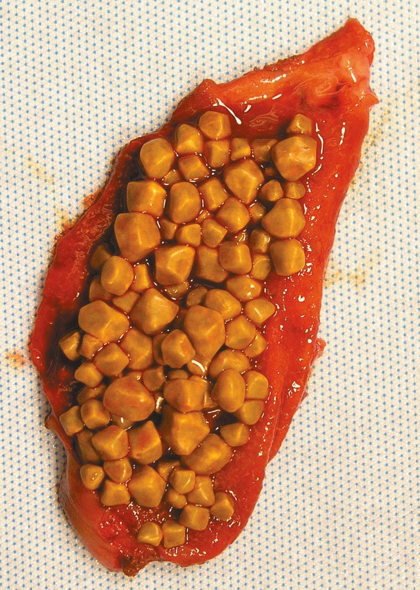
Stone composition and the inflammatory cascade in Schwartz
- In Western countries about 80% of gallstones are cholesterol stones and 15–20% black pigment stones, with brown stones a small minority, both pigment types being commoner in Asia [10].
- Pure cholesterol stones are under 10% of all stones and usually single, large and smooth; most cholesterol stones are mixed but at least 70% cholesterol by weight, multiple, faceted or multilobed, whitish-yellow to green or black, and more than 90% are radiolucent unless a high calcium carbonate content makes them radio-opaque [10].
- Cholesterol, highly non-polar, is held in solution as a vesicle complex with bile salts and lecithin; supersaturation is almost always caused by cholesterol hypersecretion rather than reduced bile salt or phospholipid secretion, and once the solubilising capacity of the triangular phase diagram is exceeded, cholesterol crystallises out [10].
- Pigment stones contain under 20% cholesterol and are dark with calcium bilirubinate; black stones (small, brittle, sometimes spiculated and often radio-opaque) form from supersaturation of unconjugated bilirubin in haemolysis (spherocytosis, sickle cell disease) or cirrhosis, whereas brown stones, usually under 1 cm, soft and mushy, form in the gallbladder or ducts when bacterial β-glucuronidase from organisms such as E. coli deconjugates bilirubin, which precipitates with calcium and dead bacterial bodies; in Asia they follow stasis from Ascaris lumbricoides or Clonorchis sinensis, in the West they are primary duct stones behind strictures or other stones [10].
- Untreated, about two-thirds of patients with biliary colic develop chronic non-infective inflammation, ranging from a near-normal gallbladder with minor mucosal inflammation to a shrunken non-functioning organ with transmural fibrosis, the mucosa atrophying and protruding into the muscle coat as Aschoff-Rokitansky sinuses [10].
- In acute cholecystitis, gallstones are present in 90–95%, a tumour obstructs the cystic duct in under 1%, and the initial inflammation is probably mediated by lysolecithin (a product of lecithin), bile salts and platelet-activating factor with prostaglandin synthesis amplifying it; the wall becomes thickened and reddish with subserosal haemorrhage and patchy mucosal necrosis, in about 5–10% progressing to ischaemia and necrosis, though more often the stone dislodges and the inflammation resolves, and secondary bacterial contamination occurs in only 15–30% [10].
- Perforation is usually contained in the subhepatic space by omentum, but free perforation with peritonitis, intrahepatic perforation with abscess, and perforation into duodenum or colon with cholecystoenteric fistula occur, and gas-forming organisms produce emphysematous cholecystitis with gas in the lumen and wall [10].
Clinical features
- Symptomatic gallstones classically present with biliary colic: dull, continuous epigastric or right-upper-quadrant pain, often after a fatty meal (from cholecystokinin-mediated gallbladder contraction against an obstructed cystic duct), lasting minutes to hours and frequently starting at night, with associated nausea and vomiting [1][3].
- Only about 50% of patients report a clear association with food [3].
- Attacks typically occur within 1 to 2 hours postprandially or wake the patient from sleep, last between 30 minutes and 6 hours, and radiate to the back [4].
- Pain lasting more than 4–6 hours, or accompanied by fever, suggests progression to acute cholecystitis [3][9].
Why the physical examination separates colic from cholecystitis
Patients with uncomplicated symptomatic cholelithiasis typically manifest no signs on examination even during an attack, because the pain is visceral, without the inflammation that would produce local peritonitis and somatic pain; significant right-upper-quadrant tenderness therefore points to acute cholecystitis or another inflammatory process rather than to simple biliary colic [4]. In gallstone colic the pain begins suddenly across the upper abdomen and patients are often unable to say which side is worse; mild tachycardia is common but temperature is usually normal, and about one-fifth become jaundiced [8].
- On examination, acute cholecystitis produces right-upper-quadrant tenderness with inspiratory arrest on palpation of the subcostal region (Murphy's sign) although this is non-specific [1][2].
- Elicited properly, the examiner palpates just below the tip of the ninth costal cartilage and asks the patient to inspire deeply, so the descending liver and attached gallbladder strike the hand [8].
- Against cholescintigraphy as the reference standard, a positive Murphy sign has a sensitivity of 97% but a specificity of only 48% [4].
- A palpable mass may reflect omentum walling off the inflamed gallbladder [1].
Other named signs
- Two further eponymous signs are described.
- Zachary Cope's sign is an inflammatory mass becoming palpable several days into the illness as tenderness begins to subside, still very tender, moving little with respiration, and usually indicating an empyema [8].
- Boas' sign is hyperaesthesia of the dermatome over the back of the chest, detected by lightly drawing a pin down it, reflecting the referral of gallbladder pain to the tip of the scapula [8].
- Where the obstructed gallbladder does not become infected, a large mucocele may develop that reaches down to the level of the umbilicus [8].
- Bowel sounds remain normal unless the gallbladder has infarcted or ruptured to cause biliary peritonitis, a rare complication [8].
Chronic cholecystitis
- The chronic form presents as an indigestion-like upper abdominal pain beginning gradually 15–30 minutes after a meal and lasting 30–90 minutes, moving into the right hypochondrium as it becomes severe and radiating through to the back, characteristically worse after a fatty meal.
- Appetite remains good and weight is steady or increasing, and postprandial belching gives the description "flatulent dyspepsia" [8].
- Attacks are irregular, lasting weeks or months with pain-free intervals of varying length [8].
- Tenderness is found in the right hypochondrium just below the tip of the ninth rib, where the edge of rectus abdominis crosses the costal margin, and deep palpation behind the costal margin on inspiration may be needed to detect it [8].
- The familiar "five Fs" (female, fair, fat, fertile and forty) describe many patients but enough are male, thin, dark and of any age that the mnemonic deserves scant attention as a diagnostic aid [8].
Jaundice suggests a gallstone migrating to obstruct or compress the common bile duct (choledocholithiasis) or, rarely, extrinsic compression of the common hepatic duct by a stone impacted in the gallbladder neck (Mirizzi syndrome) [1]. A palpable, non-tender gallbladder in a jaundiced patient (Courvoisier's sign) is unlikely to be due to gallstones, because chronic inflammation renders the gallbladder thick-walled and non-distensible, and instead suggests distal malignant obstruction [1][8].
The pain, its variants and hydrops
- Biliary colic is constant, builds over the first half hour after a meal and lasts 1–5 hours, sits in the epigastrium or right upper quadrant radiating to the right upper back or between the scapulae, comes on abruptly at night or after a fatty meal with nausea and sometimes vomiting, and leaves the patient well between discrete attacks; in a series of 107 patients the subxiphoid and right subcostal areas were the commonest sites but the left subcostal region was not unusual [10].
- Atypical presentations are common, a meal association is present in only about 50%, pain may be primarily in the back or the left upper or right lower quadrant, and bloating and belching may accompany attacks, so peptic ulcer, reflux, herpes zoster, abdominal wall hernia, inflammatory bowel and diverticular disease, pancreatitis, liver disease, renal stones and pleuritic or cardiac pain must be excluded even when stones are present [10].
- Pain lasting more than 24 hours suggests an impacted cystic-duct stone or acute cholecystitis; an impacted stone without cholecystitis produces hydrops, in which bile cannot enter but the epithelium continues to secrete mucus, distending the gallbladder with clear-white mucinous material (palpable but usually not tender) which may persist harmlessly but can progress to wall oedema, infection and perforation, so early cholecystectomy is generally indicated [10].
- About 80% of patients with acute cholecystitis give a history compatible with chronic cholecystitis; the attack begins like colic but the pain does not subside, persisting for days and more severe, with fever, anorexia, nausea and vomiting and reluctance to move from focal peritonitis, and a mass of gallbladder and adherent omentum is occasionally palpable though guarding may hide it [10].
- Leucocytosis is typically 12,000–15,000/mm³ but a normal count does not exclude the diagnosis, a count above 20,000 suggests gangrene, perforation or cholangitis, bilirubin may be mildly raised (<4 mg/dL) with mild rises in alkaline phosphatase, transaminases and amylase, and severe jaundice suggests duct stones or Mirizzi's syndrome; the elderly and diabetic may present subtly with delayed diagnosis and more treatment-related morbidity, and the differential includes peptic ulcer, pancreatitis, appendicitis, hepatitis, Fitz-Hugh–Curtis perihepatitis, myocardial ischaemia, pneumonia, pleuritis and herpes zoster [10].
Etiology
- Recognised risk factors include female sex, increasing age (particularly >40 years), obesity, pregnancy, rapid weight loss (including after bariatric surgery), a high-calorie/high-cholesterol diet, family history, diabetes mellitus, ileal disease or resection (depleting the bile-acid pool), total parenteral nutrition, prolonged fasting, and drugs such as oral contraceptives, octreotide and thiazides [1][3][9].
- Vagotomy is a further listed risk factor, and TPN is specifically associated with pigmented stones [7].
- Chronic haemolytic conditions predispose to pigment stones, and native North Americans are particularly liable to develop gallstones [1][8].
- After bariatric surgery, gallstones develop in approximately 30% of patients during the period of rapid weight loss [4].
- Acalculous cholecystitis occurs in the absence of stones, typically in critically ill patients, and those recovering from major surgery, trauma or burns, with biliary stasis and ischaemia implicated; diagnosis is often delayed and mortality is high [1][3].
- The primary pathology is bile stasis from narcotics and fasting, leading to distension and ischaemia, compounded by increased bile viscosity secondary to dehydration, ileus and transfusion [7].
- Risk factors are old age, burns and trauma, prolonged total parenteral nutrition, critical illness, immunosuppression and diabetes [3].
Risk factors and natural history in Schwartz's figures
- Autopsy studies find gallstones in 10–15% of adults, with higher prevalence in Native American and Latin American populations; predisposing conditions include pregnancy, non-HDL hyperlipidaemia, Crohn's disease, hereditary spherocytosis, sickle cell disease and thalassaemia, terminal ileal resection and gastric or duodenal surgery that disturb neurohormonal regulation, rapid weight loss after bariatric surgery, and somatostatin analogues and oestrogen-containing contraceptives; women are three times as likely to form stones and first-degree relatives have twice the prevalence [10].
- About 80% of incidentally diagnosed asymptomatic patients remain symptom-free, 2–3% per year develop colic, and once symptomatic 3–5% per year develop complications, so prophylactic cholecystectomy is rarely indicated except for those isolated from medical care for long periods, populations at increased risk of gallbladder cancer, and porcelain gallbladder, a rare premalignant condition that is an absolute indication even when asymptomatic [10].
- Cholesterolosis, cholesterol accumulating in lamina propria macrophages, locally or as polyps, giving the studded "strawberry gallbladder", and adenomyomatosis (cholecystitis glandularis proliferans, with hypertrophic smooth muscle, mucosal glands growing into the muscle layer, fundal granulomatous polyps, wall thickening and septa) can both cause typical biliary symptoms and are treated by cholecystectomy when symptomatic, as is sludge with typical biliary pain in the absence of stones [10].
Diagnosis
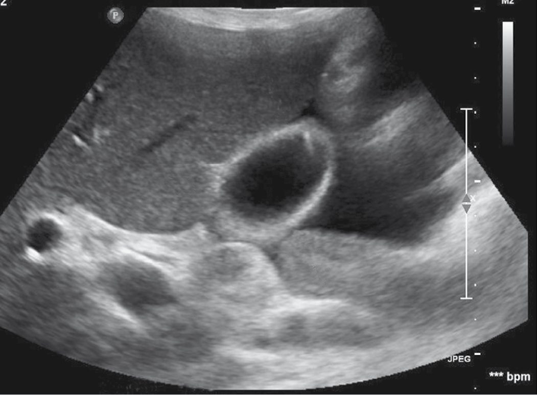
- Ultrasonography (USG) is the first-line and most sensitive/specific imaging modality, identifying stones and features of acute cholecystitis such as gallbladder wall thickening (>3 mm), pericholecystic fluid and a sonographic Murphy's sign; sensitivity is approximately 85% and specificity 95% [1][3][9].
- Gallstones appear as rounded hyperechoic shapes casting a hypoechoic acoustic shadow, and move with changes in position; ultrasound is 95% sensitive for detecting stones and is the best initial test for jaundice or right-upper-quadrant pain [4][7].
- Gallbladder wall thickening beyond 4 mm together with pericholecystic fluid gives a sensitivity of 88% and specificity of 80% for acute cholecystitis, although in the Maingot's authors' experience the presence or absence of these findings correlates poorly with the degree of inflammation found at subsequent operation [4].
- Ultrasound also reports the diameter of the common bile duct and can incidentally diagnose non-biliary disease such as hepatic lesions, hepatic steatosis, pancreatic masses or renal tumours [4].
Normal calibres and laboratory patterns
- Normal reference sizes are a common bile duct of 6 mm or less (up to 10 mm after cholecystectomy), a gallbladder wall of 4 mm or less, and a pancreatic duct of 4 mm or less; a duct dilated beyond 6 mm suggests a stone with obstruction [7].
- Mild elevations of ALP, bilirubin, transaminases and leukocytosis support the diagnosis; profound jaundice is uncommon in isolated cholecystitis and should raise suspicion of cholangitis or Mirizzi syndrome [3].
- In acute cholecystitis the white cell count is often raised while liver function tests, especially the direct bilirubin, should be relatively normal, since there is no biliary obstruction, an elevated direct bilirubin instead suggests choledocholithiasis, and a raised amylase or, more specifically, lipase suggests gallstone pancreatitis [4].
- Among the biochemical markers for choledocholithiasis, GGT has the highest sensitivity and negative predictive value while alkaline phosphatase has the highest specificity and positive predictive value [7].
The Tokyo Guidelines 2018 diagnostic criteria for acute cholecystitis require at least one local sign of inflammation (Murphy's sign, right-upper-quadrant pain/tenderness/mass), at least one systemic sign (fever, elevated CRP, elevated WBC), and, for a definite diagnosis, supportive imaging [1].
Cholescintigraphy
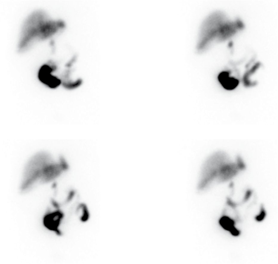
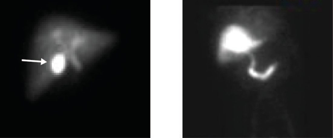
- Hepatobiliary iminodiacetic acid (HIDA) scanning shows non-visualisation of the gallbladder with prompt filling of the common bile duct and duodenum in acute cholecystitis, with sensitivity and specificity of about 95% each; normal filling occurs within 60 minutes in fasting subjects (liver uptake within 10 minutes) [2].
- Maingot's cites a sensitivity greater than 95% and a specificity of 90%, and positions it as the confirmatory test where history, examination and ultrasound leave doubt, which is uncommon [4].
- Adding cholecystokinin stimulation converts it into the CCK-cholescintigraphy test, the most sensitive test for cholecystitis; indications for cholecystectomy on that test are non-visualisation of the gallbladder, emptying taking longer than 60 minutes, or an ejection fraction below 40% [7].
- CT is reserved for atypical presentations, diagnostic uncertainty, or suspected complications such as perforation [1][3].
- If jaundice with deranged liver function tests is present, MRCP is used to exclude choledocholithiasis [1].
- Where symptoms are atypical in a patient with gallstones, a wider workup, upper gastrointestinal contrast radiography or endoscopy, CT, or cardiac and pulmonary evaluation, is appropriate to exclude significant non-biliary disease [4].
- NICE reduces the diagnostic workup to a four-step ladder, with liver function tests and ultrasound as the entry point for anyone with suspected gallstone disease and for anyone whose abdominal or gastrointestinal symptoms have not responded to previous management [5].
- MRCP is considered only if ultrasound has not detected common bile duct stones but the bile duct is dilated and/or liver function test results are abnormal [5].
- Endoscopic ultrasound is considered only if MRCP does not allow a diagnosis to be made [5].
- Where conditions other than gallstone disease are suspected, patients are referred for further investigation instead [5].
- Two divergences from the textbook workup are worth stating explicitly.
- First, cholescintigraphy does not appear anywhere in CG188, neither the plain HIDA scan nor the CCK-stimulated ejection-fraction test that the North American texts treat as the most sensitive test for cholecystitis and the diagnostic basis for functional gallbladder disorder.
- Second, ERCP has no diagnostic role: it should be reserved for therapy, not used as a diagnostic test for bile duct stones, with preoperative on-table cholangiography an alternative strategy in units offering laparoscopic bile duct exploration [6].
The commissioning guide describes the presentation that should trigger the pathway: epigastric or right upper quadrant pain, frequently radiating to the back, lasting several minutes to hours and often occurring at night; those patients should have liver function tests checked and be referred for ultrasonography [6]. Except in the acutely unwell, ultrasound confirmation of gallstones should be obtained before referral, and the ultrasound report (including bile duct findings) should be provided with the referral [6].
Test performance for acute cholecystitis
Ultrasound is the most useful initial test with a sensitivity and specificity of 70–90%, showing stones, wall thickening, pericholecystic fluid and a sonographic Murphy's sign; HIDA scanning helps atypical cases, non-filling of the gallbladder after 4 hours confirms cystic duct obstruction with sensitivity above 90%, and clear filling rules the diagnosis out, and CT, frequently obtained for undifferentiated pain, shows the same features but is somewhat less sensitive than ultrasound [10]. Stones found incidentally on CT or plain films in asymptomatic patients should be left in place, whereas typical symptoms with stones seen on a CT obtained for other reasons justify intervention [10].
Scoring and Severity
- The Tokyo Guidelines 2018 grade acute cholecystitis into three severity tiers.
- Grade III (severe) is associated with dysfunction of any organ system (cardiovascular, neurological, respiratory, renal, hepatic or haematological).
- Grade II (moderate) is associated with any one of: WBC >18,000/mm³, a palpable tender right-upper-quadrant mass, symptom duration >72 hours, or marked local inflammation (gangrenous cholecystitis, pericholecystic or hepatic abscess, biliary peritonitis, emphysematous cholecystitis).
- Grade I (mild) meets neither of these, occurring in a healthy patient with mild inflammatory changes, making cholecystectomy a low-risk procedure [1].
- The American Association for the Surgery of Trauma (AAST) Emergency General Surgery grading system similarly stratifies cholecystitis into five grades, from localised inflammation (Grade I) to pericholecystic abscess, bilioenteric fistula and peritonitis (Grade V) [3].
- These grading systems, combined with patient comorbidity (e.g.
- Charlson Comorbidity Index, ASA class) and institutional capability, guide the Tokyo Guidelines management algorithm [1].
Rome III criteria for functional gallbladder disorder
Where there are no stones, the diagnosis rests on a symptom checklist rather than a severity grade. Episodes of pain must be located in the epigastrium and/or right upper quadrant and meet all of the following [4]:
| Criterion | Requirement |
|---|---|
| 1 | Episodes lasting 30 minutes or longer |
| 2 | Recurrent symptoms occurring at different intervals, not daily |
| 3 | The pain builds up to a steady level |
| 4 | The pain is moderate to severe enough to interrupt daily activities or lead to an emergency department visit |
| 5 | The pain is not relieved by bowel movements |
| 6 | The pain is not relieved by postural change |
| 7 | The pain is not relieved by antacids |
| 8 | Exclusion of other structural disease that would explain the symptoms |
- Patients must additionally have normal liver function tests and no gallstones on ultrasound [4].
- The strictness of the list is deliberate: it is intended to limit the number of cholecystectomies performed for this indication and raise the probability that the operation resolves the symptoms [4].
- Diagnosis is confirmed by CCK-stimulated cholescintigraphy, with an ejection fraction below 35–40% indicative; patients below that threshold have markedly better symptomatic outcomes after cholecystectomy, and Rome III advises that those with an ejection fraction above 40% be evaluated more carefully for alternative causes of pain [4].
- Sabiston sets the threshold at less than 35%, and reports that more than 85% of patients improve symptomatically after cholecystectomy, better response being predicted by typical biliary colic symptoms and a reduced ejection fraction, and worse response by nausea as the dominant symptom [3].
Gallbladder polyps stratified by size
| Polyp size | Cancer risk and management |
|---|---|
| Under 10 mm | Low risk of cancer; observe with surveillance ultrasound for growth, initially yearly. Multiple small polyps usually signify cholesterolosis, and the gallbladder is removed only for biliary colic |
| 10–20 mm | Higher risk of malignancy, but when cancer is present it is usually early stage; treated with laparoscopic cholecystectomy without liver resection |
| Over 20 mm | Almost always malignant; radical cholecystectomy with lymph node dissection |
- Table reformats the size-based recommendations [4].
- Polyps are typically an incidental finding affecting approximately 5% of the population, with a higher percentage in Asian populations, and may be adenocarcinoma, benign adenoma, or cholesterolosis, a clinically insignificant accumulation of cholesterol on the gallbladder wall [4].
- Porcelain gallbladder, calcification of the entire gallbladder wall, carries an associated risk of gallbladder cancer: earlier studies estimated 12% to 60%, but recent series suggest 7% or less, and while radiographic evidence of it was traditionally an indication for cholecystectomy, some authors now recommend surveillance imaging in the absence of another indication such as biliary colic [4].
- The early dogma that porcelain gallbladder contraindicated laparoscopy has also largely been abandoned [4].
Treatment and Management
- Asymptomatic gallstones generally require no intervention; prophylactic cholecystectomy is reserved for specific higher-risk situations: large (>3 cm) stones, concomitant choledocholithiasis, chronic haemolytic disease, gallbladder polyps >1 cm, porcelain gallbladder, high-risk ethnic/geographic populations for gallbladder cancer, transplant recipients, and bariatric surgery patients [1].
- Over a 10-year period roughly two-thirds of patients with asymptomatic stones remain symptom-free [2].
- Maingot's puts the case the other way round: patients with incidentally discovered stones have a less than 20% chance of ever developing symptoms, so the risks of prophylactic operation outweigh the benefit in almost all patients, with prophylaxis justified in sickle cell disease, open bariatric surgery, long-term total parenteral nutrition, and possibly therapeutic immunosuppression after solid organ transplantation [4].
- Prophylactic cholecystectomy has occasionally been advocated in diabetics; there is no evidence to support that policy, but good evidence to support early cholecystectomy once a diabetic patient becomes symptomatic, because diabetics present with acute cholecystitis more often and withstand complications less well [4].
- Removing the gallbladder at the time of open bariatric surgery abolishes gallstone-related morbidity easily, but the practice has largely been abandoned in the laparoscopic era, because the added morbidity of a laparoscopic cholecystectomy in a patient with morbid obesity appears to exceed the later risk of stone-related complications [4].
- For symptomatic gallstones, cholecystectomy is the treatment of choice [1].
- Once symptoms begin there is a greater than 80% chance of further symptoms or a complication, and because there are no effective medical therapies for cholelithiasis, any patient who develops symptoms should be offered laparoscopic cholecystectomy unless comorbidity makes the operative risk prohibitive [4].
- Initial non-operative management of acute cholecystitis includes nil-by-mouth status, intravenous fluids, analgesia, and broad-spectrum antibiotics effective against Gram-negative aerobes (e.g. cefazolin, cefuroxime or ciprofloxacin), since the concentration of antibiotic in serum matters more than biliary concentration once the cystic duct is obstructed [1].
- Common causative organisms include E. coli, Klebsiella, Enterobacter and Bacteroides species; E. coli is the single commonest, followed by Klebsiella and Enterococcus [3][7].
Timing of surgery in acute cholecystitis
- Early laparoscopic cholecystectomy, performed by an experienced surgeon within 5–7 days of symptom onset, is safe and shortens hospital stay compared with delayed (interval) surgery at 6 weeks, although conversion rates are higher than in elective surgery [1][3].
- Approximately 20% of patients managed non-operatively while awaiting interval surgery fail to respond and require urgent intervention [3].
- The randomised evidence is strongly in favour of operating early.
- A trial of over 600 patients comparing early laparoscopic cholecystectomy within 24 hours against antibiotics with interval cholecystectomy at 7 to 45 days found fewer complications in the early group (12% vs 34%), a shorter overall length of stay and lower costs.
- A meta-analysis of 15 randomised controlled trials found shorter stays, lower costs and higher patient satisfaction and quality of life with early surgery, with similar perioperative mortality and morbidity [4].
- Delaying surgery allows the inflammation to become more intense and neovascularised, increasing the technical difficulty of the laparoscopic operation [4].
- There is no benefit to "cooling the patient off" with antibiotics before surgery [7].
Drainage as a temporising measure
- In critically ill or high-risk patients with severe (Grade III) cholecystitis, percutaneous cholecystostomy tube drainage under ultrasound or CT guidance, combined with antibiotics, is used to temporise, with interval cholecystectomy 6–12 weeks later once the patient has recovered [2][3].
- Endoscopic alternatives exist: a transpapillary tube inserted at ERCP, or a transduodenal tube placed under endoscopic ultrasound guidance [4].
- All are temporary solutions, if the tube is removed the patient remains at high risk of recurrent stone symptoms, so drainage should be reserved for the sickest patients in whom early surgery would pose a prohibitive risk, and interval cholecystectomy should follow recovery [4][7].
- More than 90% of patients with acalculous cholecystitis improve with cholecystostomy alone, and interval cholecystectomy is needed only if follow-up imaging remains abnormal; failure to improve within 24 hours of tube placement suggests perforation and should prompt exploration [3].
Special populations
- Symptomatic gallstones in pregnancy that cannot be managed expectantly are treated with laparoscopic cholecystectomy, ideally in the second trimester [1][2].
- Operative modifications are open (Hassan) port placement, low pneumoperitoneum, and rolling the patient to the left off the inferior vena cava, and the spontaneous abortion rate is lower than with non-operative treatment [7].
- The second-trimester convention arose because organogenesis is complete and the fundus has not yet encroached on the operative field, but there are no data suggesting first-trimester laparoscopy is more dangerous, and current society recommendations generally favour operating at any point in pregnancy as soon as symptoms arise.
- Insufflation pressure should be limited to less than 12 mm Hg, maternal end-tidal CO₂ closely monitored to prevent fetal acidosis, and laparoscopic ultrasound used in place of cholangiography to limit fetal radiation exposure [4].
- In cirrhosis, patients with Child's A or B disease should undergo laparoscopic cholecystectomy while Child's C patients are better managed medically with antibiotics [7].
- After Roux-en-Y gastric bypass the biliary tree is not easily accessible for ERCP: if the gallbladder is still present, cholecystectomy with intraoperative CBD exploration is used; if it has been removed, options are double-balloon ERCP or laparoscopic gastrostomy tube placement into the distal stomach remnant with ERCP performed through the tube [7].
The NICE management recommendations are short enough to quote in full, and three of them differ materially from the textbook position.
| NICE CG188 | Recommendation |
|---|---|
| 1.2.1 | Reassure people with asymptomatic gallbladder stones found in a normal gallbladder and normal biliary tree that they do not need treatment unless they develop symptoms |
| 1.2.2 | Offer laparoscopic cholecystectomy to people diagnosed with symptomatic gallbladder stones |
| 1.2.3 | Offer day-case laparoscopic cholecystectomy for people having it as an elective planned procedure, unless their circumstances or clinical condition make an inpatient stay necessary |
| 1.2.4 | Offer early laparoscopic cholecystectomy, to be carried out within 1 week of diagnosis, to people with acute cholecystitis |
| 1.2.5 | Offer percutaneous cholecystostomy to manage gallbladder empyema when surgery is contraindicated at presentation and conservative management is unsuccessful |
| 1.2.6 | Reconsider laparoscopic cholecystectomy for people who have had percutaneous cholecystostomy once they are well enough for surgery |
Table reproduces the recommendations of section 1.2 [5].
The clock starts at diagnosis, not at symptom onset. NICE sets one week from diagnosis; the textbook figure of 5–7 days is measured from symptom onset, and the randomised trials quote windows measured in hours from admission. A patient who has been symptomatic for five days before presenting is late by the textbook clock and on time by the NICE one [5].
Day-case surgery is the default, not the exception. NICE makes day-case laparoscopic cholecystectomy the offer for every elective case, with inpatient stay requiring a positive justification [5]. None of the textbooks frames it that way.
- Cholecystostomy is narrower than the textbook indication.
- NICE restricts it to gallbladder empyema, and requires both that surgery is contraindicated at presentation and that conservative management has already failed [5].
- The textbook indication is broader, any patient too high-risk for surgery.
- NICE also frames the follow-up as reconsidering cholecystectomy once the patient is well enough, rather than as scheduled interval surgery [5].
- The commissioning guide adds the primary-care half of the pathway.
- An incidental finding of stones in an otherwise normal gallbladder requires no further investigation or referral [6].
- Most patients with symptomatic stones present with a self-limiting attack lasting hours only, which can often be controlled in primary care with appropriate analgesia, avoiding emergency admission.
- Referral as an emergency is for pain that cannot be managed, for a patient who is otherwise unwell, or for suspicion of acute cholecystitis, cholangitis or acute pancreatitis [6].
- Further episodes of biliary pain can be prevented in around 30% of patients by a low-fat diet, because fat in the stomach releases cholecystokinin, which precipitates gallbladder contraction [6].
On drug treatment the guide is unusually blunt: there is no evidence to support hyoscine or proton pump inhibitors in the management of gallbladder symptoms, antibiotics should be reserved for patients with signs of sepsis, and there is no evidence of benefit from non-surgical definitive treatments, gallstone dissolution therapies, ursodeoxycholic acid, or extracorporeal lithotripsy [6].
- The decision to operate on symptomatic stones is made by the patient with guidance from the surgeon, weighing a 50% risk per annum of a further episode of biliary colic and a 1–2% risk per annum of a serious complication against the individual's operative risk and preference [6].
- Patients with gallstone pancreatitis should undergo definitive treatment (usually cholecystectomy, though endoscopic sphincterotomy may suit frail patients) within two weeks of recovery from the index episode [6].
- Two service-level requirements follow: providers offering cholecystectomy must be able to offer intraoperative on-table cholangiography and have arrangements for urgent access to ERCP and interventional radiology for postoperative complications, and any patient with a suspected bile duct injury should be referred to their regional tertiary hepatopancreatobiliary service [6].
Advice after treatment is that patients should avoid food and drink that triggers symptoms only until the gallbladder or stones are removed; afterwards they should not need to avoid those foods, and should seek advice from their GP if eating or drinking triggers existing symptoms or causes new ones once they have recovered [5].
Timing, conversion and cholecystostomy in Schwartz
- Non-surgical treatment with drugs or lithotripsy has had disappointing long-term results and is not part of the primary algorithm; about 90% of patients with typical symptoms are rendered symptom-free by cholecystectomy, with poorer results for dyspepsia, flatulence, belching, bloating and fat intolerance [10].
- While awaiting surgery patients avoid dietary fat and large meals; diabetics should be operated on promptly because they are prone to severe acute cholecystitis, and pregnant women who cannot be managed by diet can safely have laparoscopic cholecystectomy, preferably in the second trimester [10].
- In acute cholecystitis intravenous fluids, analgesia and antibiotics covering Gram-negative enteric organisms and anaerobes are standard because it is impossible to know who is secondarily infected; early cholecystectomy within 72 hours is preferred to delayed operation at 6–10 weeks, giving a single admission, quicker recovery, similar complications and earlier return to work, and conversion to open surgery has fallen below 5% [10].
- Patients unfit for surgery are treated with antibiotics and a cholecystostomy tube, which usually stabilises them; the tube is removed once the track has matured (about 4 weeks) and cholangiography shows a patent cystic duct, elective cholecystectomy follows at about 6–8 weeks, and failure to improve after cholecystostomy suggests gangrene or perforation requiring damage-control surgery [10].
Acalculous cholecystitis and biliary dyskinesia as Schwartz treats them
- Acalculous cholecystitis is a rare disease of the critically ill, parenteral nutrition, extensive burns, sepsis, major operations, multiple trauma and prolonged multi-organ failure, attributed to distension, stasis and ischaemia, with serosal and muscular oedema and patchy thrombosis of arterioles and venules on pathology; in the sedated patient the clinical features are masked and fever, leucocytosis and raised alkaline phosphatase and bilirubin are the prompts to investigate [10].
- Bedside ultrasound is the test of choice (distended thick-walled gallbladder, sludge, pericholecystic fluid, abscess), CT adds a search for other sources of sepsis, and HIDA is less sensitive with more false positives in prolonged fasting, parenteral nutrition or liver disease; urgent broad-spectrum antibiotics and resuscitation are followed by laparoscopic cholecystectomy if the patient can tolerate it, otherwise percutaneous cholecystostomy, after which about 90% improve, and interval cholecystectomy is not strictly required in the absence of stones [10].
- Percutaneous cholecystostomy tubes are pigtail catheters passed under ultrasound guidance through the liver (a transhepatic route minimises bile leak around the catheter) and are removed after a tube cholangiogram confirms a patent cystic duct, with interval cholecystectomy considered when fitness has improved, particularly in stone disease [10].
- Biliary dyskinesia, typical biliary pain without stones or sludge and a HIDA ejection fraction below 35%, improves or resolves after cholecystectomy in up to 90%; sphincter of Oddi dysfunction presents as episodic biliary pain with abnormal liver tests or recurrent pain after cholecystectomy, sometimes with jaundice or pancreatitis, is suggested by a dilated duct that is difficult to cannulate or delayed contrast emptying at ERCP, can be confirmed by ampullary manometry and provocation tests in specialist units, and usually responds well to sphincterotomy [10].
Surgeries
Laparoscopic cholecystectomy is the gold-standard operation, performed via four ports (a 12-mm umbilical port for specimen extraction and three smaller ports for dissection), with the patient in reverse Trendelenburg [3]. The advantages over open cholecystectomy are earlier return of bowel function, less postoperative pain, improved cosmesis, shorter hospital stay, earlier return to full activity, fewer wound infections and incisional hernias, and lower overall cost; its introduction was followed by an increase in the cholecystectomy rate, with evidence of a lower clinical threshold for operating [4].
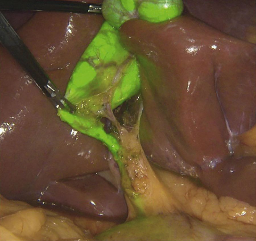
Anatomy and its variants
- The classic anatomy of the biliary tree is present in only 30% of individuals, so anomalies are the rule rather than the exception [4].
- The cystic duct may join the common bile duct at an acute angle, run parallel to it for several centimetres before inserting, insert into the right hepatic duct, or be congenitally absent [4].
- The cystic artery usually arises from the right hepatic artery and is found in the triangle of Calot, bounded by the cystic duct laterally, the common hepatic duct medially and the liver superiorly [7].
- The surgeon must be certain the artery seen is entering the gallbladder wall: occasionally the right hepatic artery loops onto the surface of the gallbladder and gives rise to a very short cystic artery, and a posterior cystic artery is easily injured if not recognised [4].
- An aberrant right hepatic duct is the commonest anomaly to cause trouble, and the most dangerous variant is a cystic duct joining a low-lying aberrant right sectoral duct; injuries to these ducts are under-reported because occlusion may be asymptomatic [4].
- Two triangles are distinguished.
- The hepatocystic triangle is the ventral aspect of the area bounded by the gallbladder wall and cystic duct, the liver edge, and the common hepatic duct; the eponymous Calot triangle contained within it is bounded by the cystic duct, cystic artery and common hepatic duct [4].
- The gallbladder lies beneath segments IV and V, cystic veins drain into the right branch of the portal vein, and the lymphatics lie on the right side of the common bile duct [7].
- Ducts of Luschka are small biliary ducts lying in the gallbladder fossa that can leak after cholecystectomy [7].
Positioning, access and exposure
- Patients are fasted for a minimum of 8 hours, given antiembolic stockings and sequential compression devices because of the reverse Trendelenburg position, and have an orogastric tube placed after induction to decompress the stomach.
- Prophylactic antibiotics are discretionary, since most patients have a very low risk of perioperative infection and antibiotics have not been shown to reduce it significantly [4].
- Pneumoperitoneum is established at the umbilicus by either a closed (Veress needle) or open technique, and surgeons should learn both and select between them by body habitus and previous surgical history [4].
- An angled 30- or 45-degree laparoscope is preferred over a 0-degree scope because it gives multiple views of the same field [4].
- The patient is placed in 30 degrees of reverse Trendelenburg with the table rotated 15 degrees to the left, allowing colon and duodenum to fall away from the liver edge [4].
Two 5-mm subcostal ports are then placed under vision, the first along the right anterior axillary line at least two finger-breadths below the costal margin and as lateral as possible while remaining anterior to the ascending colon, the second in the right subcostal midclavicular line, with a fourth epigastric working port inserted about 5 cm below the xiphoid, entering the peritoneum to the right of the falciform ligament [4]. The assistant's axillary grasper pushes the fundus laterally and cephalad, rolling the right lobe of the liver cranially, a manoeuvre complicated by a fixed cirrhotic liver or a heavy, friable, fatty one [4].
Dissection and the critical view of safety
The gallbladder infundibulum is retracted inferolaterally to open the triangle of Calot, and dissection proceeds until the critical view of safety is achieved: the lower third of the gallbladder is dissected free of the liver bed, and only two structures (the cystic duct and cystic artery) are seen entering the gallbladder, before either is clipped or divided [3][4]. Maingot's specifies at least one-third of the cystic plate should be exposed, so that two and only two structures cross the window; no structure should be divided until both are unequivocally identified [4].
- Several dissection principles reduce risk.
- Dissection begins from a known structure (the gallbladder) rather than in an unknown area, starting 4 or 5 cm proximal to the gallbladder neck and proceeding distally in a modified "top-down" fashion, and is initiated on the anatomic right side to avoid approaching the cystic duct and artery before the true gallbladder edge is defined [4].
- Electrocautery is pulled away from the gallbladder to avoid perforating it or injuring underlying structures [4].
- It is generally unnecessary and potentially harmful to dissect the cystic duct all the way down to its junction with the common bile duct [4].
- The Calot lymph node typically overlies the cystic artery and serves as a useful landmark, occasionally requiring a brief application of low-wattage coagulation as it is swept away [1][3][4].
Imaging the duct intraoperatively
- Intraoperative cholangiography or laparoscopic ultrasound may be used selectively to define anatomy or identify choledocholithiasis, particularly in cases of gallstone pancreatitis or anomalous anatomy, though routine use is not universally recommended [3][4].
- Before either, a clip is applied high on the cystic duct at its junction with the gallbladder to prevent stones migrating down during manipulation [4].
- For cholangiography the anterolateral cystic duct wall is incised, stones are milked back toward the gallbladder and out of the ductotomy, and a 4- or 5-Fr catheter is passed and secured with a loose clip or an Olsen clamp; 10 to 20 mL of water-soluble contrast is injected under real-time fluoroscopy, mixed half-strength to avoid obscuring small stones, with digital subtraction used to eliminate the vertebrae from the image [4].
- Five things are looked for: the length of the cystic duct and the location of its junction with the common bile duct, the diameter of the duct, luminal filling defects, free flow of contrast into the duodenum, and the anatomy of the extrahepatic and intrahepatic tree [4].
Laparoscopic ultrasound is the alternative. In a prospective multicentre trial of 209 laparoscopic cholecystectomy patients, ultrasound took significantly less time than cholangiography (7 ± 3 vs 13 ± 6 minutes) and was more sensitive for detecting stones, while cholangiography was better at delineating intrahepatic anatomy and defining ductal anomalies, the authors concluded the two are complementary [4].
Completing the operation
- Two clips are placed distally on the cystic duct and one toward the gallbladder before division, with a preformed endoloop preferred for a large or friable duct.
- The posterior jaw of the clip applier must be visualised beyond the structure before each clip is applied, and care taken that the common bile duct is not tented into the clip [4].
- The cystic artery is then dissected over an adequate length for three clips, after confirming it is not the right hepatic artery looping onto the gallbladder neck or an accessory or replaced right hepatic artery.
- Electrocautery must not be used for its division, since current transmitted to the proximal clips can cause necrosis and later haemorrhage [4].
- The gallbladder is separated from its hepatic bed with an electrosurgical spatula or hook under alternating medial and lateral countertraction, and before the final attachments are divided the fossa and porta hepatis are inspected for haemostasis and bile leakage [4].
- The specimen is placed in an entrapment sac if it has been perforated or is large, and extracted at the umbilical port where there are no muscle layers anterior to the fascia and where extending the incision causes less pain and gives better cosmesis than enlarging the subxiphoid one [4].
- Umbilical fascia is closed with one or two large absorbable sutures; closure of the subxiphoid fascia is optional, because the oblique trocar entry angle and its position anterior to the falciform ligament make herniation unlikely [4].
- More than 90% of patients are discharged within 24 hours and preoperatively selected fit patients may safely go home within 6 hours [4].
Contraindications and conversion
| Category | Contraindications to laparoscopic cholecystectomy |
|---|---|
| Absolute | Unable to tolerate general anaesthesia; refractory coagulopathy; gallbladder carcinoma |
| Relative | Cholangitis; diffuse peritonitis; cirrhosis and/or portal hypertension; cholecystoenteric fistula |
- Table reproduces the listed contraindications [4].
- Diffuse peritonitis with haemodynamic compromise is a surgical emergency in which laparoscopy is imprudent, because the aetiology is not secure and pneumoperitoneum may cause vascular collapse.
- Suspicion of gallbladder malignancy mandates open resection, because of concerns over adequacy of resection and the 20–30% rate of gallbladder perforation at laparoscopy with the risk of intraperitoneal dissemination [4].
- Further relative contraindications dictated by surgeon philosophy and experience include previous upper abdominal surgery with extensive adhesions, severe cardiopulmonary disease, morbid obesity and pregnancy [4].
- Conversion should be regarded as sound clinical judgment rather than a complication, and should not be delayed when the anatomy is unclear, complications arise, or reasonable progress is not being made.
- It is also indicated for massive haemorrhage, major bile duct injury, a biliary-enteric fistula, potentially resectable gallbladder carcinoma, and duct stones that can be cleared neither laparoscopically nor endoscopically [4].
- Conversion rates rise significantly after 72 hours of acute cholecystitis [4].
When dissection is unsafe due to severe inflammation, bailout options include gallbladder decompression by needle aspiration, a dome-down (retrograde) dissection, subtotal cholecystectomy (fenestrating, leaving the cystic duct stump open, or reconstituting, closing the stump) leaving the infundibulum in situ with a drain, or early conversion to open cholecystectomy via a midline or right subcostal incision [3]. Open common bile duct exploration, choledochoduodenostomy, and Roux-en-Y hepaticojejunostomy are reserved for concomitant choledocholithiasis not clearable by other means [3].
Open cholecystectomy
- The technique has changed little since Langenbuch's description in 1882, and most surgeons prefer a right subcostal (Kocher) incision, though midline and paramedian are also used [4].
- Adequate exposure of the gallbladder and hepatoduodenal ligament is the key to safety: sponges are packed between the dome of the liver and the diaphragm, the hepatic flexure retracted inferiorly and the medial segment of the left lobe superiorly, and the surgeon introduces the left index finger into the foramen of Winslow to palpate for duct calculi [4].
- Where acute inflammation or chronic scarring precludes approaching the infundibulum first, a fundus-first (top-down) technique is used, addressing the ductal and vascular structures only after the organ has been separated from the liver [4].
- Experience from laparoscopic surgery has changed one long-standing teaching: ligating the cystic duct close to its junction with the common bile duct was considered essential, but stump length is not in fact a critical factor and probably does not contribute significantly to postcholecystectomy syndrome, so it is safer to divide the duct closer to the infundibulum once the critical view is obtained and avoid dissection near the common bile duct altogether [4].
- Where severe inflammation has obliterated normal tissue planes, subtotal cholecystectomy, leaving part of the infundibulum in situ after removing all stones and suture-ligating the mucosal side of the cystic duct origin, may be the safest course [4].
- Drains are not mandatory and are indicated only where the surgeon is concerned about identifying or controlling a possible bile leak [4].
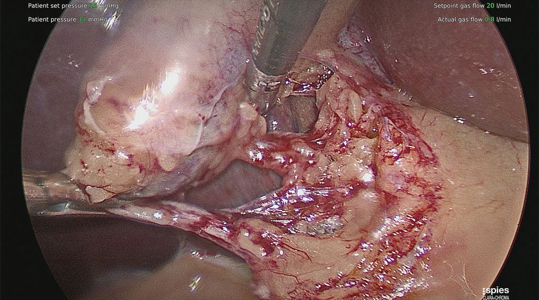
Investigational approaches
- Single-incision laparoscopic surgery (SILS) introduces all instruments through one umbilical incision, with the theoretical benefits of less pain and fewer incisional complications; in practice all instruments enter in line with the optics, making triangulation and countertraction more difficult [4].
- A randomised trial found prolonged operative times without any difference in postoperative pain scores, though with better cosmesis ratings at 12 months.
- The largest trial, a 200-patient multicentre randomisation, found similar perioperative outcomes and complication rates but more postoperative pain and a higher incidence of incisional hernia at one year than the traditional four-trocar approach (8.4% vs 1.2%), and most surgeons have consequently abandoned the technique except where cosmesis matters greatly to a particular patient [4].
- Natural orifice transluminal endoscopic surgery (NOTES) aims to eliminate abdominal incisions entirely.
- The transgastric route proved technically challenging and was abandoned, while transvaginal hybrid NOTES using a rigid laparoscope through a posterior fornix colpotomy plus one or two abdominal ports is performed in selected centres, mostly in Germany and South America, with trials showing decreased postoperative pain and similar complication rates, but it remains an investigational procedure that should only be performed under an approved research protocol [4].
History, contraindications, conversion and technique in Schwartz
- Over 750,000 cholecystectomies are performed each year in the United States; Carl Langenbuch performed the first open cholecystectomy in 1882, Philippe Mouret introduced the laparoscopic operation in France in 1987, and it more or less ended attempts at non-invasive management such as shock-wave or cholangioscopic lithotripsy and bile-salt therapy [10].
- Absolute contraindications to laparoscopy are few (haemodynamic instability, uncontrolled coagulopathy and frank peritonitis) while severe COPD or heart failure with an ejection fraction below 20% may not tolerate carbon dioxide pneumoperitoneum; acute cholecystitis, gangrene, empyema, biliary-enteric fistula, obesity, pregnancy, ventriculoperitoneal shunts, cirrhosis and previous upper abdominal surgery are now risk factors for a difficult operation rather than contraindications [10].
- Conversion is not a failure and is needed for intolerance of pneumoperitoneum, a complication that cannot be fixed laparoscopically, unclear anatomy or lack of progress over a set time (about 5% electively and 10–30% in emergency or complicated disease) and must be discussed preoperatively; mortality is about 0.1%, wound and cardiopulmonary complications are lower than after open surgery, and the historically higher duct-injury rate appears to be disappearing with experience [10].
- Preoperatively a blood count, liver tests and thromboprophylaxis are obtained, the patient empties the bladder to avoid catheterisation and an orogastric tube is placed if the stomach is distended; pneumoperitoneum is established by Hasson, optical trocar or Veress technique at the supraumbilical site (or elsewhere after previous surgery), a 30° laparoscope is used with a 10–12-mm epigastric port and 5-mm right midclavicular and flank ports, the assistant retracts the fundus over the liver toward the right shoulder, reverse Trendelenburg with right side up aids exposure, and the surgeon's left hand retracts the infundibulum laterally while dissection begins at the infundibulum just above the cystic duct take-off, reflecting peritoneum and fat toward the bile duct until the neck and proximal cystic duct are clear [10].
- The cystic artery usually runs parallel to and behind the duct, often behind Lund's node; with the critical view of safety obtained, duct and artery are clipped with two clips at the base and one on the gallbladder side, a very dilated duct is secured with an endoloop, stapler or suture, the gallbladder is taken off the liver bed watching for aberrant posterior ducts or arteries and used as a retractor for a final check of bleeding, bile staining and clip position, the specimen is removed through the epigastric or umbilical incision (enlarged if needed) in a retrieval bag, spilled stones are retrieved, and a closed-suction drain under the right lobe is placed only for severe inflammation or expected bile or blood [10].
- Open cholecystectomy, now usually a conversion or an addition to laparotomy for another reason, is done through a midline or more commonly right subcostal incision, dissecting from the fundus toward the hepatocystic triangle; when the gallbladder is partly obliterated or the anatomy cannot be identified, a partial cholecystectomy removes as much mucosa as possible, attempts closure of the cystic duct stump and drains the area widely [10].
- Routine cholangiography detects stones in about 7% of patients and outlines anatomy and injury, but the routine-versus-selective debate is unresolved; selective indications are abnormal liver tests, pancreatitis, jaundice, a large duct with small stones, a dilated duct on ultrasound or failed preoperative ERCP, the technique clips the proximal cystic duct, opens it anteriorly just below the clip, secures a catheter and injects under live fluoroscopy, an ideal study filling both hepatic ducts and emptying into the duodenum without filling defects, avoiding air bubbles and using glucagon if contrast does not reach the duodenum, and laparoscopic ultrasound is as accurate for duct stones and less invasive but needs more skill [10].
- Laparoscopic duct exploration can be a safe primary approach even in the elderly: small stones may be flushed into the duodenum with saline and glucagon, otherwise the cystic duct is balloon-dilated and stones are retrieved with a fluoroscopic wire basket or a flexible choledochoscope passed through an introducer sheath, caught under vision or pushed into the duodenum, before the cystic duct is ligated below the ductotomy; a choledochotomy is closed primarily if the duct is very large or over a T-tube [10].
- When neither ERCP nor exploration clears an impacted stone, open transduodenal sphincterotomy incises the duodenum transversely and cuts the sphincter at the 11 o'clock position to avoid the pancreatic duct, and bypass is by side-to-side choledochoduodenostomy after a Kocher manoeuvre, end-to-side choledochojejunostomy with a roughly 45-cm Roux limb when the distance is too great, or hepaticojejunostomy when the whole extrahepatic tree must be bypassed, procedures also used for strictures and for palliation of periampullary malignancy [10].
- ERCP itself uses a 90° side-viewing scope to cannulate the ampulla on the medial wall of the second part of the duodenum for cholangiography, brushings, stenting, dilatation and basket or balloon retrieval, sphincterotomy being performed whenever duct stones are present so that larger stones and any later stones can pass; Roux-en-Y anatomy or a previous hepaticojejunostomy makes it difficult but not impossible, using laparoscopic-assisted ERCP through the gastric remnant or double-balloon enteroscopy [10].
Complications
Effects and complications of gallstones include biliary colic, acute and chronic cholecystitis, empyema and mucocele of the gallbladder, gallbladder perforation, biliary obstruction with jaundice, acute cholangitis, gallstone pancreatitis, and gallstone ileus (intestinal obstruction from a stone eroding through a cholecystoduodenal fistula) [1].
Bile duct injury
- Bile duct injury complicates cholecystectomy in a small but important proportion of cases; more than 80% of bile duct injuries occur during cholecystectomy, most commonly from misidentification of anatomy rather than lack of technical skill, and are classified by the Strasberg system (modifying the Bismuth classification) according to location and extent [3].
- Most series quote a major bile duct injury rate of around 0.2% at open cholecystectomy against 0.40% or higher at laparoscopic cholecystectomy [4].
- The classic injury occurs when the common bile duct or a right hepatic duct is mistaken for the cystic duct and divided between clips, commonly attributed to the direction of traction on the gallbladder pulling the common bile duct and cystic duct into alignment so they appear to be one structure.
- Contributing factors are a short cystic duct, a large stone in Hartmann's pouch making display of the cystic duct difficult, and tethering of the infundibulum to the duct by inflammation [4].
- The ABSITE Review names excessive cephalad retraction of the gallbladder fundus as the single commonest cause [7].
- Experience is the dominant risk factor.
- In a multivariate regression analysis of 8839 laparoscopic cholecystectomies containing 15 bile duct injuries, surgeon experience was the only significant factor associated with an adverse outcome, the model predicting a 1.7% chance of injury on a surgeon's first case and 0.17% on the fiftieth [4].
- A partial injury recognised intraoperatively should have immediate primary repair, possibly with a T-tube; complete transection is rare and end-to-end repair technically challenging, warranting help from an experienced hepatobiliary surgeon [4].
- An intraoperative injury involving less than 50% of the circumference can probably be repaired primarily; otherwise hepaticojejunostomy or choledochojejunostomy is needed, and the duodenum should not be used because it will not reach [7].
- Postoperative presentation follows a defined algorithm.
- Persistent nausea and vomiting or jaundice after laparoscopic cholecystectomy should prompt ultrasound for a fluid collection.
- A bilious collection is drained percutaneously and investigated by ERCP, with sphincterotomy and stenting sufficient for a cystic duct stump leak, small hepatic or common bile duct injuries, or a duct of Luschka leak, while complete transection requires hepaticojejunostomy or choledochojejunostomy [7].
- Where no collection is present but the hepatic ducts are dilated, a completely transected common bile duct is likely, managed initially with a PTC tube.
- Lesions causing early symptoms within 7 days are repaired by hepaticojejunostomy, while those presenting later are repaired 6–8 weeks after injury because the tissue is too friable for surgery after the first week [7].
Other operative complications
- Complications of laparoscopic removal largely mirror those of open surgery, haemorrhage, bile duct injury, bile leak, retained stones, pancreatitis, wound infection and incisional hernia, with two additional families specific to the approach: pneumoperitoneum-related (gas embolism, vagal reaction, ventricular arrhythmias, hypercarbia with acidosis) and trocar-related (injury to the abdominal wall, intra-abdominal organs or major vessels) [4].
- The protective shield on disposable trocars is not insurance against perforation, particularly after previous abdominal operations; a trocar should never be aimed toward the spine or great vessels, and a hand should be used as a brake [4].
- Where trocar injury to a major vessel is suspected the patient must be opened immediately without removing the trocar until the vessel is isolated, whereas a Veress needle injury to a viscus or vessel generally permits the operation to be completed with close postoperative monitoring [4].
Intraoperative gallbladder perforation with bile or stone spillage occurs in almost one-third of patients and is a nuisance rather than a reason to convert; in a series of 250 consecutive patients with a mean follow-up of 41 months, spillage produced no increase in infection, hospital stay, postoperative disability or long-term complications, the only difference being an operating time about 10 minutes longer [4]. Escaped cholesterol stones pose little infective threat, but pigment stones frequently harbour viable bacteria and can cause later intra-abdominal abscess or port-site abscess, so vigilance in avoiding perforation, a careful search for escaped stones, liberal irrigation and use of a retrieval bag for large or friable gallbladders are all recommended [4].
- Postcholecystectomy syndrome (recurrence of upper abdominal pain and dyspepsia, with or without jaundice) occurs in 10–15% of patients and may reflect retained stones, bile leak, a long cystic duct remnant, or sphincter of Oddi dysfunction [3].
- Laparoscopic cholecystectomy carries approximately 0.1–0.5% mortality and 2–3% morbidity, with reported risks of bleeding (~1%), conversion to open surgery, bile leak (~1%) and bile duct injury (0.3–0.7%) [3][9].
- After cholecystectomy the total bile salt pool falls [7].
Late biliary strictures and hemobilia
- Ischaemia following laparoscopic cholecystectomy is the most important cause of late postoperative biliary strictures, with chronic pancreatitis, gallbladder cancer and bile duct cancer the other causes.
- A stricture without a history of pancreatitis or biliary surgery is cancer until proven otherwise, investigated by MRCP and, if malignancy is not excluded, ERCP with brush biopsies, and treated where benign by choledochojejunostomy as the best long-term solution [7].
- Hemobilia is a fistula between the bile duct and, most commonly, the hepatic arterial system, presenting classically with haematemesis, jaundice and right-upper-quadrant pain, most often after percutaneous instrumentation such as a PTC tube or after liver trauma.
- Upper endoscopy is the first test and shows blood at the ampulla of Vater, and treatment is angioembolisation with operation reserved for failure [7].
- The highest incidence of positive bile cultures occurs with postoperative strictures, usually E. coli and often polymicrobial [7].
Gallstone ileus and Mirizzi syndrome
- Gallstone ileus is a misnomer for a mechanical obstruction caused by a large stone fistulising from the dependent gallbladder into the adjacent duodenum, usually in older patients, from inflammation or pressure necrosis; the commonest obstruction site is the terminal ileum before the caecum, but a stone lodging in the duodenum causes gastric outlet obstruction, called Bouveret syndrome [3][7].
- The classic presentation is an elderly patient with some history of biliary disease, no previous surgery and no hernia, presenting with sudden mechanical small bowel obstruction, sometimes with only episodic discomfort because the stone obstructs intermittently [3].
- Rigler's triad on plain film, pneumobilia, high-grade small bowel obstruction and a radiopaque stone in the right lower quadrant, is pathognomonic, and CT shows the same findings with better anatomic detail; pneumobilia is ubiquitous because the fistula that let the stone through also admits air [3].
- Treatment is a longitudinal enterotomy on the antimesenteric border a few centimetres proximal to the stone, milking the stone back out, with resection if the impaction site shows ischaemia; approximately 10% of patients have multiple large stones, so the remaining small intestine must be palpated [3].
- Concomitant cholecystectomy should be avoided given the intense inflammation that created the fistula and the age of these patients, with one-stage repair reserved for healthy patients without severe right-upper-quadrant inflammation and interval cholecystectomy considered only for recurrent symptoms [3][7].
- Air in the biliary system most commonly follows previous ERCP with sphincterotomy, but also occurs with cholangitis and with erosion of the biliary system into the duodenum [7].
Gallstone ileus and gallstone pancreatitis
- Gallstone ileus follows erosion of a large stone through the gallbladder wall into the bowel through a cholecystoenteric fistula; the stone travels until it meets a fixed narrowing, the pylorus or proximal duodenum (Bouveret syndrome), a surgical anastomosis or the ileocaecal valve, and accounts for under 1% of intestinal obstructions [10].
- Plain films show an obstructive gas pattern but may miss a radiolucent stone, ultrasound is limited by bowel gas, and CT is highly sensitive and specific and localises the obstruction; very proximal stones may be retrieved endoscopically, more distal ones by laparoscopic or open enterolithotomy through an enterotomy that is repaired or resected according to size, stones that have passed the ileocaecal valve are likely to pass spontaneously, and whether to add cholecystectomy and fistula closure at the same operation or later remains debated but should be considered to reduce recurrence [10].
- Gallstone pancreatitis arises from transient or persistent obstruction of the pancreatic duct by a stone passing through or impacted in the ampulla, probably through raised ductal pressure leaking enzymes into the gland; management is supportive with bowel rest, hydration and analgesia, antibiotics are not indicated without infected necrosis, when the pancreatitis is mild the stone has probably passed and cholecystectomy with cholangiography should follow in the same admission because of the high recurrence rate and greater morbidity of further attacks, and ERCP with sphincterotomy is reserved for severe pancreatitis with a persistently obstructing stone when supportive measures fail, balanced against ERCP-induced pancreatitis [10].
Carcinoma of the gallbladder
- Gallbladder cancer is the sixth commonest GI malignancy in the West, 2–4% of malignant GI tumours with about 4000 US cases a year, two to six times commoner in women with a peak in the seventh decade, found in about 0.4% of random autopsies and incidentally in 0.3–3% of cholecystectomies for stones; overall US incidence is about 1.5 per 100,000, rising to 7.1 in Native American women with gallstones and 27.3 in native Chilean women, with Korean populations also at increased risk [10].
- Cholelithiasis is the most important risk factor, up to 85% of patients have stones, though under 3% of stone carriers develop cancer and the 20-year risk is under 0.5% overall and 1.5% in high-risk groups, with stones over 3 cm carrying a 10-fold risk, symptomatic and cholesterol stones higher risk, polyps over 10 mm a 25% risk of malignancy (solitary, sessile or rapidly growing polyps, with stones or age over 50, also warrant cholecystectomy even if asymptomatic), porcelain gallbladder about a 10% risk (once an absolute indication, now with observation regarded as acceptable given the low rate, though resection remains reasonable especially if symptomatic), and choledochal cysts, primary sclerosing cholangitis, anomalous pancreaticobiliary junction and carcinogens such as azotoluene and nitrosamines as other associations [10].
- Between 80% and 90% are adenocarcinomas (papillary, nodular or tubular; the papillary under 10% but with better outcomes because found localised), spreading by lymphatics, first to the cystic duct node, then pericholedochal and hilar nodes, then peripancreatic, duodenal, periportal, coeliac and superior mesenteric nodes, by venous drainage directly into segments IVb and V, and by direct invasion; because the wall lacks a muscularis mucosae and submucosa and lymphatics lie only in the subserosa, tumours that have not breached the muscle carry minimal nodal risk, but only 10–25% are found while still confined to the gallbladder [10].
- Symptoms are indistinguishable from cholecystitis and stones, common misdiagnoses being chronic or acute cholecystitis, duct stones, hydrops and pancreatic cancer; ultrasound shows an irregular thickened wall (>3 mm) with hypervascularity or a mass replacing the gallbladder with 70–100% sensitivity, CT assesses nodes and local invasion, MRCP completes biliary, vascular, nodal and hepatic assessment, EUS stages and biopsies, and tissue diagnosis is not required before cholecystectomy if the tumour looks resectable [10].
- Simple cholecystectomy is adequate for T1 tumours (lamina propria or muscle) with near-100% 5-year survival; T2 tumours (perimuscular connective tissue) need open extended cholecystectomy with resection of segments IVb and V and lymphadenectomy of the cystic duct, pericholedochal, portal, right coeliac and posterior pancreaticoduodenal nodes, since half have nodal disease, giving over 70% 5-year survival against 25–40% after simple cholecystectomy; T3 tumours beyond the serosa or into liver or adjacent organs without peritoneal or nodal spread need extended right hepatectomy with possible caudate resection and lymphadenectomy, port sites must be excised when a T2–T3 tumour is found after laparoscopic cholecystectomy because of recurrence there, and T4 tumours invading major vessels or two or more extrahepatic structures are typically unresectable [10].
- Palliation of jaundice or duodenal obstruction remains the commonest operation, jaundice now usually managed by endoscopic or percutaneous stents; chemoradiotherapy in any setting gives marginal benefit of a few months without a standard recommendation [10].
- Overall 5-year survival is under 5% with median survival about 6 months, but incidental early cancers exceed 80% (T1 85–100%), resectable T3–T4 disease 20–50%, and distant metastasis at presentation gives median survival of 1–3 months; recurrence is commonest in the liver and coeliac or retropancreatic nodes, follow-up is essentially palliative for pruritus and cholangitis from obstruction, carcinomatosis and pain, and death usually follows biliary sepsis or liver failure [10].
Prognosis
- The natural history of asymptomatic gallstones is favourable: longitudinal follow-up shows that over 20 years only about 18% of patients develop biliary pain, with the annual probability of symptoms declining from 2% in the first 5 years to 0.5% by the third 5-year period [1].
- Maingot's states the same natural history as a less than 20% lifetime chance of ever becoming symptomatic [4].
- Once symptomatic, patients tend to have recurring episodes, and complicated gallstone disease develops in 3–5% of symptomatic patients per year [2]; Maingot's puts the chance of continuing symptoms or a complication after the first episode at greater than 80% [4].
- Cholecystectomy is curative in more than 90% of patients with symptomatic cholelithiasis, leaving them symptom-free [3].
- Recovery is quick: more than 95% of patients are back to a normal routine by the one-week clinic review, and most return to work immediately after it [4].
- In a cohort of almost one million patients from the National Hospital Discharge Surveys 2000–2005, patients undergoing laparoscopic cholecystectomy for acute cholecystitis had a low conversion rate (9.5%) with lower morbidity (16% vs 36%) and unadjusted mortality (0.4% vs 3%) than those undergoing open cholecystectomy [4].
References
- Bailey & Love's Short Practice of Surgery, 28th ed., Ch. 71
- Schwartz's Principles of Surgery: ABSITE and Board Review, Ch. 32
- Sabiston Textbook of Surgery, 22nd ed., Ch. 88 Biliary System
- Maingot's Abdominal Operations, 13th ed., Ch. 62, Cholelithiasis and Cholecystitis
- NICE Clinical Guideline CG188: Gallstone disease: diagnosis and management (2014), 1.1, 1.2, 1.3; 1.1.1; 1.1.2; 1.1.3; 1.1.4; 1.2.1, 1.2.2, 1.2.3, 1.2.4, 1.2.5, 1.2.6; 1.2.3; 1.2.4; 1.2.5; 1.2.6; 1.4.1, 1.4.2, 1.4.3 www.nice.org.uk
- Royal College of Surgeons of England / Association of Upper Gastrointestinal Surgeons: Commissioning Guide — Gallstone Disease (2013, reviewed 2016), 1.1; 1.1, 1.2; 1.2; Figure 2; Introduction www.rcseng.ac.uk
- The ABSITE Review, 2022, Ch. Biliary System
- Browse's Introduction to the Symptoms and Signs of Surgical Disease, 6th ed., Ch. 15, The abdomen
- Oxford Handbook of Clinical Surgery, 5th ed., Ch. 9
- Schwartz's Principles of Surgery, 11th ed., Ch. 32, Gallbladder and the Extrahepatic Biliary System
- Sabiston Textbook of Surgery, 22nd ed., Ch. 11 Advances and Training Considerations in Laparoscopic Surgery