



Chronic Pancreatitis
Summary
- Chronic pancreatitis is characterised by persistent inflammation and irreversible fibrosis with atrophy of the pancreatic parenchyma, producing chronic pain and progressive exocrine and endocrine insufficiency [1].
- It affects 5–8 per 100,000 adults, is more common in men, and carries a higher risk among Black patients than White patients [1].
- European, North American and Japanese studies put the incidence at 2 to 10 new cases per 100,000 per year with a prevalence around 13 per 100,000, rising to 100–200 per 100,000 in parts of southern India; the male-to-female ratio is 4:1 and mean age of onset about 40 years [2].
- Alcohol is the leading cause in the Western world [1][3].
- Management progresses from lifestyle modification and enzyme replacement through endoscopic therapy to surgical drainage or resection depending on ductal anatomy and the presence of a dominant inflammatory mass [1].
- NICE NG104 gives chronic pancreatitis a longer section than acute, with 25 recommendations, and its emphasis falls on long-term surveillance rather than on acute decision-making.
- The single recommendation most likely to surprise a surgical reader is 1.3.8: consider surgery (open or minimally invasive) as first-line treatment in adults with painful chronic pancreatitis that is causing obstruction of the main pancreatic duct [4].
- This is a first-line recommendation, not one contingent on failed endoscopy.
The guideline also opens the diagnosis: think about chronic pancreatitis as a possible diagnosis for people presenting with chronic or recurrent episodes of upper abdominal pain, and refer accordingly [4]. As in acute disease, do not assume that a person's chronic pancreatitis is alcohol-related just because they drink alcohol, other causes include genetic factors, autoimmune disease (in particular IgG4 disease), metabolic causes, and structural or anatomical factors [4].
Definition
- Chronic pancreatitis is a chronic inflammatory condition of the pancreas resulting in irreversible destruction of the pancreatic secretory parenchyma and progressive fibrosis, in contrast to the acute, largely reversible inflammation of acute pancreatitis [1][3].
- The Marseilles-Rome classification of 1988 defines it as recurrent or persistent abdominal pain associated with irreversible and ongoing inflammatory destruction of exocrine parenchyma and eventually the islets, reserving "acute pancreatitis" for single or repeated episodes associated with potentially reversible lesions [5].
- In practice, however, the distinction is rarely made on tissue sampling, there is no consensus on the definition of irreversible morphological change, and certain forms of chronic pancreatitis occur without pain at all [5].
Morphological subtypes
The same classification divides chronic pancreatitis into morphological subtypes that may coexist in one patient. Chronic obstructive pancreatitis shows exocrine atrophy with duct stenosis caused by tumour, pseudocyst, or scarring from a prior acute attack. Chronic calcifying pancreatitis shows intraductal calcification and protein plugs, often with atrophy, stenotic ducts and areas of acute inflammation or pseudocyst. Chronic inflammatory pancreatitis consists of dense mononuclear infiltration. Retention cysts and pseudocysts occur in both the calcifying and obstructive forms and may become infected, and fibrosis may develop without any symptoms [5].
Singer–Chari histopathological subtypes and autoimmune pancreatitis
Singer and Chari's classification by histopathology and aetiology places most patients in chronic calcific (lithogenic) pancreatitis, in which stone formation and parenchymal calcification follow alcohol in the majority but also hereditary and tropical disease, so calcification should not be taken as proof of alcohol abuse; chronic obstructive pancreatitis follows compression or occlusion of the proximal duct by tumour, gallstone, post-traumatic scar or inadequate duct calibre (as in pancreas divisum), producing diffuse fibrosis, dilated main and secondary ducts and acinar atrophy; and chronic inflammatory pancreatitis shows diffuse fibrosis, acinar loss and a mononuclear infiltrate [6]. Its non-obstructive, diffusely infiltrative variant with lymphocytes, plasma cells or eosinophils and raised autoantibody titres is autoimmune pancreatitis (AIP), associated with Sjögren's syndrome, rheumatoid arthritis and type 1 diabetes, divided into type I with systemic or multi-organ involvement and type II confined to the pancreas; compressive stenosis of the intrapancreatic bile duct with obstructive jaundice is frequent in both, serum β-globulin or IgG4 is raised, steroid therapy is uniformly successful including for the duct compression, and although biopsy confirms the diagnosis, presumptive steroids are usually given when clinical and laboratory findings such as IgG4 support it; CFTR mutations that dislocate the transmembrane protein have been found in AIP, with steroids restoring its localisation and normal chloride and bicarbonate secretion, and the differential includes lymphoma and plasmacytoma [6].
Pathophysiology
- Pain results from a combination of ductal obstruction/hypertension (from strictures or stones), parenchymal disease, and obstructive pancreatopathy, with retroperitoneal inflammation causing persistent neural involvement [3][7].
- Pancreatic stellate cells (PSCs), specialised quiescent fibroblasts at the base of acinar cells, are central to fibrogenesis: once activated by chronic necroinflammation, PSCs differentiate into myofibroblasts that synthesise collagen I and III, fibronectin, laminin and matrix metalloproteinases, driving progressive fibrosis [1].
- Alcohol increases protein concentration in pancreatic juice and promotes lithostathine and glycoprotein 2 secretion, leading to protein plug and stone formation within the duct, obstruction, and predisposition to acinar autodigestion; alcohol metabolites (fatty acid ethyl esters, reactive oxygen species, acetaldehyde) also directly injure acinar organelles [1].
The sentinel acute pancreatitis event
- Maingot's frames fibrosis (the central histological feature marking the evolution from acute to chronic disease) through the SAPE hypothesis: a sentinel acute pancreatitis event primes the pancreas for fibrogenesis [5].
- Local inflammatory cytokines released during the acute attack activate circulating macrophages that infiltrate the gland along with the normally quiescent resident stellate cells; during the healing phase anti-inflammatory mediators, particularly TGF-β, drive those stellate cells and tissue macrophages to synthesise and deposit fibrogenic matrix proteins [5].
- After a mild self-limited episode the parenchyma may return to normal, but damage may not fully resolve after a severe attack with significant necrosis.
- The local environment is then permanently altered by persistent anti-inflammatory and profibrogenic cell populations kept activated by ongoing toxic-metabolic, oxidative or mechanical stress, and the pancreas enters repeated cycles of inflammation and progressive fibrosis [5].
- Two hypotheses compete to explain the pain.
- One is capsular stretch from ductal or organ hypertension, supported by the favourable results of ductal drainage in patients with dilated ducts and by the success of resection in selected patients.
- The other, possibly complementary, is that the pain is a neuropathy caused by repeated inflammatory insults to retroperitoneal sensory nerves, supported by evidence of neuroplasticity in nociceptive dorsal root ganglia in chronic inflammatory states, with upregulation of nociceptors such as TRPV1 by proteolytic enzymes including trypsin [5].
Genetics
- Genetic contributors include mutations in PRSS1 (cationic trypsinogen, hereditary pancreatitis), SPINK1 (a trypsinogen-activation inhibitor), and CFTR (impairing bicarbonate/chloride secretion and concentrating pancreatic enzymes within the duct) [1][3].
- At least half of hereditary pancreatitis kindreds carry germline PRSS1 mutations, the arginine-to-histidine (R122H) substitution being commonest; inheritance is autosomal dominant with high penetrance, and the mutant trypsin is unusually stable and resists autolytic inactivation, predisposing to premature and extended intrapancreatic trypsin activation [2][5]. SPINK1 is a natural protease inhibitor that co-localises with trypsinogen in zymogen granules and binds activated trypsin as a buffer against inappropriate early activation; mutations, notably N34S, raise the risk particularly with two mutated alleles, while a single mutated allele appears to increase the risk of alcohol-associated and tropical pancreatitis [5]. CFTR encodes a chloride channel localised in the pancreas to centroacinar and proximal lobular duct cells; over 90% of cystic fibrosis patients are pancreatic-insufficient and severe fibrosis is common while acute pancreatitis is rare, and the mechanism of CFTR-associated pancreatitis is viscous, low-volume, low-bicarbonate secretion producing duct sludge, obstruction and enzyme hyperconcentration [5].
- CFTR inheritance is autosomal recessive with incomplete penetrance, so a family history of cystic fibrosis or pancreatic disease is usually absent in CFTR-associated pancreatitis [5].
- Mutations in anionic trypsinogen (PRSS2), the calcium-sensing receptor (CASR), and the trypsin-degrading enzyme chymotrypsin C have also been reported [5].
Clinical features
- Pain is the primary manifestation, initially precipitated by oral intake, increasing in intensity, frequency and duration as disease progresses, though 5–10% of patients remain pain-free [1][3].
- The pattern is variable: some patients have recurrent attacks of moderate to severe pain with periods of relative or complete quiescence, while in others the pain is persistent and disabling; during exacerbations it is increased by food intake and often accompanied by nausea and vomiting [5].
- The site depends on where the disease is focused, head disease gives epigastric and right subcostal pain, left-sided disease gives left subcostal and back pain, and radiation to the shoulder can occur; the pain is often dull and gnawing with severe flare-ups superimposed on background discomfort [2].
- Nausea, vomiting, anorexia and weight loss occur, particularly as disease advances [3].
Measuring the disability
Bailey & Love points to three practical markers of severity that are not laboratory values: the number of hospital admissions for acute exacerbations, analgesic use and abuse, and the destruction of the patient's lifestyle by pain, analgesic dependence, weight loss and inability to work, with pain preventing sleep and time off work frequent [2].
- Exocrine insufficiency requires at least 90% loss of functioning gland before manifesting as steatorrhoea (soft, greasy, foul-smelling, floating stools), diarrhoea and malabsorption, occurring in 80–90% of patients with long-standing disease; protein malabsorption contributes to anorexia and weight loss, and fat-soluble vitamin deficiency can cause bleeding tendencies, osteopenia and osteoporosis [1][3].
- A practical trap is that the classical steatorrhoea can be obscured by narcotic-associated constipation [5].
- Loss of exocrine function leads to steatorrhoea in more than 30% of patients [2].
- Endocrine insufficiency, from loss of β-islet cells, produces abnormal glucose tolerance in two-thirds of patients and overt (type 3c) diabetes in 30–80% depending on disease duration [1][3].
- It typically appears late, usually after exocrine insufficiency and not before about 90% of the parenchyma has been replaced by fibrosis, because islet cells are relatively spared until late, histologically, islets persist within areas of extensive fibrotic replacement of exocrine tissue [5].
- Diabetes is more common in alcohol-associated chronic calcifying pancreatitis, with 80% of those individuals showing endocrine insufficiency within 10 years of developing severe exocrine insufficiency [5].
- Crucially, because the diabetes of chronic pancreatitis involves indiscriminate destruction of all cell types within the islets, counter-regulatory glucose control is considerably more labile than in either type 1 or type 2 diabetes [5].
- Both endocrine and exocrine insufficiency occur later and less frequently in gene-mutation-associated disease [5].
Jaundice or cholangitis occurs in 5–10% of patients from fibrotic compression of the distal common bile duct, and duodenal obstruction from scarring of the pancreatic head can cause severe nausea and vomiting [1]. Occasionally the first manifestation is extrapancreatic: intestinal or biliary obstruction from compression by a pseudocyst or progressive peripancreatic fibrosis, or gastrointestinal haemorrhage from blood lost into the pancreatic duct (hemosuccus pancreaticus), rupture of a pseudoaneurysm into a pseudocyst, or splenomesenteric vein thrombosis [5].
The head mass, "burnout" and internal fistulae
- In Beger's series, patients with an inflammatory head mass (n = 138) differed from those without (n = 141): daily severe pain 67% vs 40%, cholestasis 46% vs 11%, duodenal obstruction 30% vs 7%, vascular involvement 15% vs 8%, but diabetes 18% vs 30% [6].
- The pain may decrease or disappear over years as exocrine and endocrine deficiency emerge, "burned-out" pancreatitis, correlating with progression to severe destruction of the gland [6].
- Pseudocysts are multiple in 17%, may be multilobulated or extend into other compartments, and can become infected, compress or thrombose the superior mesenteric–portal or splenic veins, erode visceral arteries to cause intracystic haemorrhage or pseudoaneurysm, or perforate; any presumed pseudocyst without a documented attack of acute pancreatitis needs investigation of its cause [6].
- Pancreatic ascites (free extravasation from a disrupted duct that does not wall off) and pancreatic pleural effusion from fluid tracking into the chest are internal pancreatic fistulae seen more often in chronic than acute pancreatitis, occurring together in 14% and as effusion alone in 18%; patients present with progressive abdominal swelling despite weight loss, rarely pain or nausea, CT shows ascites with chronic pancreatitis or a partly collapsed pseudocyst, and paracentesis or thoracentesis yields non-infected fluid with protein above 25 g/L and markedly raised amylase, serum amylase also rising by reabsorption across the parietal membrane [6].
Etiology
- Alcohol is the most common cause, accounting for 60–80% of cases [1][2][3], though only 3–7% of heavy drinkers actually develop chronic pancreatitis, implying other cofactors are required [1]; Maingot's puts it at fewer than 5% of alcoholics and Bailey & Love at 5–10% [2][5].
- A dose-and-duration relationship has been repeatedly documented, with a threshold for risk at approximately 50 g (four drinks) per day [5].
- Idiopathic and genetic causes together comprise more than half of cases when considered together, with idiopathic disease alone accounting for up to 20–30% [1][2][7].
- Other causes include ductal obstruction (trauma, pancreas divisum, peri-ampullary tumours, post-ERCP stricture), metabolic disorders (hypercalcaemia, hypertriglyceridaemia), hereditary pancreatitis (PRSS1, CFTR mutations), autoimmune pancreatitis (usually IgG4-associated), and tropical pancreatitis (associated with cassava ingestion and SPINK1 mutations in equatorial regions, particularly India) [1][3].
- Smoking increases the risk of alcohol-induced chronic pancreatitis and independently accelerates disease onset, pancreatic calcification and diabetes [1]; tobacco confers increased risk independent of alcohol use [5].
TIGAR-O: risk modifiers rather than causes
Because no uniform pathogenic trigger has been identified, Maingot's argues the concept of risk modifiers rather than causes is more appropriate, and adopts Whitcomb's TIGAR-O framework, Toxic/metabolic, Idiopathic, Genetic, Autoimmune, Recurrent acute, Obstructive [5]. Chronic pancreatitis is not simply a "drunkard's disease" but is better attributed to genetic, environmental, anatomic, immunologic and other poorly understood susceptibility factors interacting to initiate and perpetuate the pathology [5].
- Two entries in that framework deserve qualification.
- Several medications implicated in acute pancreatitis probably play no role in the chronic form, and hypercalcaemia and hyperlipidaemia are likewise linked to acute but not chronic disease [5], a position Bailey & Love does not share, listing hyperlipidaemia and hypercalcaemia as causes of chronic pancreatitis [2].
- And the evidence linking pancreas divisum specifically to chronic pancreatitis is largely circumstantial and may reflect referral bias, though it may contribute in the presence of certain genetic risk factors [5].
- Autoimmune pancreatitis has two histologic variants: type 1 (IgG4-related, with dense periductal lymphoplasmacytic infiltrate, storiform fibrosis and obliterative venulitis) and type 2 (pancreas-specific, neutrophilic, not IgG4-associated); type 1 predominates and typically presents with a "sausage-shaped" pancreas on imaging that can mimic pancreatic adenocarcinoma [1].
- Also known as lymphoplasmacytic sclerosing pancreatitis, it is characterised by diffuse glandular enlargement with CD4- or CD8-positive lymphocytes and IgG4-positive plasma cells; inflammatory infiltrates concentrate in duct rather than acinar zones, so a duct-origin autoantigen has been postulated, and diffuse ductal narrowing rather than dilation is usually observed [5].
- Most patients report little pain and prior acute attacks are unusual; serum IgG4 is elevated in about two-thirds, and around 20% have another autoimmune condition, Crohn's disease, ulcerative colitis, Sjögren syndrome, primary biliary cirrhosis or primary sclerosing cholangitis [5].
- Tropical pancreatitis begins at a young age with a high incidence of diabetes and stone formation, proposed mechanisms including malnutrition, ingestion of cyanogenic glycosides in cassava, and exposure to hydrocarbons from kerosene or paraffin lamps [2].
Diagnosis
Serum amylase is usually normal in chronic pancreatitis [3]; a rise is seen only in the early stages, and tests of pancreatic function merely confirm insufficiency or that more than 70% of the gland has been destroyed [2]. Laboratory investigation is of limited value overall: an acute exacerbation may be paralleled by a transient rise in amylase or lipase, but these may be normal as acinar cell mass is progressively destroyed, while elevation of bilirubin and alkaline phosphatase may indicate bile duct obstruction [5].
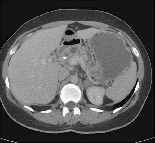
- Cross-sectional imaging is central: CT findings include a dilated pancreatic duct (68%), parenchymal atrophy (54%) and pancreatic calcifications (50%), with sensitivity 56–95% and specificity 85–100% [1].
- Plain abdominal radiography may show pancreatic calcification [2][3].
- Calcification is seen very well on CT but not on MRI, while a normal-looking pancreas on CT or MRI does not rule out chronic pancreatitis [2].
- CT findings depend on the morphological type, the duration of disease and the presence of complications.
- Early ductal or parenchymal changes may be subtle, while advanced disease shows readily apparent irreversible architectural change, including the focal enlargement with dense calcification in the head known as the inflammatory head mass, which appears more common in European than American cohorts [5].
- In autoimmune pancreatitis calcifications are almost uniformly absent and the pancreas is usually diffusely enlarged, though a focal mass-forming variant occurs; in obstructive forms the duct is dilated upstream of the stenosis with atrophic acinar parenchyma [5].
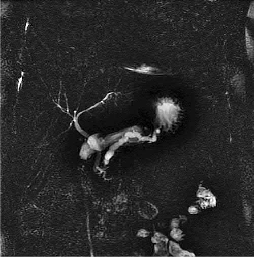
- MRI/MRCP, particularly with secretin stimulation, is useful for evaluating ductal strictures and pancreatic duct disruption [1][3]; intravenous secretin during the study may demonstrate a stricture not apparent on standard MRCP [2], and secretin stimulation also allows some assessment of exocrine function [5].
- ERCP, historically the diagnostic gold standard, is now reserved for cases where other imaging is contraindicated or inconclusive, or for therapeutic intervention on strictures, stones, or pseudocysts [1]; its classic features are marked main duct dilation, intraluminal filling defects from stones, clubbing of side branches, and areas of duct stricture [5].
- Histologically proven chronic pancreatitis can nonetheless occur with entirely normal pancreatography [2].
Endoscopic ultrasound and minimal-change disease
- Endoscopic ultrasound (EUS) is the most accurate technique for diagnosing early or minimal-change disease, using the Rosemont criteria, which combine major parenchymal features (hyperechoic foci with shadowing; honeycombing lobularity), minor parenchymal features, major ductal features (main duct calculi), and minor ductal features to categorise findings as consistent with, suggestive of, indeterminate for, or normal for chronic pancreatitis [1][7].
- Even a "normal" Rosemont classification has a poor negative predictive value, with up to 55% of such cases ultimately proving to have chronic pancreatitis histologically [1].
- Bailey & Love lists the sonographic features, stones, visible side branches, cysts, lobularity, an irregular main duct, hyperechoic foci and strands, main duct dilatation, and hyperechoic duct margins, and holds that four or more is highly suggestive of the diagnosis [2].
- Maingot's is more sceptical of EUS as an early-diagnosis tool.
- It adds little in advanced disease; various systems using up to 11 endosonographic criteria have been proposed but there is no gold-standard grading system and no agreement on the threshold number of abnormalities, so its value in early diagnosis remains uncertain [5].
- Its more practical utility is in suspected autoimmune pancreatitis, where EUS-directed core needle biopsy demonstrating the pathognomonic lymphoplasmacytic infiltrate can rule out malignancy and spare a patient with a mass-forming variant an operation [5].
- Functional testing includes faecal elastase-1 (>200 μg/g normal, 100–200 μg/g mild-to-moderate insufficiency, <100 μg/g severe insufficiency) and the 72-hour faecal fat test (>7 g/day defines steatorrhoea) [1].
- These are most reliable precisely in the patients in whom the diagnosis is already clinically obvious, and are of questionable accuracy in the subtle cases where objective documentation would be most useful [5].
- Elevated fasting glucose or HbA1c suggests pancreatic diabetes, and formal oral glucose or arginine tolerance testing may be helpful before pancreatic resection, particularly if autologous islet transplantation is contemplated [5].
- In suspected autoimmune disease, serum IgG4 is the key test, with rheumatoid factor, antinuclear antibody, CRP and ESR less specific [5].
Genetic testing
- The role of genetic testing is controversial.
- It is most reasonable to screen for PRSS1 mutations in patients with a strong family history, given autosomal dominant inheritance and the high risk of pancreatic cancer, a risk dramatically elevated further by tobacco use.
- However, hereditary pancreatitis patients without PRSS1 mutations may carry the same elevated cancer risk, and there is no evidence that serial imaging leads to earlier diagnosis or improved prognosis.
- Identifying CFTR or SPINK1 mutations may reassure patients with idiopathic disease by providing an explanation, but in the absence of therapy directed at the specific functional defect the clinical value is debatable; genetic counselling is highly advisable both in deciding whether to test and in interpreting the result [5].
Functional tests and the Cambridge morphological grades
- The pancreatic endocrine product that correlates most strongly with chronic pancreatitis is the pancreatic polypeptide response to a test meal, blunted or absent in severe disease, though a normal response does not exclude early disease [6].
- Direct measurement of exocrine secretion requires duodenal aspiration of juice after a nutrient (Lundh test meal) or hormonal (CCK or secretin) stimulus (endoscopic cannulation of the duct is neither risk-free, comfortable nor more sensitive) while indirect tests measure metabolites of compounds digested by pancreatic enzymes: the bentiromide test, in which ingested N-benzoyl-L-tyrosyl-p-aminobenzoic acid yields urinary PABA, the Schilling test, faecal fat, chymotrypsin or elastase, and [14C]-olein absorption [6].
- The Cambridge classification grades morphology as normal (no abnormal side branches; normal gland), equivocal (normal main duct with one of: fewer than three abnormal side branches, main duct 2–4 mm, gland enlarged more than twice normal, heterogeneous parenchyma), mild (normal main duct with two or more of those), moderate (main-duct changes; small cysts under 10 mm, duct irregularity, side-branch changes, focal acute pancreatitis, echogenic duct walls, contour irregularity) and severe (any of the above plus cysts, intraductal filling defects, calculi, duct obstruction or stricture, severe irregularity or contiguous organ invasion); on ERCP alone the grades run from suggestive (normal main duct, fewer than three abnormal branches) through mild (three or more) and moderate (abnormal main duct, more than three branches) to severe (plus a large cavity, obstruction, dilatation or irregularity, or filling defects) [6].
Scoring and Severity
- The Rosemont EUS classification (above) functions as the principal diagnostic/severity framework, stratifying findings into "consistent with," "suggestive of," "indeterminate for," and "normal" for chronic pancreatitis based on combinations of major and minor parenchymal and ductal criteria [1].
- The older Cambridge classification, agreed at a 1983 conference in Cambridge, England, grades chronic pancreatitis as equivocal, mild, moderate or marked according to combinations of features seen in the main and side-branch pancreatic ducts on CT and ductography [5].
- Neither resolves the persistent problem of patients with symptoms suspicious for chronic pancreatitis in whom imaging is negative, some of whom have functional abdominal pain disorders rather than pancreatic disease, and some of whom have genuine early or "minimal change" disease, the subject of successive Japan Pancreas Society consensus workshops in 1995 and 2001 [5].
- Surgical decision-making is further stratified by pancreatic duct calibre: a main pancreatic duct ≥7 mm defines "large duct disease," suitable for a decompressive (drainage) procedure, while a non-dilated duct defines "small duct disease," generally requiring a resectional procedure [1].
- Maingot's sets the large-duct threshold at a lumen exceeding 7 to 8 mm and adds a third morphological category between them, the dominant inflammatory head mass, sometimes called the "pacemaker" of chronic pancreatitis, since disease is often driven predominantly by pathology in the head [5].
- The ABSITE Review sets its threshold for a drainage procedure at ducts greater than 6 mm with a disease-free pancreatic head [8].
Treatment and Management
Medical management centres on cessation of alcohol and smoking, treatment of predisposing metabolic factors (e.g. hypertriglyceridaemia), and a low-fat diet, since disease progression can be slowed but not reversed [1][3]. Bailey & Love organises medical treatment under four headings, treat the addiction (helping the patient stop alcohol and tobacco, involving a dependency counsellor or psychologist); alleviate abdominal pain (eliminating obstructive factors in duodenum, bile duct or pancreatic duct, escalating analgesia stepwise, referring to a pain specialist, and considering CT- or EUS-guided coeliac axis block for intractable pain); nutritional and pharmacological measures (a diet low in fat and high in protein and carbohydrate, enzyme supplementation with meals, correction of fat-soluble vitamin and B12 malabsorption, micronutrient therapy with methionine, vitamins C and E and selenium, steroids only in autoimmune pancreatitis, medium-chain triglycerides in severe fat malabsorption because they are absorbed directly without digestion, and reduction of gastric secretion); and treat diabetes mellitus [2].
- Pain management follows a stepwise approach: NSAIDs first-line, then tramadol, then potent long-acting narcotics for severe pain, with adjuncts (tricyclic antidepressants, SSRIs, SNRIs, gabapentin) considered and multidisciplinary pain-team involvement recommended given addiction risk [1].
- Long-acting narcotics supplemented by short-acting formulations for breakthrough pain may be more effective than short-acting agents alone, though narcotic dependency is a common consequence, and psychosocial support such as counselling is essential to successful longitudinal management [5].
- Because in some patients pain burns out as the disease reaches end stage but the natural history is highly variable and relapsing pain may take years to relent if it ever does, a conservative watch-and-wait approach is rarely acceptable [5].
Enzyme replacement, and the paradox of coated versus uncoated
- Pancreatic exocrine insufficiency is treated with pancreatic enzyme replacement therapy (e.g.
- Creon/pancrelipase), generally around 90,000 USP units of lipase per day divided across meals, given with proton pump inhibitors to improve efficacy of uncoated enzymes [1][3].
- For steatorrhoea, enteric-coated preparations are most useful, and patients must be instructed to time ingestion in relation to meals to optimise mixing [5].
- For pain in early, small-duct or minimal-change disease the logic inverts: high doses of non-coated enzyme preparations appear to help, because activated enzymes in the duodenum decrease cholecystokinin-mediated stimulation of the pancreas, but they must be protected from gastric acid by suppression therapy, and trials using enteric-coated delayed-release formulations for this indication showed no benefit [5].
- Several randomised trials suggest a five-component antioxidant regimen reduces the frequency and intensity of painful episodes [5].
- Autoimmune pancreatitis confirmed by elevated IgG4 or biopsy may be treated with an 8-week tapering course of corticosteroids, including resolution of jaundice from mechanical biliary obstruction [5].
Endocrine insufficiency (type 3c diabetes) requires management by an endocrinologist experienced with the higher risk of severe hypoglycaemia in this population [1].
Neurolysis
- Neurolysis may be considered for patients who have failed medical management and lack anatomy amenable to endoscopic or surgical intervention.
- The commonest procedure is coeliac plexus block under radiological or endoscopic guidance, injecting steroid with local anaesthetic into the coeliac ganglion and, if temporary relief is obtained, following with permanent neurolysis using 100% alcohol; results in chronic pancreatitis have been mixed, with transient improvement typically no more than 6 months [5].
- Thoracoscopic splanchnicectomy has also been used, but as with other forms of neurolysis permanent resolution of pain is unusual [5].
Endoscopy versus surgery
- Endoscopic therapy (ERCP with duct dilation and stenting, stone extraction, sometimes preceded by extracorporeal shock wave lithotripsy) is used for symptomatic ductal obstruction, biliary obstruction (with plastic stenting for cholangitis or malnutrition), and pseudocyst drainage, malignancy must first be excluded, particularly for a pancreatic duct stricture [1].
- Technical success is reliably achieved in selected patients, but the effectiveness of endotherapy over time is often less than 50% for pain improvement or reduced attack frequency; multiple procedures are often needed, strictures and stones recur frequently, and the substantial fraction who fail generally require surgery anyway [5].
- Long-term presence of a stent within the pancreatic duct may worsen inflammatory strictures, though most series find a few patients achieve durable relief after stent removal [5].
- Bailey & Love adds that a stent placed across a dominant stricture should be left no more than 4–6 weeks because it will block, that the complication rate is high, and that fewer than two-thirds of patients get pain relief, though those who do may benefit from a subsequent surgical bypass [2].
The ESCAPE trial and earlier randomised trials found early surgical ductal drainage superior to an endoscopy-first approach for pain relief, with fewer reinterventions [1]. Two randomised trials directly comparing surgery with endoscopic stenting reported long-lasting superiority of the surgical approach for pain relief, quality of life over time, and other endpoints, and patients suitable for endotherapy are usually also candidates for surgery, absent medical contraindications [5].
When to operate
- The optimal timing of surgery remains debated.
- Most practitioners hold off until medical and endoscopic options are exhausted, while others advocate moving more expeditiously on the argument that early definitive intervention may forestall a permanent, refractory retroperitoneal neuropathy [5].
- One retrospective Dutch Pancreatitis Study Group evaluation of 266 surgically treated patients found that surgery performed within 3 years of symptom onset was independently associated with more significant postoperative pain relief and less endocrine insufficiency [5].
NICE puts surgery first for one specific anatomy and defers to endoscopy for another, without staging the two against each other as a sequence.
| NICE NG104 | Recommendation |
|---|---|
| 1.3.8 | Consider surgery (open or minimally invasive) as first-line treatment in adults with painful chronic pancreatitis that is causing obstruction of the main pancreatic duct |
| 1.3.9 | Consider extracorporeal shockwave lithotripsy for adults with pancreatic duct obstruction caused by a dominant stone if surgery is unsuitable |
| 1.3.10 | Offer EUS-guided drainage, or endoscopic transpapillary drainage for pancreatic head pseudocysts, to people with symptomatic pseudocysts (for example, those with pain, vomiting or weight loss) |
| 1.3.11 | Consider the same drainage for non-symptomatic pseudocysts meeting one or more of: associated with pancreatic duct disruption; creating pressure on large vessels or the diaphragm; at risk of rupture; suspicion of infection |
| 1.3.12 | Consider surgical (laparoscopic or open) drainage of pseudocysts that need intervention if endoscopic therapy is unsuitable or has failed |
Table reproduces the recommendations on managing complications [4].
- Note the asymmetry.
- For main duct obstruction, surgery is the first-line offer and lithotripsy is the fallback where surgery is unsuitable.
- For pseudocysts, endoscopic drainage is the offer and surgery is the fallback where endoscopy is unsuitable or has failed.
- NICE also identifies four specific criteria that justify draining a pseudocyst that is causing no symptoms at all, duct disruption, pressure on large vessels or the diaphragm, risk of rupture, and suspicion of infection.
- On nutrition NICE is emphatic that everyone is at risk: be aware that all people with chronic pancreatitis are at high risk of malabsorption, malnutrition and deterioration in quality of life [4].
- Protocols agreed with the specialist pancreatic centre should identify when specialist dietitian advice is needed (on food, supplements and long-term pancreatic enzyme replacement therapy) and when to start those interventions, with assessment by a dietitian considered for anyone diagnosed [4].
- For neuropathic pain related to chronic pancreatitis, NICE refers to its guideline on neuropathic pain in adults rather than making a pancreatitis-specific recommendation [4].
- People with pancreatic ascites and pleural effusion should be considered for referral to a specialist pancreatic centre [4].
- And on smoking, be aware of the link between smoking and chronic pancreatitis, and advise people with chronic pancreatitis to stop smoking in line with the NICE guideline on tobacco [4].
- A structured follow-up schedule is the part of NG104 with no textbook counterpart.
- Monitoring by clinical and biochemical assessment for exocrine insufficiency and malnutrition, agreed with the specialist centre, at least every 12 months (every 6 months in under-16s) adjusting treatment of vitamin and mineral deficiencies accordingly [4].
- A bone density assessment every 2 years for adults [4].
- HbA1c monitoring at least every 6 months, because the lifetime risk of diabetes is as high as 80% and rises with duration of pancreatitis and the presence of calcific disease [4].
- People with type 3c diabetes assessed every 6 months for the potential benefit of insulin therapy [4].
- And on cancer: be aware that people with chronic pancreatitis have an increased risk of pancreatic cancer, with the lifetime risk highest (around 40%) in those with hereditary pancreatitis, for whom annual monitoring for pancreatic cancer should be considered [4].
The handover to primary care is specified too: information passed to GPs should include how the person should take their pancreatic enzyme replacement therapy, including dose escalation as necessary, and that they should be offered HbA1c testing at least every 6 months and bone mineral density assessment every 2 years [4].
Endoscopic results, lithotripsy and the Forsmark algorithm
- In a prospective randomised trial of endoscopic therapy for idiopathic recurrent pancreatitis, 53% of controls had further attacks against 11% of treated patients; a single ESWL session in 35 patients with duct stones, with 86 ERCP sessions to complete clearance, gave significant symptom relief in 80% at 2.4 years, and endoscopic intraductal lithotripsy is available in specialist centres, but recurrent stone formation leaves the long-term role of lithotripsy in calcific disease uncertain [6].
- Patients with sphincter of Oddi dyskinesia are at high risk of post-ERCP pancreatitis after biliary sphincterotomy, reduced by a prophylactic pancreatic stent or rectal indomethacin, and prolonged pancreatic stenting is usually avoided because of duct injury [6].
- Schwartz reproduces Forsmark's algorithm: confirm the diagnosis (history; MRI/MRCP, EUS, CT; tube- or endoscopy-based secretin test if imaging is equivocal), exclude and treat cancer or IPMN, pseudocyst, bile duct and duodenal obstruction; medical therapy measures pain and its impact, refers for structured smoking and alcohol cessation, supplements vitamin D and calcium with baseline bone densitometry, starts analgesia with tramadol escalating slowly, adds pregabalin, gabapentin, SSRIs, SNRIs or tricyclics for persistent pain or rising opioid need, tests faecal elastase or serum trypsin and HbA1c or glucose tolerance for insufficiency, and gives steroids for autoimmune disease; anatomy then directs treatment, an inflammatory head mass to DPPHR (Beger, Frey or Berne) or Whipple with the patient told that data support surgery's superiority, a dilated duct (≥6 mm) to endoscopic sphincterotomy, dilatation, stenting, lithotripsy and stone extraction with modified Puestow or Frey for failure, and small-duct disease (≤6 mm) to continued medical therapy, "V-plasty" or total pancreatectomy with islet autotransplantation [6].
- Nealon and Thompson's 1993 study showed that duct decompression can delay or prevent the progression of chronic obstructive pancreatitis (the only therapy shown to do so) and Nealon and Matin described pain syndromes predictive of surgical response, pain from ductal hypertension being most readily relieved by decompression [6].
Surgeries
- Surgical intervention is indicated for intractable pain, biliary or pancreatic duct obstruction, duodenal obstruction, pseudocyst/pseudoaneurysm formation, or inability to exclude malignancy [1].
- In over 90% of patients the main indication is pain, with occasional operations to relieve biliary or gastrointestinal obstruction, drain a symptomatic pseudocyst internally, or address vascular complications such as gastric variceal haemorrhage from splenic vein thrombosis [5].
- Choice of operation depends on ductal calibre and the presence of a dominant inflammatory mass.
- These operations are technically demanding and carry significant morbidity and mortality; even in appropriately selected patients, durable pain relief is achieved in at most 85% at 5 years [5].
Large duct disease
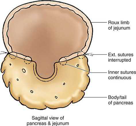
- For large duct disease (duct ≥7 mm) from stones or strictures, the side-to-side Roux-en-Y pancreaticojejunostomy (modified Puestow / lateral pancreaticojejunostomy) is standard: the anterior pancreatic duct is unroofed from tail to near the ampulla, stones extracted, and a Roux limb anastomosed laterally; this preserves parenchyma and relieves pain in about 80% of cases, though 30% recur (usually 3–5 years later) [1][3].
- Puestow's original description included resection of the pancreatic tail, which the 1960 Partington–Rochelle modification eliminated [5].
- The ABSITE Review notes that most patients improve, and that the operation suits ducts over 6 mm with a pancreatic head free of disease [8].
- The original Duval procedure combined distal pancreatectomy, splenectomy and end-to-side pancreaticojejunostomy, and the Puestow–Gillesby procedure added a longitudinal element with distal resection [3].
- Two technical points from Maingot's shape the operation.
- The gastroduodenal artery is suture-ligated at both the superior and inferior border of the pancreatic head before the ductotomy, to prevent intraoperative haemorrhage during incision of the head and duct and postoperative bleeding at the anastomosis [5].
- And the ductotomy must extend across the neck into the head where the gastroduodenal artery traverses the pancreas, and as far laterally along the tail as possible, so the entire dilated segment is unroofed.
- The Roux limb is 40 to 50 cm, with at least 40 cm between the pancreaticojejunostomy and the enteroenterostomy to prevent reflux of enteric contents [5].
- In most series, 75% to 80% of patients with diffusely dilated ducts and no dominant mass achieve durable pain relief over 5 to 10 years, with low perioperative morbidity and preserved endocrine and exocrine function since no parenchyma is removed.
- Failures are usually from inappropriate selection (an underappreciated extent of disease with significant head fibrosis) or from ongoing fibrosis with progressive neuropathic pain [5].
The dominant head mass
Lateral pancreaticojejunostomy has limited applicability without diffuse duct dilation: an isolated drainage procedure in patients with complex inflammatory changes in the head, body or tail gives poor outcomes, with quick recurrence of pain and progression to exocrine insufficiency [5]. Four operations are used with frequency today, pancreaticoduodenectomy with or without pyloric preservation, and three forms of duodenum-preserving pancreatic head resection: the Beger, Berne and Frey procedures [5].
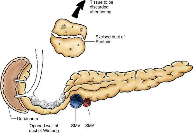
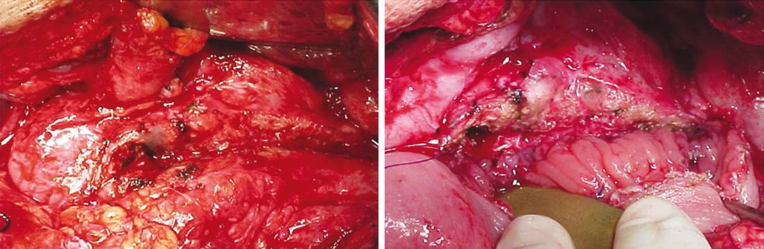
- For an enlarged pancreatic head (≥5 cm) with severe inflammation precluding safe pancreaticoduodenectomy, the Frey procedure combines local coring-out ("excavation") of the pancreatic head with a longitudinal pancreaticojejunostomy, leaving a 1-cm rim of tissue along the duodenal margin.
- This achieves complete or satisfactory pain control in about 95% of patients, though roughly one-third develop endocrine or exocrine insufficiency [1][3].
- Its distinguishing technical feature is that no tunnel is created behind the pancreatic neck, the entire length of the pancreas is exposed anteriorly, the gastroduodenal artery ligated, the gallbladder removed, the cystic duct cannulated with a Bakes dilator so the bile duct can be palpated through the head, and the head excavated down to the level of the portal vein, leaving a rim of tissue around the bile duct at the duodenal margin [5].
- It was developed specifically to address the disadvantage of the Beger operation, that it does not deal with disease coexisting in the body and tail, to which late Beger failures have been attributed [5].
- The Beger procedure (duodenum-preserving pancreatic head resection) removes the pancreatic head while preserving duodenal continuity, with reconstruction via a Roux limb anastomosed to the pancreatic remnant; randomised trials show pain relief equivalent to pancreaticoduodenectomy and the Frey procedure [1][3].
- First described in 1972, it grew from the premise that pancreaticoduodenectomy is unnecessarily radical for benign pathology and that a more limited resection preserving the duodenum would avoid delayed gastric emptying and insulin-dependent diabetes.
- The neck is divided over the portal vein, the head rotated out of the retroperitoneum, and the gland cored out with electrocautery leaving a rim attached to the duodenum with the bile duct intact within it [5].
- The Berne procedure is a further modification adopting the Frey operation's technical safety advantage of not transecting the neck off the portal vein: the anterior surface of the head mass is palpated and cored out, and a Roux limb sewn to the residual rim, with no lateral pancreaticojejunostomy [5].
| Operation | Reported outcomes |
|---|---|
| Beger DPPHR, 603 consecutive patients | Pancreatic fistula 3.3%, delayed gastric emptying 1.5%, perioperative mortality 0.7%; 91.3% free of pain at median 5.7 years |
| Beger DPPHR vs pylorus-preserving pancreaticoduodenectomy, 40 patients | At 6 months, 75% pain-free after DPPHR vs 40% after PPPD; weight gain 4.1 kg vs 1.9 kg. At median 7 years, identical quality of life, pain scores, and rates of exocrine and endocrine insufficiency |
| DPPHR (Beger and Frey) vs PPPD, 85 patients over 5 years | Operative time 360 vs 435 minutes favouring DPPHR; no difference in morbidity, mortality, long-term quality of life, pain control, or endocrine/exocrine function |
| Frey local resection with longitudinal pancreaticojejunostomy | Mortality under 1%, morbidity 19% to 32%; excellent pain relief in about 75%, with pain scores and insufficiency rates identical to Beger over follow-up as long as 9 years |
| Frey vs PPPD, prospective randomised | Postoperative morbidity 17% vs 30% favouring Frey, similar pain improvement, better overall quality of life for Frey; at median 7 years no differences; at 15 years pain comparable but Frey patients scored 100 in physical status and working ability against 60 and 50 for PPPD |
| Berne modification, 160 patients, mean 5.3 years | Preserved endocrine function relative to preoperative tests, with significant and durable improvement in pain and quality of life |
- Table reformats the reported outcome data [5].
- No randomised trial has shown any one technique clearly superior.
- The numbers are small and the outcome metrics variable and imperfect, so no consensus has emerged, European surgeons have tended to favour duodenum-preserving approaches and American surgeons pancreaticoduodenectomy, and in one survey of 59 American Pancreas Club members only 34 had ever performed a duodenum-preserving head resection and only 23 continued to do so regularly [5].
Maingot's therefore tailors the operation to the morphology rather than to a hierarchy: a dominant head mass with a dilated main duct but no biliary dilation is best served by a Frey procedure; a dominant head mass without main duct dilation and without biliary obstruction suits the Berne modification; and biliary obstruction, or imaging features suspicious for malignancy, should probably lead to pancreaticoduodenectomy rather than any form of duodenum-preserving resection [5].
For a single stricture proximal to the papilla, or a focal inflammatory mass without significant ductal dilation, pancreaticoduodenectomy (Whipple procedure) or pylorus-preserving pancreaticoduodenectomy may be used, contraindicated if more than one obstruction is present in the duct [1][3]. One caution specific to operating in a pancreatitic field: small areas of fat necrosis or fibrosis from prior attacks are easily mistaken for metastatic deposits at exploration [5].
Small duct disease and total pancreatectomy
- For diffuse glandular involvement without ductal dilation ("small duct disease"), total pancreatectomy provides the most durable pain relief but invariably causes diabetes; islet autotransplantation at the time of total pancreatectomy can reduce this burden, with roughly one-third of patients achieving insulin independence, another third requiring intermittent insulin, and the remainder fully insulin-dependent, while 90% experience pain relief or reduction [1].
- Total pancreatectomy is reserved as a salvage operation given the resultant "brittle" diabetes [3].
- These patients present a particular challenge because there is no discrete target for either endoscopic or surgical intervention [5].
- The islet isolation process is worth understanding.
- The resected pancreas is cooled to 4°C in an organ-preserving solution, transected at the neck and the duct cannulated, the ductal system perfused with cold purified collagenase, and the gland sectioned and physically shaken in a small digestion chamber at 37°C, digestion monitored continuously by microscopy of samples of the digestate and stopped once acinar tissue has separated from the islets but before the islets begin to fragment
- The islets are then partially purified by gradient density centrifugation on a cold dextrose gradient, washed, resuspended in albumin-rich transplant medium, and injected into the portal circulation either by ultrasound-guided percutaneous transhepatic catheter or by direct operative cannulation of the portal vein [5].
- Maingot's is measured about the results.
- At 5 years in 166 patients from one centre, 74% were narcotic-independent, all had stable glycaemic control, and 27% had long-term insulin independence, but complete long-term insulin independence is achieved in only a relatively small minority, and pain is persistent or recurrent in about half of patients even after total pancreatectomy [5].
- Transplanted islets migrate across the liver sinusoids and reside in the parenchyma, exhibiting some peri-islet fibrosis; no chronic hepatic fibrosis or cirrhosis has been reported but the concern exists [5].
- The procedure is costly, requires expertise difficult to replicate, and its indications remain controversial, with the strongest argument perhaps in the limited subset of patients with hereditary pancreatitis who otherwise carry a significant long-term risk of pancreatic cancer [5].
- As salvage after a failed prior operation, a series of 64 completion total pancreatectomies with islet autotransplantation after a previous pancreaticoduodenectomy, Frey, Puestow or Berne procedure achieved narcotic independence in 44% and insulin independence in 20%, with quality of life improved in all domains [5].
Surgical pain-control adjuncts include splanchnicectomy (EUS-guided or thoracoscopic) and radiofrequency ablation, though celiac neurolysis has not shown sustained benefit in chronic pancreatitis [1][3]. For minimal-change disease with pain, bilateral thoracoscopic splanchnicectomy or coeliac ganglionectomy may be used [8].
Biliary, duodenal and cystic complications
- Biliary strictures from chronic pancreatitis are treated with temporary plastic stenting for cholangitis, Roux-en-Y hepaticojejunostomy where malignancy is excluded and significant scarring precludes head resection, or definitively by pancreaticoduodenectomy when malignancy cannot be excluded; a Frey or Beger procedure can also relieve biliary obstruction by removing the compressive head parenchyma [1].
- The ABSITE Review's threshold for operating on a common bile duct stricture is pain, jaundice, progressive cirrhosis or cholangitis, diagnosed by MRCP and treated by hepaticojejunostomy or choledochojejunostomy, always after making sure the stricture is not pancreatic cancer [8].
- Duodenal stenosis (in up to 1.2% of patients) is treated with gastrojejunostomy after nutritional optimisation [1].
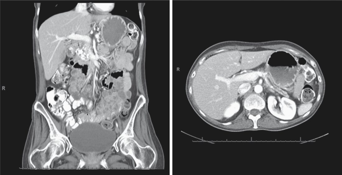
- Symptomatic pancreatic pseudocysts (developing in 30–40% of chronic pancreatitis patients, with only 10% resolving spontaneously given persistent ductal obstruction) are managed by endoscopic (EUS-guided) or surgical (cystogastrostomy, cystojejunostomy) drainage [1].
History of drainage, and who benefits from head resection
- Duval and, separately, Zollinger described caudal Roux-en-Y pancreaticojejunostomy in 1954, an operation that almost invariably failed through restenosis and segmental obstruction from progressive scarring; in 1958 Puestow and Gillesby described the "chain of lakes" and proposed longitudinal decompression of the body and tail into a Roux limb (four of their 21 cases side-to-side), and two years later Partington and Rochelle described the simpler side-to-side longitudinal Roux-en-Y pancreaticojejunostomy universally known as the Puestow procedure [6].
- Pain relief after Puestow-type decompression is 75–85% for the first few years but recurs in more than 20% after 5 years from progressive disease even with abstinence [6].
- Beger described the duodenum-preserving head resection in 1980 and Frey and Smith the local head resection with longitudinal pancreaticojejunostomy in 1987; Frey and Amikura's 50 patients followed over 7 years had complete or substantial relief in 87% with no mortality and 22% complications, Frey's 1994 modification limited the posterior resection to the back wall of the opened duct of Wirsung and the uncinate duct to avoid penetrating the posterior capsule, Ho and Frey in 2001 described excavating the head core alone, and Gloor's Bern group proposed excavation as an alternative to DPPHR in portal hypertension (the Berne modification) which Köninger's randomised trial found identical to the classic Beger at 2 years after an initial reduction in morbidity [6].
- Johns Hopkins data suggest that aetiology predicts the benefit of resection or hybrid procedures: in 35 Frey patients those with more than 80% fibrosis had 100% pain relief against 60% with under 10% fibrosis, and in 60 Frey or Whipple patients 89% with "toxic" (alcohol or tobacco) disease had prolonged relief against 39% with hereditary or idiopathic disease, independent of fibrosis, so the latter may be better served by total pancreatectomy with islet autotransplantation [6].
- Comparative trials: Klempa's and Büchler's level 1 studies gave DPPHR shorter stay, more weight gain and less diabetes and exocrine failure than the Whipple with equal pain control over 3–5 years, echoed by Keck; Izbicki's 61-patient trial found fewer complications after Frey than pylorus-preserving Whipple (19% vs 53%) and better global quality of life (71% vs 43%) with equal pain control at 2 years, Farkas found the same for his organ-preserving head resection, and Bachmann's 15-year follow-up found survival and function superior after Frey; Izbicki's 74-patient trial of DPPHR against Frey at 8.5 years showed no differences in quality of life, pain, late mortality or insufficiency [6].
- Islet yield from a sclerotic gland depends on the degree of fibrosis, so patient selection matters; total pancreatectomy with islet autotransplantation gives impressive improvement in quality of life and pain, with significantly better outcomes in children than adults because hereditary and idiopathic causes predominate in the young [6].
- Distal pancreatectomy for a pseudocyst, with or without splenectomy, is challenging after previous pancreatitis, and internal drainage of the communicating duct or the cyst itself should be considered instead [6].
Complications
- Recognised complications include pseudocyst formation, biliary duct obstruction, gastric outlet or duodenal obstruction, and gastrointestinal bleeding secondary to pseudoaneurysm formation or variceal bleeding from splenic vein thrombosis (occasionally requiring splenectomy) [3].
- Chronic pancreatitis is the commonest cause of splenic vein thrombosis, which can produce bleeding from isolated gastric varices formed as collaterals, treated by splenectomy where those varices bleed in isolation [8].
- All the complications of acute pancreatitis can occur with chronic pancreatitis, and infection is not infrequent, possibly related to the diabetes [2].
- Long-term exocrine and endocrine pancreatic insufficiency are near-universal in advanced disease [1]; pancreatic insufficiency, which generally refers to exocrine function, requires loss of over 90% of function, is diagnosed by faecal fat testing, and is treated with a high-carbohydrate, high-protein, low-fat diet plus enzyme supplementation [8].
- Patients with chronic pancreatitis carry an estimated 4% lifetime risk of developing pancreatic cancer [3], and any focal mass arising in a background of chronic pancreatitis should be regarded with suspicion and biopsied before treatment [1].
- The risk is markedly higher in hereditary pancreatitis and in pancreatitis of young onset, particularly if the patient smokes [2].
Prognosis
- Chronic pancreatitis is a progressive, irreversible disease; while cessation of the causative agent (alcohol, smoking) can slow progression, established fibrosis does not reverse [1].
- Decompressive surgery for large-duct disease provides durable pain relief in the majority of patients, though roughly 30% experience recurrence within 3–5 years [1].
- Maingot's caps realistic expectation at 85% durable pain relief at 5 years even with appropriate selection, and notes that a single unifying model of pathogenesis remains elusive, that both endoscopic and surgical procedures are technically demanding with substantial morbidity, and that high-level evidence comparing interventions in large series is lacking, which is why chronic pancreatitis is best managed in experienced centres where multidisciplinary teams individualise treatment [5].
- Groups managing severe diffuse disease have found that patients with depression or ongoing substance abuse have a poorer overall outcome, underscoring the value of multidisciplinary management incorporating pain specialists and psychiatric support [1].
References
- Sabiston Textbook of Surgery, 22nd ed., Ch. 92
- Bailey & Love's Short Practice of Surgery, 28th ed., Ch. 72
- Oxford Handbook of Clinical Surgery, 5th ed., Ch. 9, as the Partington–Rochelle modification without distal pancreatectomy
- NICE Guideline NG104: Pancreatitis (2018, updated 2020), 1.1.7; 1.1.11; 1.3.1; 1.3.2; 1.3.3; 1.3.4, 1.3.5; 1.3.8; 1.3.8, 1.3.9, 1.3.10, 1.3.11, 1.3.12; 1.3.13; 1.3.14; 1.3.15; 1.3.20; 1.3.21; 1.3.22, 1.3.23; 1.3.24, 1.3.25 www.nice.org.uk
- Maingot's Abdominal Operations, 13th ed., Ch. 71, Chronic Pancreatitis
- Schwartz's Principles of Surgery, 11th ed., Ch. 33, Tables 33-12 and 33-15
- Schwartz's Principles of Surgery: ABSITE and Board Review, Ch. 33
- The ABSITE Review, 2022, Ch. Pancreas