



Cholangitis
Summary
- Acute cholangitis is an ascending bacterial infection of the biliary tree that develops secondary to obstruction and increased intraluminal pressure, most commonly caused by choledocholithiasis [1].
- First described by Jean Martin Charcot in 1877, it classically presents with the triad of fever, jaundice and right-upper-quadrant pain, though this is present in fewer than half of patients [1][2].
- Untreated, cholangitis can progress to septic shock with altered mental status (Reynolds pentad), which carries a mortality approaching 100% without prompt treatment, making biliary decompression a surgical emergency in severe cases [1].
- Maingot's calls it the most rapidly fatal complication of gallstones, with mortality approaching 100% in patients who fail conservative therapy and do not undergo the needed drainage intervention [3].
There is no NICE guideline on acute cholangitis. The relevant UK guidance is the British Society of Gastroenterology's updated guideline on the management of common bile duct stones, which contains a short section on cholangitis, and NICE CG188's recommendations on managing common bile duct stones, which cover the underlying cause but not the septic episode [4][5].
Definition
- Cholangitis is infection and inflammation of the bile ducts arising from bacterial colonisation of stagnant, obstructed bile, with ascending spread into the biliary tree and, in severe cases, the bloodstream [1][6].
- It differs from simple bile duct obstruction (e.g. by a stone) in that infection is superimposed on the obstruction, converting a mechanical problem into a life-threatening septic one [7].
- Maingot's defines it as biliary tract bacterial infection occurring in the setting of biliary tree obstruction, both elements are required [3].
Pathophysiology
Biliary obstruction causes bile stasis, providing an environment for bacterial colonisation and ascending infection of the biliary tree [1][7]. Bactibilia can be identified in up to 90% of patients with an obstructing stone [1].
Why bile is normally sterile, and how that fails
- Bile is normally sterile, but bacteria are frequently cultured from it once the tree is obstructed or instrumented, by a stone, a stricture or an endoprosthesis [3].
- Three mechanisms maintain sterility: bile flow itself, the sphincter of Oddi, and the bacteriostatic properties of bile; obstruction reduces these antibacterial defences and allows bacteria access [3].
- The route of infection is unclear, with ascent from the duodenum and haematogenous seeding both proposed [3].
- Once colonisation has occurred, stasis allows exponential bacterial growth, and as biliary pressure rises with obstruction, bacteria and their endotoxins leak into the systemic circulation to cause the septicaemia of cholangitis [3].
- The ABSITE Review gives the threshold as a biliary pressure above 200 mm Hg, at which cholovenous reflux produces systemic bacteraemia [8].
Counter-intuitively, patients with partial obstruction have a higher chance of developing cholangitis than those with complete obstruction, and bile duct stones cause cholangitis more often than obstructing neoplasms [3].
Microbiology
- The most common pathogens are Klebsiella, E. coli, Enterobacter, Pseudomonas and Citrobacter species [1]; typically Gram-negative organisms such as E. coli and Pseudomonas predominate [7]. E. coli is the single commonest and Klebsiella the second [8].
- Maingot's quantifies the distribution: Escherichia coli in 25–50%, Klebsiella species in 15–20%, and other Enterobacteriaceae in 5–10%, with anaerobes present in 5–10% of patients [3].
- Two clinical patterns modify this: Pseudomonas species together with skin and oral flora are associated with biliary tract interventions, while anaerobes are seen most often in the elderly after biliary surgery [3].
- Hepatocellular injury from infection and inflammation elevates serum transaminases and alkaline phosphatase.
- Untreated, the infection can progress to septic shock with mental status changes and hypotension (the Reynolds pentad) an ominous sign associated with near-uniform mortality without prompt treatment [1].
- Complications include stricture formation and hepatic abscess as late sequelae [8].
Two conditions for cholangitis
Hepatic bile is sterile and duct bile is kept sterile by continuous antegrade flow and antibacterial substances such as immunoglobulin; mechanical hindrance to flow permits ascending contamination from the bowel, and positive bile cultures are common with duct stones and other obstructions, but contamination alone does not cause cholangitis, significant bacterial contamination and biliary obstruction are both required [9]. Besides stones, the causes include primary sclerosing cholangitis, benign and malignant strictures, parasites, instrumentation, indwelling stents and partially obstructed biliary-enteric anastomoses, and the organisms most often cultured are E. coli, Klebsiella pneumoniae, Streptococcus faecalis, Enterobacter and Bacteroides fragilis [9].
Clinical features
- The classic Charcot triad (fever (often with rigors), right-upper-quadrant or epigastric pain, and jaundice) is present in less than 50% of patients, with jaundice the least common of the three findings [1][2].
- Maingot's reports a higher figure, with the complete triad present in 50–70% of patients at presentation [3].
- Leukocytosis with an abnormal liver panel is more consistently found than the full clinical triad [1].
Frequency of individual findings
| Finding | Frequency at presentation |
|---|---|
| Fever | 90% |
| Abdominal pain | 70% |
| Right upper quadrant tenderness | 65% |
| Jaundice | 60% |
| Complete Charcot triad | 50–70% |
| Altered mental status | 30% |
| Hypotension | 20% |
| Peritonitis | Uncommon |
- Table reformats the reported frequencies [3].
- Hypotension and altered mental status appear in septic patients, and are termed Reynolds's pentad when they occur alongside the Charcot triad [3].
- Progression to hypotension and altered mental status signals septic shock [1][10].
- On examination the patient is febrile and icteric, with epigastric and right hypochondrial tenderness [2].
- Older patients may present atypically with episodic pain, fever and jaundice without a full clinical picture [8].
Atypical and stent-related presentations
Gallstone cholangitis most commonly affects older women, and Charcot's triad is present in about two-thirds, but the presentation may be atypical with little fever, jaundice or pain, most often in the elderly, who may have unremarkable symptoms until the process is advanced; patients with indwelling stents are at particularly high risk yet rarely become jaundiced because a patent stent maintains flow, and the abdominal findings are indistinguishable from acute cholecystitis [9]. Imaging, ultrasound for stones, dilated ducts and possibly the site of obstruction, CT and MRI for pancreatic and periampullary masses, rarely elucidates the exact cause, so the initial diagnosis is generally clinical [9].
Etiology
- Any obstructing process of the biliary tree can precipitate ascending cholangitis, but choledocholithiasis is by far the most common cause [1].
- In the United States, secondary choledocholithiasis (stones that have migrated from the gallbladder) is the commonest cause; primary bile duct stones dominate in areas where recurrent pyogenic cholangiohepatitis is endemic, including Hong Kong and Southeast Asia [3].
- Other causes include malignant biliary obstruction (cholangiocarcinoma, pancreatic head cancer, ampullary tumours), benign biliary strictures (post-surgical, from primary sclerosing cholangitis, or chronic pancreatitis), Mirizzi syndrome, choledochal cysts, indwelling biliary stents or percutaneous transhepatic catheters, and instrumentation of the biliary tree (e.g. after ERCP, or via a prior biliary-enteric anastomosis exposing the ducts to enteric organisms) [1][6][7].
- Maingot's adds tumours metastatic to the porta hepatis or peripancreatic nodes, and lists hemobilia, parasites and congenital abnormalities of the biliary tree as rare causes [3].
- Duodenal diverticula are a further recognised cause [8].
- A compelling reason to treat choledocholithiasis promptly is that the risk of developing cholangitis is upward of 5% in admitted patients with duct stones [1].
- Duct stones are more often associated with infected bile (80%) than gallbladder stones are [2].
Diagnosis
- Ultrasound is the first screening test and commonly shows biliary dilation, though it may not localise the level of obstruction [1].
- HIDA scans should be interpreted cautiously because infection reduces hepatic secretion of the tracer [1].
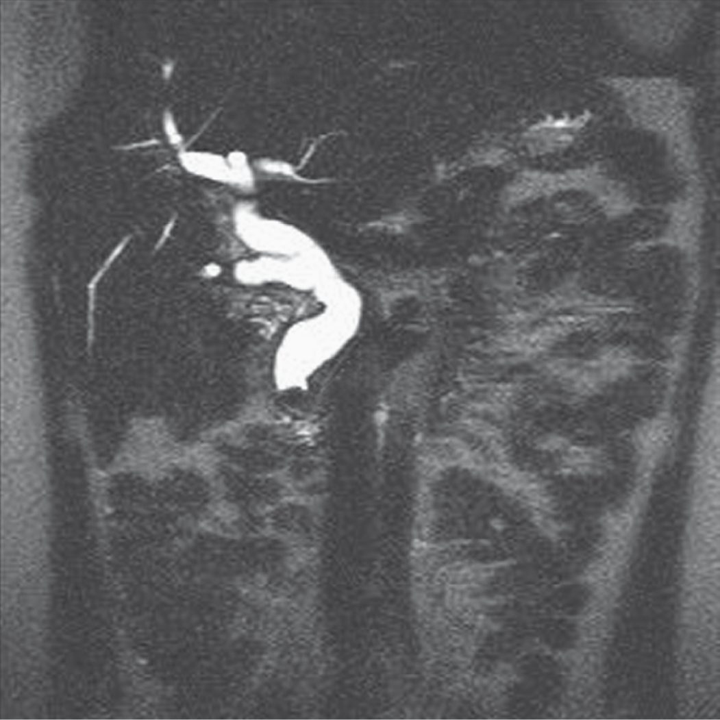
- Liver function tests and ultrasound are the initial investigations, with MRCP used to define the nature and level of obstruction [2].
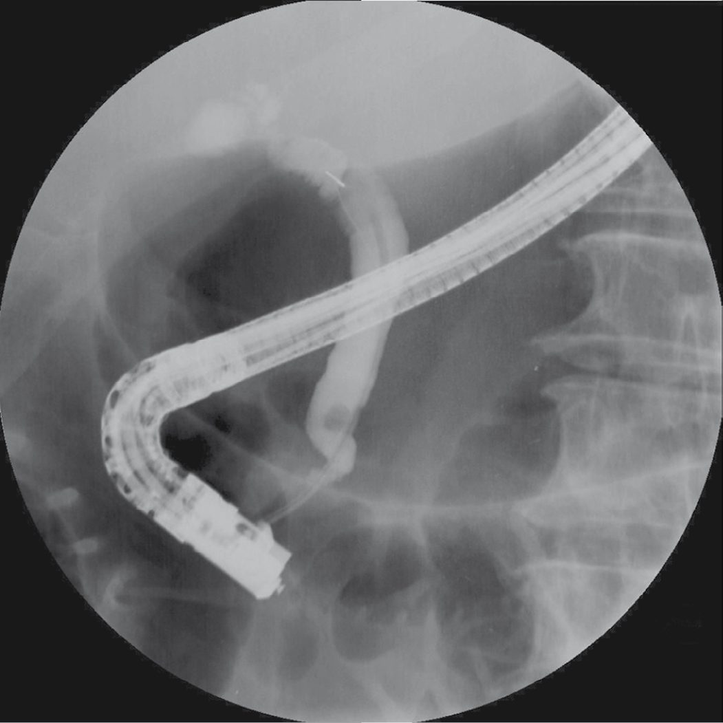
- The most valuable diagnostic and therapeutic modalities are direct cholangiography via ERCP or percutaneous transhepatic cholangiography (PTC) [1].
- Laboratory findings include leukocytosis and a cholestatic liver panel with elevated ALP and bilirubin [1].
Biochemistry
- Elevations of serum ALP, GGT and bilirubin are typical, mild increases in transaminases may be seen, and hyperamylasaemia is found in up to 30% of patients, a trap when distinguishing cholangitis from pancreatitis [3].
- Laboratory and radiological studies matter chiefly for separating cholangitis from acute cholecystitis, liver abscess and pancreatitis [3].
- Ultrasound will show a dilated common bile duct (over 6 mm, or over 10 mm after cholecystectomy) where the cause is biliary obstruction [8].
Choosing between imaging modalities in the acute setting
- Ultrasound is highly accurate for acute cholecystitis and for identifying gallstones, but its ability to establish choledocholithiasis is only about 50%, with reported figures ranging from 30% to 90%; the presence of duct stones can be inferred from associated duct dilatation, but a normal ultrasound without dilatation does not exclude either choledocholithiasis or cholangitis [3].
- CT is better at determining the level of obstruction and has 94% accuracy for choledocholithiasis where duct calculi are suspected [3].
- MRCP has sensitivity and specificity approaching ERCP and is useful for delineating anatomy, but its use in acute cholangitis is limited [3].
- ERCP is highly accurate at revealing the cause of obstruction and allows therapy in the same session, but given its well-defined life-threatening complications and the availability of non-invasive alternatives, it should not be used solely as a diagnostic tool in acute cholangitis [3].
Scoring and Severity
- Management is guided by disease severity.
- Mild acute cholangitis usually responds to antibiotics alone, though there should be a low threshold for biliary drainage if the patient fails to improve.
- Moderate cholangitis requires early duct clearance by endoscopic or percutaneous techniques.
- Severe cholangitis (with organ dysfunction, i.e. approaching the Reynolds pentad) requires haemodynamic and respiratory stabilisation followed by prompt biliary drainage [1].
In practice the operative severity marker is the response to 24 hours of resuscitation and antibiotics. Biliary sepsis resolves with conservative therapy in most patients, allowing time for detailed non-invasive delineation of the anatomy by CT or MRI to determine the cause and level of obstruction; urgent decompression is needed in the 10–15% who fail to respond within 24 hours [3].
The BSG defines the threshold for urgent intervention by clinical trajectory rather than by a numerical score: patients with acute cholangitis who fail to respond to antibiotic therapy, or who have signs of septic shock, require urgent biliary decompression [4].
The evidence on timing is quantitative even where the recommendation is not. A prospective study of 199 patients admitted with acute cholangitis found that for each day ERCP was delayed, length of stay increased by 1.44 days (95% CI 1.01 to 1.92), and identified an increased requirement for vasopressors in patients whose ERCP was performed more than 72 hours after presentation [4].
Treatment and Management
- Once cholangitis is suspected, adequate hydration, correction of coagulopathy, exclusion of diabetes, and broad-spectrum antibiotics should be started promptly [2].
- Rehydration and intravenous antibiotics are instituted immediately, with the severity of disease then assessed to guide the timing of drainage [1].
- Supportive measures begin without delay and include fluid resuscitation, correction of electrolyte deficits and coagulopathy, and analgesia [3].
Antibiotic choice and duration
- Empirical broad-spectrum antibiotics covering the common pathogens are started while blood cultures and, where available, bile cultures are sent [3].
- Aminoglycosides with ampicillin are no longer considered the ideal regimen because of Gram-negative resistance and nephrotoxicity; effective alternatives are combinations of extended-spectrum cephalosporins, extended-spectrum penicillins, metronidazole and ampicillin; fluoroquinolones as a single agent or with metronidazole; and ureidopenicillins alone or with metronidazole [3].
- Anaerobic cover matters more in the elderly and in those who have had biliary manipulation [3].
- Antibiotics are usually given for 7 to 10 days even where decompression has been achieved in the interim, but that convention has been challenged.
- A retrospective study of 80 patients successfully treated for cholangitis by ERCP and followed for 6 months compared 41 patients given antibiotics for 3 days or less, 19 for 4 to 5 days, and 20 for more than 5 days.
- The groups were well matched and the rate of recurrent cholangitis (24%) did not differ between them, leading the authors to conclude that 3 days may be sufficient once adequate drainage has been achieved and fever is abating [3].
Drainage: the mainstay
- Most patients improve with medical therapy and endoscopic or percutaneous biliary drainage; if these are unavailable or unsuccessful, surgical drainage via common bile duct exploration with T-tube placement is performed, though this is now rarely required [1][2].
- Drainage of the biliary tree is the mainstay of therapy, with timing and route varying by response to antibiotics, cause of obstruction, and comorbidity; where decompression is not achieved, liver abscesses are inevitable [3].
- Definitive treatment of the underlying cause (e.g. cholecystectomy for gallstone disease) is deferred until the patient is stabilised, the septic episode is treated, and the diagnosis is confirmed [1].
- Once relief of obstruction is required, endoscopic papillotomy/sphincterotomy is the preferred technique, followed by stone removal with a Dormia basket, or placement of a stent or nasobiliary drain for flushing if stone removal is not possible.
- If endoscopic drainage fails, PTC drainage can be performed, with subsequent percutaneous choledochoscopy [2].
- If the cholangitis is caused by an infected PTC tube, the tube itself is changed [8].
Evidence that endoscopy beats surgery
| Comparison | Endoscopic drainage | Alternative |
|---|---|---|
| 1992 randomised trial | 10% mortality | 32% mortality with surgical decompression |
| 83-patient randomised trial | 10% mortality | 30% mortality with surgical decompression |
| 65 endoscopic vs 40 surgical | No deaths | 5 deaths after traditional surgery |
| Patients aged 80 or over | 16.7% morbidity, 5.6% mortality | Surgical 87.5% and 25%; percutaneous 36.4% and 9.1% |
- Table reformats the comparative outcome data [3].
- With a success rate of 90% to 98% and low morbidity and mortality, ERCP with bile duct clearance is superior to the alternatives and is the modality of choice for decompressing the biliary tree in acute cholangitis, particularly where the cause is choledocholithiasis.
- Compared with percutaneous drainage it also has lower morbidity, shorter hospitalisation and higher definitive success rates [3].
Selecting the endoscopic technique
- Options range from nasobiliary catheters and biliary stents to sphincterotomy with stone extraction [3].
- In patients who have responded to antibiotics, sphincterotomy with duct clearance is preferred; drainage catheters are used in those with ongoing sepsis and multiple large stones, and in critically ill or coagulopathic patients, where sphincterotomy raises concerns about bleeding and lengthens the procedure [3].
- A randomised comparison of nasobiliary catheters against biliary stents found both equally effective, but stents more comfortable and not liable to accidental removal [3].
Percutaneous transhepatic drainage is reserved for patients in whom the papilla is inaccessible or ERCP has failed, and for those with suspected hilar cholangiocarcinoma, hepatolithiasis or intrasegmental cholangitis; it succeeds in 90% of patients with biliary obstruction but carries higher morbidity (30–80%) and mortality (5–15%) than endoscopic techniques, and coagulopathy must be corrected first [3].
Cholecystectomy after the episode
- The ABSITE Review's position is that cholecystectomy is generally needed before discharge to prevent further episodes [8].
- The randomised evidence supports operating on fit patients.
- In a trial randomising patients after common duct clearance, 47% allocated to a wait-and-see approach developed biliary symptoms compared with 2% allocated to cholecystectomy within 6 weeks of the endoscopic procedure, and 37% of the wait-and-see group eventually needed cholecystectomy [3].
- A second trial randomised 98 elderly patients (mean age 80) to open cholecystectomy with operative cholangiography and, if necessary, duct exploration, or to endoscopic sphincterotomy alone.
- Immediate morbidity (23% vs 16%) and mortality (4% vs 6%) did not differ significantly, but at a mean 17 months biliary symptoms had recurred in three surgical patients, none of whom needed repeat surgery, against ten endoscopic patients, seven of whom required further biliary surgery [3].
- Non-randomised and retrospective studies put the risk of subsequent biliary problems at 4–12% in patients with duct stones [3].
- The exception is patients in whom duct stones originate from intrahepatic stones, where cholecystectomy may not prevent future biliary complications [3].
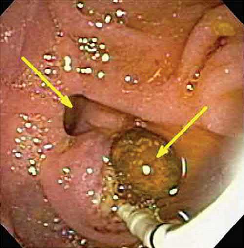
For urgent decompression the BSG recommends endoscopic stone extraction and/or biliary stenting, with percutaneous radiological drainage considered as the alternative if ERCP is not possible [4].
Where the cholangitis accompanies pancreatitis of suspected or proven biliary origin, or there is persistent biliary obstruction, biliary sphincterotomy and endoscopic stone extraction are recommended within 72 hours of presentation [4]. The BSG is explicit that there is no evidence supporting ERCP within 24 hours rather than within 72 hours, while noting that clinicians should remain alert to individuals with severe sepsis for whom urgent ERCP within 24 hours may be optimal [4].
- Two supporting recommendations shape how that ERCP is delivered.
- Tolerability and likelihood of therapeutic success are higher in selected patients if ERCP is performed under propofol sedation or general anaesthesia, and hospitals looking after these patients should have ready and prompt access to anaesthesia-supported ERCP, on site or through a clinical network [4].
- To reduce post-ERCP pancreatitis, rectal diclofenac or indometacin at a dose of 100 mg should be given at the time of ERCP to all patients without a contraindication to NSAIDs [4].
Prophylactic antibiotics before ERCP are a separate question from treating established cholangitis: in the absence of specific risk factors for sepsis, sclerosing cholangitis, communicating pancreatic cysts, hilar strictures, liver transplantation, cholangioscopy, or a failed attempt to drain an opacified bile duct, the BSG suggests they can safely be avoided [4].
- Where duct stones cannot be extracted, NICE limits stenting to a holding measure: if the bile duct cannot be cleared with ERCP, biliary stenting should be used to achieve drainage only as a temporary measure until definitive endoscopic or surgical clearance [5].
- The BSG agrees, recommending short-term use of a stent followed by further endoscopy or surgery, and restricting a stent as sole treatment to a selected group with limited life expectancy and/or prohibitive surgical risk [4].
- The reason is quantified: in one Italian series of long-term stents, patients whose stents were changed on demand had a 36% cholangitis rate with 8% associated mortality over a mean 14 months, against an 8% cholangitis rate and 2% mortality in those changed electively at three-monthly intervals [4].
On definitive treatment of the cause, cholecystectomy is recommended for all patients with common bile duct stones and gallbladder stones unless there are specific reasons for considering surgery inappropriate; where operative risk is prohibitive, biliary sphincterotomy and endoscopic duct clearance alone is suggested as an acceptable alternative [4].
Drainage route, mortality and aftercare in Schwartz
- ERCP with sphincterotomy shows the level and cause of obstruction, allows bile culture, removes stones and drains the ducts with catheters or stents as needed; PTC, EUS or surgical drainage are used when ERCP is unavailable, chosen by the type and site of obstruction and local expertise, and cholecystostomy tubes are not indicated because the source of infection is extrinsic to the gallbladder [9].
- Patients can deteriorate rapidly and may need intensive care and vasopressors, but most respond to decompression and support; overall mortality is now about 5%, much higher with renal failure, cardiac impairment, hepatic abscess or malignancy [9].
- After gallstone cholangitis elective cholecystectomy is recommended about 6 weeks after resolution; cholangitis from other causes is managed by treating the specific obstruction without necessarily removing the gallbladder, and patients with indwelling stents usually need repeated imaging and stent exchange to limit recurrence [9].
Surgeries
Surgery for cholangitis is now rarely used given the effectiveness of minimally invasive drainage. Open surgery for acute cholangitis, used for almost 100 years, carries mortality rates of up to 40%; in emergency situations it may be limited to choledochotomy, decompression and T-tube insertion, whereas in patients already drained by other means it offers definitive treatment of the underlying disease with low mortality when performed electively [3].
- When required, choledochotomy is performed via laparotomy: the common bile duct is opened longitudinally, stones are removed, on-table choledochoscopy confirms clearance, and a T-tube is placed with the duct closed around it, the long limb brought out to allow external bile drainage [2].
- Once bile drains clear and the patient recovers, a cholangiogram is performed; if residual stones remain, the T-tube tract is allowed to mature over roughly 6 weeks so that a radiologist can perform percutaneous stone extraction along the tract (the Burhenne technique) [2].
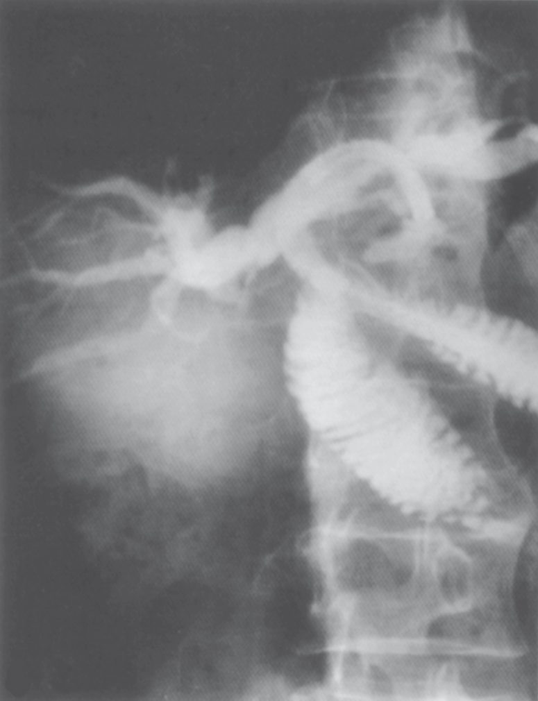
- For recurrent pyogenic cholangitis (Oriental cholangiohepatitis/hepatolithiasis), definitive surgery aims to remove all stones, bypass or resect strictures, and provide durable biliary drainage; intrahepatic strictures may require resection, strictureplasty, or hepaticojejunostomy, with the Roux limb sometimes brought out as an accessible stoma for future choledochoscopy [11].
Hepatolithiasis and recurrent pyogenic cholangitis
- Hepatolithiasis is defined as stones in ducts proximal to the confluence of the hepatic ducts, regardless of whether stones are also present in the gallbladder or common bile duct, and is more refractory to surgical treatment than most other benign biliary diseases [3].
- Its relative incidence is approximately 1% in Western countries against 20% in Taiwan, 18% in South Korea and 40% in China.
- Originally regarded as confined to Southeast Asia and called oriental cholangiohepatitis or Hong Kong disease, migration has made it a Western problem too, and the North American experience now includes a significant number of Caucasian and Latin American patients whose disease is attributed to primary sclerosing cholangitis, choledochal cysts and iatrogenic biliary strictures [3].
- Pathogenesis involves bile infection, biliary stasis, low-protein and low-fat diets with malnutrition, and parasitic infection; brown pigment (calcium bilirubinate) stones are commonest and cholesterol stones second [3].
Treatment is centred on decompressing abscesses, removing stones, dealing with recurrences, and anticipating malignancy; more than two-thirds of patients undergo multiple surgical procedures and 10% ultimately require liver transplantation for liver failure [3]. Where stones and strictures are confined to a single segment or lobe (and there is a predisposition for the left lobe) hepatic resection is generally recommended, and it is particularly important in patients with parenchymal atrophy and intrahepatic ductal stricture who may harbour concomitant cholangiocarcinoma [3].
The strictured duct must be included in the resection. In 44 patients undergoing hepatectomy for hepatolithiasis, divided by the presence or absence of intrahepatic biliary stricture and followed for a median 65 months, residual or recurrent stones occurred in 36% with stricture against 11% without, late cholangitis in 54% against 6%, initial failure in 50% against 31%, and intrahepatic stricture recurred in 46% of the stricture group against none of the other, recurring at the primary site in two-thirds [3].
- Where resection is not feasible because of diffuse multifocal disease, surgery and percutaneous transhepatic cholangioscopic lithotripsy achieve complete stone clearance in 84–100% and 72–92% respectively, but stone recurrence is high at 33–40% [3].
- Endoscopic treatment may be possible where stones lie predominantly in the extrahepatic ducts or at the primary convergence with minimal intrahepatic stenosis [3].
- Hepaticojejunostomy to prevent biliary-enteric regurgitation is controversial, some authors reporting increased biliary complications in this setting.
- Adding a cutaneous stoma to the Roux limb creates an access point for future treatment, and a more appealing alternative is the Hutson loop, tacking the jejunal loop of the anastomosis to the abdominal wall and marking it with staples or a metal ring so it can be entered percutaneously [3].
| Percutaneous series | Outcome |
|---|---|
| 79 patients, transhepatic choledochoscopy (Hong Kong) | 76.8% success, 21.5% complications, cholangitis within 3–5 years in one-third |
| Transhepatic cholangioscopic lithotripsy | 100% biliary clearance, mean two sessions, 6.7% complications, one recurrence over mean 75 months |
| 53 patients, percutaneous intracorporeal electrohydraulic lithotripsy | 92% complete clearance, 9% recurrent obstructive symptoms at mean 5 years |
| 42 patients, University of Toronto 1986–2005 | 17 (46%) operated for lobar atrophy or single-lobe stones; operated patients needed less reintervention; cholangiocarcinoma incidence 12% |
Table reformats the reported series [3]. Recurrent calculi are more common where bile duct strictures are present, so addressing the strictures is a mandatory part of treatment [3].
Cholangiohepatitis as Schwartz describes it
- Cholangiohepatitis (recurrent pyogenic cholangitis), endemic to East Asia and seen in Asian populations in the United States, Europe and Australia, affects both sexes equally in the third and fourth decades; bacterial contamination with E. coli, Klebsiella, Bacteroides or Enterococcus faecalis, often with the parasites Clonorchis sinensis, Opisthorchis viverrini and Ascaris lumbricoides, deconjugates bilirubin into sludge that, with dead bacterial bodies, forms brown pigment stones whose nucleus may contain an adult Clonorchis, an ovum or an ascarid, throughout the biliary tree [9].
- The resulting partial obstructions cause repeated cholangitis, strictures, further stones, abscesses and eventually secondary biliary cirrhosis; relapsing right upper quadrant pain, fever and jaundice are characteristic and lead without treatment to malnutrition and hepatic insufficiency [9].
- Ultrasound may show duct stones, pneumobilia from gas-forming organisms, abscesses and occasionally strictures, and the gallbladder is thickened in about 20% but rarely contains stones; ERCP or MRCP define stones and strictures, ERCP (or PTC) decompresses the septic patient, abscesses are drained percutaneously, and the long-term aim of extracting stones and debris and relieving strictures may take several procedures, a hepaticojejunostomy in refractory cases, or occasionally resection of the involved liver, recurrence is common and prognosis poor once hepatic insufficiency develops [9].
Complications
- Untreated or inadequately treated cholangitis can progress to septic shock and multi-organ failure via the Reynolds pentad, with near-uniform mortality if unaddressed [1].
- Renal failure is the commonest serious complication, related to the sepsis [8].
- Pus may accumulate within the biliary tree, and hepatic (pyogenic) abscesses may develop as a direct consequence of ascending cholangitis, particularly with biliary tract obstruction from stones or malignancy [2][11]; where biliary decompression is not achieved, liver abscess formation is inevitable [3].
- Stricture formation and hepatic abscess are recognised late complications of recurrent cholangitis [8].
- Recurrent episodes, as seen in recurrent pyogenic cholangitis, can progress to secondary biliary cirrhosis and carry an increased risk of cholangiocarcinoma [11].
- In hepatolithiasis specifically, the natural course leads to biliary cirrhosis, portal hypertension and liver failure, and is complicated by cholangiocarcinoma in about 10% of patients [3].
- Septic shock appearing more than 24 hours after laparoscopic cholecystectomy should raise the possibility of an accidental clip across the common bile duct with subsequent cholangitis [8].
Prognosis
- With prompt recognition, antibiotics and timely biliary decompression, most patients with mild-to-moderate cholangitis respond well.
- Severe cholangitis with the Reynolds pentad is an ominous presentation with a very high mortality if treatment is delayed [1].
- Overall mortality is quoted at 5–10% [8], rising toward 100% in patients who fail conservative therapy and are not drained [3].
- In recurrent pyogenic cholangitis, surgical management directed at stone clearance and adequate drainage is highly successful in the absence of malignant transformation, though the chronicity of infection and inflammation places patients at long-term risk of cholangiocarcinoma [11].
References
- Sabiston Textbook of Surgery, 22nd ed., Ch. 88
- Bailey & Love's Short Practice of Surgery, 28th ed., Ch. 71
- Maingot's Abdominal Operations, 13th ed., Ch. 63, Choledocholithiasis and Cholangitis
- British Society of Gastroenterology: Updated guideline on the management of common bile duct stones (CBDS). Gut 2017;66:765–782, Endoscopic management of CBDS; Endoscopic management of CBDS; moderate-quality evidence, strong recommendation; Management of 'difficult' ductal stones; Management of CBDS in specific clinical settings; Management of CBDS in specific clinical settings; high-quality evidence, strong recommendation; Management of cholangitis; Management of cholangitis; moderate-quality evidence, strong recommendation gut.bmj.com
- NICE Clinical Guideline CG188: Gallstone disease: diagnosis and management (2014), 1.3; 1.3.3 www.nice.org.uk
- Browse's Introduction to the Symptoms and Signs of Surgical Disease, 6th ed., Ch. 15
- Oxford Handbook of Clinical Surgery, 5th ed., Ch. 9
- The ABSITE Review, 2022, Ch. Biliary System
- Schwartz's Principles of Surgery, 11th ed., Ch. 32, Gallbladder and the Extrahepatic Biliary System
- Schwartz's Principles of Surgery: ABSITE and Board Review, Ch. 32
- Sabiston Textbook of Surgery, 22nd ed., Ch. 89