



Incisional Hernia
Summary
- Incisional hernia is a defect in the musculofascial layers of the abdominal wall arising at the site of a previous surgical incision, reported in roughly 10-20% (up to 50% in some series) of laparotomy wounds and around 1-5% of laparoscopic port sites [1][2].
- Sabiston puts the figures at 12% of major open abdominal incisions and 3% of major minimally invasive incisions, with the greatest risk in the first 5 years [3].
- It is the most common "secondary" ventral hernia and one of the most common procedures performed by general surgeons, with over 100,000 repairs performed annually in the United States alone and rates of ventral hernia repair overall exceeding 600,000 [2][3].
- Recurrence after repair remains a persistent challenge, reported as high as 40% in some ventral hernia repair series even with modern mesh techniques [3].
- There is no NICE guideline on incisional or ventral hernia repair.
- The NICE clinical guideline "Hernia: diagnosis and management of hernia" was referred by the Department of Health in March 2015, deferred in June 2015 in favour of another topic, and discontinued on 21 February 2018 as "not currently planned to be recommissioned" [4].
- The only published NICE guidance touching this territory concerns a preventive intervention at a stoma site rather than repair of an established hernia, and it is restrictive [5].
Definition
- An incisional hernia is a hernia that arises through a defect in the musculofascial layers of the abdominal wall at the site of a previous surgical scar, and may therefore occur anywhere a laparotomy or major port-site incision has been made [1].
- It is a hernia through a scar in the abdominal wall caused by a previous surgical operation or injury, scar tissue being inelastic and stretching progressively when subjected to constant stress [6].
- In the ventral hernia classification, incisional hernias are the "acquired" group, as opposed to the "spontaneous" umbilical and epigastric hernias [3].
Pathophysiology
- An incisional hernia typically begins as an early postoperative disruption of the musculofascial closure of a wound; this may progress rapidly to full-thickness wound dehiscence (classically heralded by serosanguineous discharge around the sixth postoperative day), but more often passes unnoticed if the overlying skin has healed, with a clinically apparent bulge appearing weeks, months, or even years later [1].
- Normal wound healing produces long-term collagen deposition and remodelling, but the resulting mature scar reaches only about 80% of the tensile strength of native fascia, so every laparotomy incision represents an area of relative, permanent weakness that can be exacerbated by repeated strain [3].
- Fascial strength increases rapidly between the eighth postoperative day and the second month, corresponding to the proliferative phase of wound healing and the period of active collagen synthesis, which is why at least a slowly-absorbable suture should be used for closure [3].
- Multiple, clinically unsuspected fascial defects are frequently found along the same incision at the time of repair, even when only a single hernia was apparent preoperatively [1].
Loss of domain
- In extremely large defects (generally >10 cm), 25% or more of the abdominal viscera may reside chronically within the hernia sac ("loss of domain") producing chronic venous/lymphatic congestion and dilation of the herniated bowel, hyperlordosis of the lumbar spine from disruption of the abdominal core, and altered pulmonary mechanics from the drop in intra-abdominal pressure that follows extra-abdominal migration of the viscera [2].
- Quantitatively, the Sabbagh formula calculates the index of loss of domain as the ratio of the incisional hernia volume to the sum of the abdominal cavity volume and the incisional hernia volume, both measured from CT.
- A value of 20% or more is generally considered loss of domain [3].
- These giant hernias develop through a chronic process of linea alba disruption and lateral retraction of the abdominal muscles, followed by muscle atrophy and fibrosis; as viscera protrude into the sac, intra-abdominal pressure falls and the diaphragm flattens, the dorsal muscle groups lose their counterbalance and spinal strain follows, and in some cases portal and mesenteric venous stasis develops with bowel wall oedema and digestive symptoms [3].
Anatomy of the rectus sheath and the biology of the incision in Schwartz's account
- Above the arcuate line the posterior rectus sheath is formed by part of the internal oblique aponeurosis and the transversus sheath and the anterior sheath by the remaining internal oblique and the external oblique, whereas below it all lateral aponeuroses pass anteriorly and the lower rectus has no posterior aponeurotic cover; the superior epigastric artery, continuing the internal thoracic, runs on the posterior surface of the anterior sheath to anastomose with the inferior epigastric from the external iliac at the umbilicus, lymph above the umbilicus drains to the axilla and below to the groin (periumbilical lymphatics also run along the falciform ligament to hepatic nodes), sensation is T4–L1 and motor supply T6–T12, and torso rotation pairs the ipsilateral internal with the contralateral external oblique [7].
- Transverse incisions may carry fewer incisional hernias but more wound infections than longitudinal ones with no difference in early or late complications overall, and closure of a midline classically uses bites 1 cm from the edge and 1 cm apart, but two European randomised trials showed fewer hernias with 5–8 mm bites 5 mm apart, while prophylactic mesh reduces short-term hernia formation with long-term mesh complications and ideal position and material still unsettled [7].
- Up to 20% of midline incisions eventually herniate, vertical and upper abdominal incisions carry higher risk than transverse and lower ones, and hernias may appear up to 10 years later though most arise early [7].
Clinical features
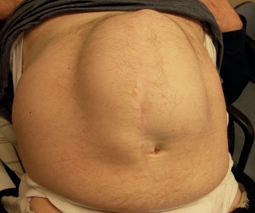
- Most patients present with a palpable bulge underlying a previous abdominal incision, ranging from a localized swelling involving part of the scar to a diffuse bulge along its full length, or several discrete hernias along one incision [1][2].
- Discomfort varies, and cosmetic concern is common as the hernia enlarges [2].
- Episodes of intestinal obstruction are common, related to coexisting internal adhesions, but frank strangulation is comparatively rare because most incisional hernias have a shallow, wide neck.
- Strangulation risk is highest, as with any hernia, when the fibrous defect is small relative to the sac [1].
- Chronic pressure on overlying skin from a large or long-standing hernia can lead to "paper-thin" skin or ulceration [1][2].
- The typical finding is a lump with an expansile cough impulse beneath an old scar, with the defect in the abdominal wall often palpable.
- Incisional hernias are not infrequently irreducible, the defect being plugged with adherent omentum [6]. If the lump does not reduce and has no cough impulse it may not be a hernia at all, but rather a tumour deposit, a chronic abscess or haematoma, or a foreign-body granuloma.
- All of these except recurrent tumour appear shortly after the initial surgery [6].
- The first signs usually appear in the first year after surgery but sometimes many years later, and patients may not recall the wound complication (a haematoma or infection) that weakened the closure in the first place [6].
Port-site hernia, Spigelian hernia, rectus diastasis and rectus sheath haematoma in Schwartz's account
- Port-site hernias occur in under 1% on meta-analysis, present early or years later, commonly as Richter's hernias with a high risk of strangulation, arise mostly at the umbilical port and through ports over 5 mm in adults (any size in children), and are repaired open by enlarging the skin incision, reducing the hernia and closing all fascial layers [7].
- Spigelian hernias form at the arcuate line where the posterior sheath is absent, track laterally beneath the external oblique aponeurosis, present with mid-to-lower abdominal pain and swelling, are irreducible in up to 20% and so are repaired, open or laparoscopically, by approximating the transversalis fascia to the rectus sheath [7].
- Rectus diastasis is separation of more than 2 cm above the umbilicus with intact linea alba (not a true hernia, so it never incarcerates) favoured by obesity, pregnancy (older age, multiparity, repeated caesarean), connective tissue disease and prior surgery, reduced by postpartum exercise, shown as a fusiform bulge on straining, confirmed by ultrasound or CT, improved by weight loss and exercise, and repaired by open or laparoscopic plication only for functional disability or cosmesis because recurrence is high and mesh adds complications [7].
- Rectus sheath haematoma follows disruption of an inferior epigastric branch, commonly near the arcuate line where the vessels are fixed and shear, after trocar trauma, coughing, sneezing, exercise or anticoagulation, presents with acute pain and a palpable mass that worsens on rectus contraction (unlike intraperitoneal disease) and does not change on contraction (Fothergill's sign), is confirmed by ultrasound or contrast CT with a type and screen, haemoglobin and coagulation profile, and is managed by observation for small stable haematomas, admission with serial haemoglobin, compression, bed rest, reversal of anticoagulation and transfusion for larger or bilateral ones, angiographic embolisation for shock, enlargement or deterioration, and surgical evacuation with vessel ligation only when embolisation fails [7].
Etiology
- Risk factors are commonly grouped into patient factors (obesity, malnutrition, immunosuppression or steroid therapy, diabetes, smoking, chronic cough, ascites, advanced age, connective tissue/collagen disorders, cancer), wound factors (infection, poor-quality tissue, wound tension, dehiscence), and surgical/technical factors (inappropriate suture material, poor closure technique) [1][2].
- Wound infection has the strongest documented association with subsequent incisional hernia, which is why most surgeons advocate early reopening and drainage of an infected surgical wound [2].
- Inadequate fascial closure technique is considered the most common preventable cause: the traditional "1 cm bite, 1 cm apart" technique produces more tissue ischaemia than a "small bites" technique (5-8 mm bites, 5 mm apart), and recent evidence favours the smaller-bite approach with a slowly absorbable 2/0 suture and a suture-to-wound-length ratio of at least 4:1 [1][8][9].
- This is the finding of the STITCH randomised trial, which established that a small-bites suture technique is more effective than traditional large bites with 2-0 slowly absorbable suture for midline laparotomy closure [3].
- Drains brought out through the primary wound (rather than a separate stab incision) increase hernia risk by preventing full fascial apposition [1].
- Two specific settings deserve separate mention.
- A traumatic abdominal wall hernia occurs when blunt forces disrupt the abdominal wall.
- There are approximately 15,000 annually, an incidence of under 1% in blunt trauma, and in over a third of cases there is a concomitant bowel injury requiring immediate laparotomy [3].
- The open abdomen, where the fascia is not reapproximated after laparotomy, following damage-control surgery, treatment of abdominal compartment syndrome, or a planned second look, leaves the patient at risk of fluid and protein loss, fistula, and eventual ventral hernia.
- Negative-pressure dressings achieve fascial closure 89% of the time compared with 59% for non-negative-pressure approaches, and in trauma closure is preferred within 48 hours [3].
Diagnosis
- Diagnosis is usually made on physical examination, with the hernia sac palpable and, depending on morphology, an attempt made to define the fascial edges.
- Examination should assess the entire length of the incision, as occult additional defects are common [2].
- It is critical to note all past surgical incisions, and to examine the patient both standing (documenting abdominal wall asymmetry) and supine, asking for a Valsalva manoeuvre and a straight bilateral leg raise to assess for fascial defects and the extent of abdominal wall weakness [3].
- Operative reports from any previous repair should be obtained where possible, because details of mesh placement and type, and whether a complex approach such as component separation has already been attempted, are invaluable for operative planning [3].
CT is used for complex or large hernias to determine the number and size of defects, characterize the contents, assess for loss of domain, and plan the operative approach, including any need for component separation [1][10]. CT also differentiates true incisional hernia from rectus abdominis diastasis, in which the midline aponeurosis remains intact [9].
Interparietal hernias after complex repair
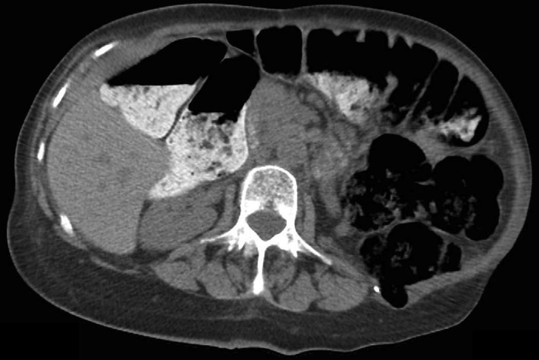
- Two interparietal hernias occur specifically as complications of posterior component separation and are diagnosed only if actively suspected. Posterior rectus sheath herniation follows Rives-Stoppa or transversus abdominis release when missed fenestrations or excessive tension break down the posterior sheath closure.
- Any patient with signs of bowel obstruction after a retrorectus or retromuscular repair should be promptly evaluated for it. Semilunar line herniation follows inadvertent division of the anterior lamella of the internal oblique during attempted division of the transversus abdominis fibres, from improper intraoperative identification of the semilunar line.
- CT is a necessary step in both, and referral to a centre specialising in complex abdominal wall surgery is recommended given the technical difficulty of recurrent repair after a transversus abdominis release [11].
Scoring and Severity
The European Hernia Society classification, developed by Muysoms and colleagues, is the most widely accepted system for both midline (subxiphoid, epigastric, umbilical, infraumbilical, suprapubic) and lateral (subcostal, flank, iliac, lumbar) incisional hernias, and grades hernias by location and width in centimetres [3]. "Complex" or "giant" incisional hernias, generally defined by a fascial defect exceeding 10 cm and/or loss of domain, represent the most severe end of the spectrum and require specialized abdominal wall reconstruction techniques [2].
- The EHS lateral classification divides the area lateral to the rectus into four compartments (L1 subcostal, L2 flank, L3 iliac, L4 lumbar) bounded by the subcostal margin superiorly, the iliac crest inferiorly, the lateral border of the rectus sheath medially, and the lumbar region laterally.
- Flank hernias lie lateral to the rectus sheath, whereas lumbar hernias lie lateral to the anterior axillary line [11].
- Flank hernias are almost always incisional, while lumbar hernias can occur as primary hernias through the superior (Grynfeltt-Lesshaft) or inferior (Petit) lumbar triangles [11].
- Procedures involving a lumbar incision such as nephrectomy carry a postoperative herniation incidence reported as high as 30% [11].
- Despite their frequently asymptomatic, fat-containing presentation, incarceration is common: one systematic review reported a 31% incarceration rate in primary lumbar hernias, and another estimated 9% present acutely and need emergency intervention, so repair of lateral hernias should be considered once diagnosed
- Incisional lumbar and flank hernias account for under 20% of all incisional hernia repairs and only 420 primary lumbar hernias have been reported in the literature.
- Current society guidelines recommend referral to specialised hernia centres [11].
Risk of emergency presentation can be estimated from morphology: a ventral hernia width-to-neck ratio greater than 2.5 is associated with an increased risk of emergency surgery [3].
Treatment and Management
Asymptomatic incisional hernias, particularly in elderly, frail, or high-comorbidity patients, may not require treatment, and an abdominal binder can provide symptomatic relief and may limit further enlargement [1][10]. Repair should always be individualized in discussion with the patient, and referral to a surgeon with a specialist interest in abdominal wall reconstruction should be considered for large or complex hernias [1].
Prehabilitation, where the textbooks disagree
For patients proceeding to elective repair, Bailey & Love recommends preoperative "prehabilitation" (smoking cessation, weight loss, and core-strengthening exercise) noting that a 7% total body weight loss produces a meaningful improvement in metabolic state, and that each 5 kg of weight loss is estimated to create roughly one extra litre of intra-abdominal space in men (0.5 L in women) [1].
- Sabiston's dedicated chapter on preoperative management reaches almost the opposite conclusion, and states it as an explicit recommendation.
- In a randomised trial of 118 patients undergoing ventral hernia repair, an early advantage for prehabilitation was not sustained: at 2 years there was no difference in hernia-free and complication-free patients between prehabilitation and standard counselling (72.9% versus 66.1%, p = 0.42), patients who completed the programme did not sustain their weight loss, and about half of patients in both groups gained weight from baseline.
- Only 22% of the prehabilitation group met the 7% total body weight loss goal.
- Critically, five patients in the prehabilitation group (8.5%) versus one in the standard counselling group (1.7%) underwent emergency repair for acute incarceration during the study period [12].
- An observational study of 191 hernia patients found 55% of those enrolled in a prehabilitation programme were lost to follow-up, and only 9 of 80 (11%) met the weight loss goal and went on to repair [12].
Sabiston's summary recommendations are worth stating in full because they overturn several long-taught rules [12]:
| Question | Recommendation |
|---|---|
| Hard BMI or weight-loss cutoffs | Strongly discouraged; the decision should be individualised because some patients will never achieve significant weight loss |
| HbA1c threshold before repair | No evidence to recommend delaying hernia repair until HbA1c is lowered to an arbitrary cutoff; a hernia-specific study of over 2,000 patients found no clinically significant difference in wound complications, reoperation, readmission, length of stay or mortality between HbA1c under 8% and 8% or above |
| Smoking | Continue to encourage cessation, but the surgeon may proceed even if the patient has failed to quit, depending on complexity and technique; a registry analysis found similar surgical site infection rates in current smokers and never-smokers (4.1% versus 4.1%), with the excess confined to surgical site occurrences driven by seroma |
| mTOR inhibitors (sirolimus, everolimus) | Stop 4 to 6 weeks before elective surgery, in consultation with the transplant team |
| Calcineurin inhibitors and antimetabolites | Continue perioperatively, monitoring trough levels daily |
| Botulinum toxin A | Use in a trial context only, it is off-label in the lateral abdominal wall, carries an FDA black box warning about systemic spread, adds significant cost, and a registry study found no difference in fascial closure rates with or without it (86% versus 85.2%, p = 0.934) |
| Tissue expanders and progressive pneumoperitoneum | Valuable adjuncts for achieving soft tissue coverage; a meta-analysis of progressive pneumoperitoneum in 1,216 patients pooled 86% fascial closure but a 12% complication rate, including two hollow viscus perforations and five deaths |
| Preoperative chlorhexidine shower | No evidence supports prehospital chlorhexidine to reduce wound infection after hernia repair; a hernia-specific study of 3,924 patients found higher surgical site occurrences (OR 1.34) and infections (OR 1.46) in the chlorhexidine group |
- Table reformats Sabiston's evidence-based recommendations on preoperative optimisation [12].
- The underlying reasoning is that a significant amount of effort and time is required to achieve meaningful weight loss, and during that time the patient is exposed to the risk of an emergency operation, which carries worse outcomes than the elective setting.
- Risk calculators such as CeDAR and ORACLE can help frame that trade-off with the patient [12].
Mesh selection and position
- Prosthetic mesh is recommended for essentially all incisional hernia repairs in clean fields, since primary suture repair (even overlapping techniques such as Mayo repair or the layered da Silva closure) carries an unacceptably high recurrence rate.
- Mesh may still be used in a clean-contaminated field (e.g., after elective bowel resection) with appropriate prophylaxis, but is generally avoided in frank contamination, where a temporary absorbable mesh or delayed repair may be preferred [1][3].
- Mesh selection depends on planned location and contamination risk: permanent synthetic mesh (polypropylene, polyester, PTFE) is standard for clean, definitive repair; biologic mesh (decellularized collagen matrix) is sometimes used in contaminated fields but is associated with higher recurrence and cost without proven superiority; and rapidly absorbable synthetic mesh (e.g., polyglactin) offers a cheap temporary option in contaminated fields where fascial closure is desired without permanent material [3].
- Sublay (retrorectus) mesh placement has been shown in meta-analysis to have significantly lower recurrence and surgical-site infection rates than onlay, inlay/bridging, or intraperitoneal underlay placement in open repair [3][9].
The emergency presentation
Acutely incarcerated or strangulated hernias require urgent surgery; the goals are to relieve obstruction and resect necrotic bowel, with resuscitation and broad-spectrum antibiotics started in the emergency department [3]. Mesh use is decided by the intraoperative wound class rather than by a blanket rule [3]:
| Wound class | Finding | Repair |
|---|---|---|
| I (clean) | Intestinal obstruction, no strangulation, no resection | Synthetic mesh recommended |
| II (clean-contaminated) | Strangulated bowel resected, no gross enteric contamination | Mesh does not increase 30-day wound morbidity and decreases recurrence |
| III-IV (contaminated / dirty-infected) | Bowel necrosis and/or perforation | Primary repair indicated; if too large for primary repair, skin closure may be the only option |
Table reformats the surgical wound classification and its consequences for mesh [3]. Component separation should never be attempted in the emergency setting (it is reserved for elective situations) and biologic mesh, once advocated for contaminated cases, has fallen out of favour given its high cost and unacceptable recurrence rates [3].
Surgeries
Open repair, general principles, the previous incision is reopened along its full length to expose any unsuspected defects. The sac is opened, contents reduced, adhesions divided, and redundant sac excised. Repair should cover the whole length of the previous incision, approximate the musculofascial layers with minimal tension, and use mesh to reduce recurrence risk [1].
- Retromuscular sublay ("Rives-Stoppa") repair, the posterior rectus sheath is opened at the linea alba on each side, the retrorectus space developed laterally to the semilunar line (protecting the neurovascular bundles), and the posterior sheath closed.
- Mesh is placed in this highly vascularized retrorectus plane, deep to the rectus muscle, and the anterior sheath is then closed over it.
- This is now the preferred open technique for medium-to-large or multiple defects, with reported recurrence rates of 7-11% in contemporary series [2][3].
Preperitoneal sublay repair, mesh is placed in the preperitoneal plane rather than the retrorectus space; this avoids incising any fascial layer and is not bound by the semilunar line, but the preperitoneal dissection can be technically difficult because the peritoneum is thin [3].
Onlay repair, after fascial closure, skin flaps are raised between the anterior rectus fascia and subcutaneous tissue to allow wide mesh overlap (at least 5 cm) placed anterior to the closed fascia; it avoids intraperitoneal dissection but requires large lipocutaneous flaps, carrying a higher rate of wound complications and mesh infection than sublay techniques [1][3].
- Intraperitoneal underlay repair (open or laparoscopic, IPOM), mesh (coated with an antiadhesive barrier, or ePTFE) is placed deep to the peritoneum in direct contact with viscera.
- A laparoscopic approach lyses adhesions first, reduces hernia contents, and fixes a mesh with wide (>4-5 cm) overlap using transfascial sutures and/or a "double crown" of tacks.
- Primary closure of the defect before mesh placement does not affect recurrence but may reduce seroma formation [2][3].
Bridging/inlay repair, used when fascial edges cannot be approximated and mesh must bridge the gap directly; this carries the highest recurrence rate of any mesh position and the poorest functional result because fascial continuity is not restored, but is sometimes the only option in massive defects, reoperative surgery, or after treatment of abdominal compartment syndrome [3].
Anterior component separation (Ramirez, 1990), the external oblique aponeurosis is divided 1-2 cm lateral to the rectus sheath along the length of the defect bilaterally, releasing up to 10 cm of fascial advancement per side to allow midline closure. Large lipocutaneous flaps predispose to wound complications, and active smoking and poorly controlled diabetes (HbA1c ≥7) are considered relative contraindications by most centres [2].
Posterior component separation / transversus abdominis release (TAR) (Novitsky and Rosen), extends the retrorectus (Rives-Stoppa) dissection by incising the posterior lamella of the internal oblique aponeurosis medial to the neurovascular bundles and dividing the transversus abdominis fibres, creating a large posterolateral pocket for mesh placement without the wound morbidity of anterior component separation. A hernia defect-to-rectus-width ratio greater than 0.5 favours choosing TAR over a standard Rives-Stoppa repair [2][3].
- Laparoscopic incisional hernia repair (LIHR), first described by LeBlanc in 1993; associated with lower rates of surgical site infection than open repair (reported as low as 2.3% vs 9.2%) and shorter hospital stay, though evidence on recurrence and postoperative pain compared with open repair is mixed, and most laparoscopic repairs leave the defect unclosed (bridged), often producing a persistent, benign postoperative bulge that must be distinguished from true recurrence [13].
- Enterotomy is the most feared complication, occurring in a similar proportion of laparoscopic and open repairs (7.9% vs 7.3% in one series).
- An unrecognized enterotomy carries substantially higher mortality (7.7%) than one recognized intraoperatively (1.7%) [13].
Lateral, flank and lumbar hernias
- Repair requires a solid understanding of posterior abdominal wall anatomy.
- A robotic transabdominal preperitoneal approach is typical for suitable candidates, taking advantage of the thicker preperitoneal fat lateral to the semilunar line and excluding mesh from the viscera.
- The patient is positioned supine and tilted, or in lateral decubitus with the bed flexed to open the space between the anterior superior iliac spine and the costal margin.
- Lateral dissection continues into the pararenal space to the quadratus lumborum, and the investing fascia of the quadratus lumborum and psoas must be left intact to protect the ilioinguinal, iliohypogastric, genitofemoral and lateral femoral cutaneous nerves, with the ureter and gonadal vessels identified and preserved.
- Transfascial suture fixation is avoided in favour of fibrin sealant, to prevent entrapment of surrounding neurovascular structures [11].
Parastomal hernia
- Approximately 100,000-120,000 new ostomies are created annually in the United States, and nearly half of these patients develop a parastomal hernia; recurrence rates after repair approach 70%.
- The fundamental difficulty is that a sound hernia repair obliterates the defect, which cannot be done here because a fascial aperture must remain for the conduit.
- Most parastomal hernias can be managed non-operatively, though a third of patients will need an operation, and the best treatment is reversal of the ostomy where that is possible.
- Re-siting the ostomy has fallen out of favour because of the risk of a de novo incisional hernia at the old site plus a parastomal hernia at the new one, and primary repair should be used only in the acute setting where the clinical goal is management of threatened bowel [3].
- The two common mesh techniques are the keyhole repair, in which a slit and aperture in the mesh allow the conduit through, and the Sugarbaker repair, in which the conduit is lateralised and mesh placed over both conduit and hernia.
- The Sugarbaker repair has lower recurrence rates with similar rates of overall complications, reoperation, stoma outlet obstruction, mesh infection and postoperative bleeding.
- A modified Sugarbaker technique using transversus abdominis release and retromuscular mesh has been described but lacks long-term data, and synthetic mesh in apposition to bowel may cause erosion or obstruction [3].
NICE has evaluated the preventive use of mesh at the time of stoma formation, and restricted it. "The evidence on the safety of reinforcement of a permanent stoma with a synthetic or biological mesh to prevent a parastomal hernia shows there are serious but well-recognised complications. The evidence on efficacy is limited in quantity and quality. Therefore, this procedure should not be used unless special arrangements are in place for clinical governance, consent, and audit or research." [5]. The guidance was published on 26 June 2019 and migrated unchanged from interventional procedures guidance IPG654 [5].
- Clinicians wishing to do the procedure must inform their trust's clinical governance leads, ensure patients understand the safety and efficacy of the procedure and the uncertainties about both, provide clear written information to support shared decision making, and audit and review the clinical outcomes of every patient having the procedure [5].
- All adverse events involving the mesh must be reported to the MHRA [5].
- Further research should report patient selection, mesh type, mesh-associated complications and long-term outcomes at a minimum of 3 years, and in participating centres clinicians should encourage patients to take part in the NIHR CIPHER study [5].
- Note what this does and does not say.
- HTG519 covers prophylactic mesh at stoma creation, not repair of an established parastomal hernia (for which there is no NICE guidance at all) so the textbook account of keyhole versus Sugarbaker repair is unaffected by it.
- What HTG519 changes is the pre-emptive step: a UK surgeon cannot simply add a prophylactic mesh at stoma formation on the strength of the textbook rationale, because "special arrangements" means governance approval, documented consent, and audit or enrolment in research [5].
The article's own finding, that wound infection has the strongest documented association with a subsequent incisional hernia, is the reason the UK surgical site infection guideline belongs to this topic twice over: once for preventing the hernia, and again for protecting the mesh that repairs it.
- Mesh makes the prophylaxis decision automatic.
- NICE directs that antibiotic prophylaxis be given before clean surgery involving the placement of a prosthesis or implant, as well as before clean-contaminated and contaminated surgery [14].
- A mesh is an implant, so a mesh repair never falls into the category where prophylaxis is withheld, and that category is real: do not use antibiotic prophylaxis routinely for clean non-prosthetic uncomplicated surgery [14].
- Timing and redosing are specified, and the redosing rule is the one most often missed in a long repair.
- Consider a single intravenous dose on starting anaesthesia, given earlier where a tourniquet is used [14]. Give a repeat dose when the operation lasts longer than the half-life of the antibiotic given, taking the agent's pharmacokinetics and infusion time into account [14].
- The choice of agent comes from the local formulary, weighing adverse effects [14].
- Several long-standing theatre habits are explicitly not recommended.
- Do not use hair removal routinely; if hair must be removed, use electric clippers with a single-use head rather than a razor [14].
- Do not use mechanical bowel preparation routinely to reduce infection risk [14].
- The operating team should remove hand jewellery, artificial nails and nail polish before operating [14].
Recurrence figures, components separation and mesh classes in Schwartz's account
- Simple suture repair recurs in up to 54%, and a Cochrane review found open mesh repair cut recurrence from 33% to 16% at the price of more infection; Ramirez's 1990 components separation divides the external oblique aponeuroses bilaterally and may incise the posterior rectus sheath to gain up to 10 cm of medial advancement, its early skin-flap infections reduced by flap-sparing and endoscopic techniques (which mobilise less), it matches mesh repair for recurrence without implanting mesh, and with added mesh recurrence falls to 4–10% [7].
- Mesh may be placed as overlay, interlay (bridging), sublay or intraperitoneal underlay, and a systematic review favours sublay for recurrence and wound complications, the plane being developed between rectus and posterior sheath with the anterior sheath closed if tension allows [7].
- Permanent synthetics are polypropylene, polyester (polyethylene terephthalate) and expanded PTFE; degradable synthetics such as Vicryl lose support and recur but suit temporary closure of contaminated fields, with Gore BioA and Phasix degrading more slowly; biologic meshes are decellularised porcine, bovine or human collagen scaffolds for infected fields of uncertain efficacy; composite meshes pair a synthetic parietal face with a visceral barrier of polyglactin, collagen, cellulose, titanium, omega-3 or hyaluronate against adhesion, erosion and fistula; and large-pore lightweight mesh incorporates well with better elasticity, less pain and possibly less infection [7].
- Laparoscopic repair (LeBlanc and Booth, 1993) places ports laterally for midline and contralaterally for lateral defects, reduces contents and leaves the sac, and fixes mesh with circumferential transfascial sutures for 4–5 cm overlap plus optional tacks; a meta-analysis of 11 studies and another of six randomised trials found equal recurrence, fewer wound infections and less drainage but more bowel injury than open repair, with follow-up short, and robotic repair's cost-effectiveness remains unclear [7].
- Desmoid tumours arise in 10–15% of FAP patients (more in Gardner's), become the leading cause of death after prophylactic colectomy, follow incision, pregnancy, hormones and trauma in sporadic cases, are diagnosed by core or incisional biopsy, do worse when large, in the young or extra-abdominal, and are now observed when asymptomatic (16% of 106 abdominal wall desmoids needed surgery within 3 years and 29 regressed), resected with mesh reconstruction when symptomatic, invasive or growing without re-resecting a positive margin at high morbidity, and treated with radiation, hormones, NSAIDs, doxorubicin, carboplatin or imatinib when unresectable; other abdominal wall tumours (sarcoma, dermatofibrosarcoma protuberans, schwannoma, melanoma) need core biopsy, MRI, chest CT for high-grade disease and resection with 1 cm margins (2 cm for dermatofibrosarcoma) including the fascia, mesh or flap closure, and adjuvant or neoadjuvant therapy for tumours over 5 cm or high grade [7].
Complications
- Complications may be early (wound-related) or late (chronic pain, recurrence).
- Surgical site infection after ventral/incisional hernia repair ranges from about 4% to 21% in the literature and is itself a risk factor for later recurrence [3].
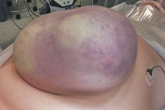
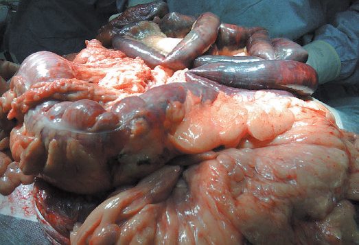
- Mesh infection occurs in roughly 1-8% of cases and, unlike standard surgical site infection (diagnosed within 90 days by CDC criteria), may not present for up to a year or more, commonly as a chronic draining sinus (68% of cases), recurrent seroma, enterocutaneous fistula, or exposed mesh [3].
- Risk factors for mesh infection include diabetes, smoking, obesity, urgent/emergency surgery, and immunosuppression.
- Open surgery and onlay mesh position carry higher infection rates than laparoscopic surgery or sublay/underlay placement [3].
- Macroporous polypropylene mesh in an extraperitoneal position has the highest salvage rate if infected (around 72%).
- PTFE and composite meshes are unlikely to be salvageable and usually require explantation [3].
- Seroma occurs in a wide range (3-52%) depending on technique, with risk increased by emergency surgery, high BMI, large skin flaps, component separation, onlay mesh, and biologic mesh [3].
"Pseudo-recurrence," a postoperative bulge from mesh laxity or bridging without true herniation, must be distinguished from genuine recurrence, particularly after laparoscopic repair [1][13]. Developing a wound complication has a significant negative effect on quality of life at one year, and patients who develop complications are at increased risk of recurrence and reoperation, the "vicious cycle of complications" that makes the index repair the one that matters [12].
Prognosis
- Recurrence rates vary widely with technique and hernia complexity, from roughly 7-11% with modern retromuscular sublay repair to as high as 40% in some series of complex ventral hernia repair overall [2][3].
- A recent population-based registry study found a 5-year recurrence rate greater than 40% in patients repaired with mesh and greater than 70% in patients repaired without it [3].
- Bridging/inlay mesh repair, used when fascia cannot be re-approximated, carries the highest recurrence of any technique [3].
- Modern sublay techniques have shifted outcomes favourably compared with historical primary suture repair and onlay mesh placement.
Lateral hernias are under-represented in outcome data because they are rare, incisional lumbar and flank hernias make up less than 20% of all incisional hernia repairs, and only 420 primary lumbar hernias have ever been reported, which is the stated reason society guidelines recommend referral to a specialised hernia centre rather than quoting an expected recurrence rate [11]. Parastomal hernia is the outlier: recurrence after repair approaches 70%, which is why ostomy reversal, where feasible, is the definitive answer rather than any repair technique [3].
References
- Bailey & Love's Short Practice of Surgery, 28th ed., Ch. 64 The abdominal wall, hernia and umbilicus
- Maingot's Abdominal Operations, 13th ed., Ch. 13 Ventral and Abdominal Wall Hernias
- Sabiston Textbook of Surgery, 22nd ed., Ch. 80 Ventral Hernias
- NICE guideline development project GID-CGWAVE0771: Hernia — diagnosis and management (deferred June 2015; discontinued February 2018, not currently planned to be recommissioned), Timeline www.nice.org.uk
- NICE HealthTech Guidance HTG519: Reinforcement of a permanent stoma with a synthetic or biological mesh to prevent a parastomal hernia (2019, migrated from IPG654), 1.1; 1.2; 1.3; 1.4; Overview www.nice.org.uk
- Browse's Introduction to the Symptoms and Signs of Surgical Disease, 6th ed., Ch. 14 The abdominal wall, hernias and the umbilicus
- Schwartz's Principles of Surgery, 11th ed., Ch. 35, Abdominal Wall, Omentum, Mesentery, and Retroperitoneum
- The ABSITE Review, 2022, Ch. 38 Hernias, Abdomen, and Surgical Technology
- Schwartz's Principles of Surgery: ABSITE and Board Review, Ch. 35 Abdominal Wall, Omentum, Mesentery, and Retroperitoneum
- Oxford Handbook of Clinical Surgery, 5th ed., Ch. 10 Abdominal wall
- Sabiston Textbook of Surgery, 22nd ed., Ch. 81 Hernias of the Abdominal Wall: Atypical Locations
- Sabiston Textbook of Surgery, 22nd ed., Ch. 79 Preoperative Management of the Hernia Patient
- Maingot's Abdominal Operations, 13th ed., Ch. 14 Perspectives on Laparoscopic Incisional Hernia Repair
- NICE Guideline NG125: Surgical site infections: prevention and treatment. National Institute for Health and Care Excellence, London, UK, 2019, updated 2020., 1.2.4; 1.2.5; 1.2.9; 1.2.10; 1.2.11; 1.2.12; 1.2.13; 1.2.14; 1.2.15; 1.2.16 www.nice.org.uk
- Bailey & Love's Short Practice of Surgery, 28th ed., Ch. 78