



Small Bowel Disorders
Summary
- The small intestine is long, mobile and comparatively rarely diseased, so its disorders present either as an emergency (bleeding, obstruction or perforation) or as a chronic failure of absorption [1].
- This page covers the conditions that fall to the general surgeon outside obstruction and inflammatory bowel disease, which have pages of their own: Meckel's diverticulum, acquired jejunal diverticula, benign and malignant small bowel tumours, enterocutaneous fistula, and short bowel syndrome.
- Malignant small bowel tumours are rare, and adenocarcinoma is the most common of them [2].
- Short bowel syndrome and loss of enteral autonomy are expected after resection of more than 70% of the native intestine [1].
Definition
Meckel's diverticulum is a true diverticulum arising from failure of closure of the omphalomesenteric duct [3]. Acquired diverticula are different in origin and site: they develop most often in the jejunum and arise from the mesenteric side of the bowel, through mucosal herniation at the point of entry of the blood vessels where there is a potential defect in the muscularis [4].
- Short bowel syndrome and intestinal failure are used interchangeably [1].
- The absolute length of remnant bowel needed to sustain nutrient absorption varies with the individual and with age, but as a rule of thumb short bowel syndrome and loss of enteral autonomy are expected after resection of more than 70% of the native intestine [1].
- A functional definition, in which insufficient absorptive capacity produces diarrhoea, dehydration and malnutrition, is more appropriate than a length, though a standard definition of 200 cm has been widely used in adults [5].
Intestinal failure may also describe patients with normal or nearly normal intestinal length but abnormal function, from Crohn's disease, motility disorders such as intestinal pseudo-obstruction and long-segment Hirschsprung's disease, or diseases of the enterocyte such as intestinal epithelial dysplasia; these are less common than short bowel syndrome but share the same consequences [1].
Pathophysiology
Meckel's diverticulum
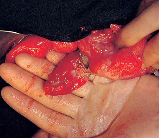
The classical teaching is a set of twos: the diverticulum lies about 2 feet from the ileocaecal valve and is present in 2% of the population, usually presenting in the first 2 years of life with bleeding [3]. Pancreatic tissue is the most common heterotopic tissue found within it, and can cause diverticulitis; gastric mucosa is the tissue most likely to make it symptomatic, bleeding being the commonest manifestation [3]. It accounts for 50% of all painless lower gastrointestinal bleeds in children under 2 [3].
Acquired jejunal diverticula
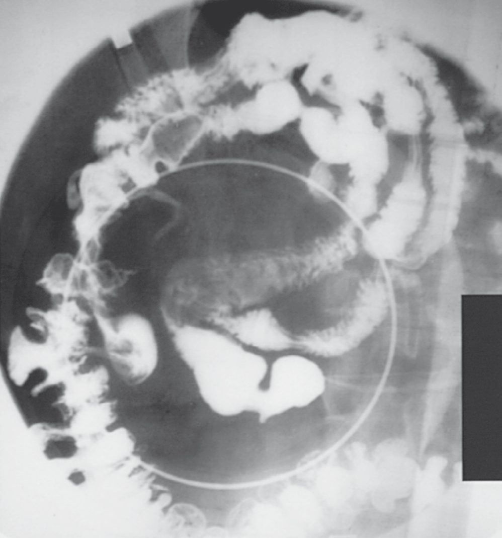
These vary in size and are frequently multiple [4]. They are commonly asymptomatic and found incidentally at surgery or on imaging, but may cause malabsorption through bacterial overgrowth, or present acutely if they become inflamed or perforate; bleeding is a rare complication, in contrast to sigmoid diverticular disease [4].
Enterocutaneous fistula
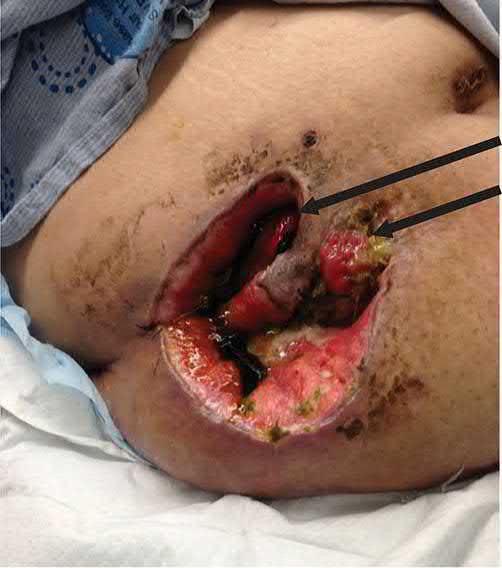
Fistulas are most commonly iatrogenic after surgery, anastomotic leakage, injury to the bowel or its blood supply, erosion by suction catheters, or laceration by wire mesh or retention sutures [1]. Other causes are predisposing conditions: Crohn's disease, malignancy, radiation enteritis, diverticulitis, intra-abdominal sepsis and trauma [1].
Factors that prevent a fistula healing are a foreign body in the tract, epithelialisation of the tract, downstream obstruction, radiation enteritis, associated infection such as abscess or sepsis, malignancy, and a short tract under 2 cm [5].
Adaptation and failure after massive resection
After massive resection, hypergastrinaemia and gastric acid hypersecretion occur and contribute significantly to diarrhoea [1]. Diarrhoea may also follow ileal resection, which disrupts the enterohepatic circulation so that excessive bile salts enter the colon and act as a cathartic [1]. Volume losses in the early phase may exceed 5 L/day [1].
Why small-bowel tumours are rare, and how fistulae and radiation injure the bowel
- The small intestine holds over 90% of the mucosal surface of the GI tract yet only 1.1–2.4% of its malignancies; proposed reasons are dilution of carcinogens in liquid chyme, rapid transit, low bacterial counts and hence few bacterial carcinogens, mucosal protection by secretory IgA and hydrolases such as benzpyrene hydroxylase, and efficient apoptosis of mutated clones [7].
- Adenocarcinomas arise from adenomas by a colorectal-type sequence, tubular adenomas least aggressive, villous most, the latter large, sessile and in the second part of the duodenum with malignant change in up to 45% by diagnosis, and GISTs are defined by gain-of-function KIT mutations that constitutively activate the receptor tyrosine kinase, the interstitial cells of Cajal that normally express KIT being the presumed cell of origin, with CD117 staining positive in 95% [7].
- Enterocutaneous fistulae draining under 200 mL/day are low-output and over 500 mL/day high-output; over 80% are iatrogenic from enterotomies or anastomotic dehiscence, the rest arising spontaneously from Crohn's disease or cancer, and low-resistance enteroenteric fistulae that bypass long segments cause malabsorption while enterovesical fistulae cause recurrent urinary infection [7].
- Acute radiation enteritis affects about 75% of patients irradiated for abdominal or pelvic cancer and chronic enteritis 5–15%; radiation kills rapidly proliferating epithelium by free-radical double-strand DNA breaks and apoptosis, producing villus blunting, crypt infiltrates and in severe cases sloughing and haemorrhage, mostly above 4500 cGy and potentiated by hypertension, diabetes, coronary disease, adhesion-fixed bowel and radiosensitising chemotherapy (doxorubicin, 5-FU, actinomycin D, methotrexate), whereas the chronic form is a progressive occlusive vasculitis with ischaemia and full-thickness fibrosis producing strictures, abscesses and fistulae [7].
- Meckel's diverticulum follows failure of obliteration of the omphalomesenteric (vitelline) duct in the eighth week, whose other remnants are an omphalomesenteric fistula, an enterocyst and a fibrous band to the umbilicus, while a persistent left vitelline artery forms a mesodiverticular band tethering the diverticulum to the mesentery; bleeding comes from ileal ulceration beside acid-secreting heterotopic gastric mucosa, and obstruction from volvulus around the umbilical band, entrapment beneath a mesodiverticular band, intussusception with the diverticulum as lead point, stricture after chronic diverticulitis, or incarceration in a Littre's hernia [7].
- Acquired diverticula are false diverticula of mucosa and submucosa herniating through weakened muscularis from smooth-muscle abnormality or dysmotility; bacterial overgrowth within them causes B12 deficiency, megaloblastic anaemia, malabsorption and steatorrhoea, periampullary duodenal diverticula distended with debris may compress the bile or pancreatic duct, and jejunoileal diverticula obstruct by intussusception or compression [7].
- After massive resection, clinically significant malabsorption follows loss of more than 50–80% of the small bowel; lifelong parenteral dependence is likely below 100 cm of residual bowel without a colon and below 60 cm with an intact colon, whereas infants have been weaned with as little as 10 cm; an intact colon absorbs large fluid loads and short-chain fatty acids, an intact ileocaecal valve prolongs mucosal contact, healthy residual bowel absorbs better than diseased, jejunal loss is tolerated better than ileal because bile-salt and B12 absorption are ileal, and hypergastrinaemic acid hypersecretion for 1–2 years inhibits the alkaline-dependent digestive enzymes [7].
Clinical features
Meckel's diverticulum presents differently by age: bleeding is the usual presentation in the first two years of life, whereas obstruction is the most common presentation in adults [3].
Jejunal diverticula are usually silent, presenting with malabsorption or as an acute abdominal emergency if inflamed or perforated [4].
Enterocutaneous fistula is usually not difficult to recognise. The typical presentation is a febrile postoperative patient with an erythematous wound; when a few skin sutures are removed, purulent or bloody discharge is noted, and leakage of enteric contents then follows [1].
Small bowel adenomas present with bleeding or obstruction and most are found in the duodenum [2]. Adenocarcinoma presents with obstruction and jaundice, a high proportion being duodenal; tumours arising from the ampulla of Vater present with jaundice, anaemia and guaiac-positive stools [2].
How the small-bowel lesions present
- Small-bowel tumours are asymptomatic until large: partial obstruction from luminal narrowing or intussusception is the commonest presentation and indolent haemorrhage the second, with a palpable mass, occult blood, jaundice from biliary obstruction or hepatic metastasis, and cachexia, hepatomegaly and ascites in advanced disease [7].
- Adenocarcinomas and adenomas favour the duodenum except in Crohn's disease, where most are ileal; periampullary lesions cause jaundice or pancreatitis; duodenal cancers are found earlier than jejunal or ileal ones, which are rarely diagnosed before locally advanced or metastatic disease; small-bowel carcinoids are usually diagnosed after metastasis, behave more aggressively than appendiceal carcinoids, and produce the carcinoid syndrome, diarrhoea, flushing, hypotension, tachycardia and right-heart endocardial and valvular fibrosis, in 25–50% of those with liver metastases, because serotonin, bradykinin and substance P are almost completely cleared on first hepatic passage; primary lymphoma is commonest in the lymphoid-rich ileum and perforates in 10%; and the small bowel holds 25–35% of GISTs, evenly distributed and more prone to overt haemorrhage than other small-bowel malignancies [7].
- Iatrogenic enterocutaneous fistulae declare themselves between the fifth and tenth postoperative days with fever, leucocytosis, prolonged ileus, tenderness and wound infection before enteric drainage appears through the wound or drains, often with an associated abscess [7].
- Acute radiation enteritis causes transient nausea, vomiting, diarrhoea and cramping that settle when treatment stops; chronic enteritis usually appears within 2 years but from months to decades later, most often as diarrhoea or partial obstruction of the terminal ileum, with complete obstruction, bleeding, abscess or fistula as other manifestations [7].
- Meckel's complications have a lifetime incidence of about 4–6%, and Cullen's Olmsted County data suggest the risk does not fall with age; bleeding is the commonest presentation in children (over half of complications under 18) and rare after 30, obstruction the commonest in adults, diverticulitis (20% of symptomatic cases) is indistinguishable from appendicitis, and carcinoid or other neoplasms are found in 0.5–3.2% of resected symptomatic diverticula [7].
- Acquired diverticula complicate in 6–10% with obstruction, diverticulitis, haemorrhage, perforation or malabsorption; periampullary diverticula are associated, without proven causation, with duct stones, cholangitis, recurrent pancreatitis and sphincter dysfunction, and intermittent pain, flatulence, diarrhoea and constipation are reported in 10–30% of patients with jejunoileal diverticula [7].
- Chylous ascites (milky triglyceride-rich fluid from intestinal lymph) presents with distension over weeks to months, or acutely in the first week after operations such as aortic aneurysm repair, retroperitoneal node dissection, caval resection and liver transplantation, with malignancy and cirrhosis the commonest Western causes and tuberculosis and filariasis the commonest in developing countries [7].
- Adult intussusception, far rarer than the paediatric idiopathic ileocolic form, usually has a pathological lead point that is malignant in up to half and presents with intermittent pain and obstruction; a retrograde form without a lead point follows Roux-en-Y gastric bypass [7].
Etiology
Causes of short bowel syndrome include congenital malformation, traumatic injury, infection and ischaemia [1].
Benign small bowel tumours
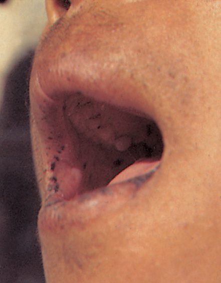
Adenomas are the commonest and most are duodenal [2]. Peutz-Jeghers syndrome is an autosomal dominant condition caused by mutation of the STK11 gene, producing benign hamartomas throughout the small and large bowel with mucocutaneous melanotic pigmentation; affected patients have an increased rate of extraintestinal malignancy, most commonly breast cancer, but only a small risk of gastrointestinal malignancy, under 3%, and prophylactic colectomy is not indicated [2].
Malignant small bowel tumours
These are rare. Adenocarcinoma is the most common malignant small bowel tumour, a high proportion of them duodenal [2]. Risk factors for duodenal cancer are familial adenomatous polyposis, Gardner's syndrome, polyps, adenomas and von Recklinghausen's disease [2].
Infective causes
- Abdominal actinomycosis is rare, caused by Actinomyces israelii, and usually develops several weeks after an apparently straightforward perforated appendicitis [4].
- An abscess develops and spreads to retroperitoneal tissues and the adjacent abdominal wall, eventually forming multiple indurated discharging sinuses; the discharge is at first thin, watery and inoffensive, later thicker and malodorous, and secondary fistulation may occur with woody induration of the tissues [4]. In contrast to tuberculosis, the mesenteric lymph nodes are not involved and the intestinal lumen is not narrowed [4].
- Haematogenous spread by the portal vein may cause multiple liver abscesses [4].
Intestinal complications are common after the development of AIDS, when opportunistic organisms cause gastroenteritis, and HIV may also cause a specific enteropathy [4].
Epidemiology of small-bowel neoplasms
- Small-bowel tumours are found in 0.2–0.3% of autopsies, far more than the operation rate, so most are asymptomatic; duodenal polyps are seen in 0.3–4.6% of gastroscopies, benign lesions (adenomas, lipomas, haematomas, haemangiomas, fibromas, lymphangiomas, neurofibromas) make up 30–50% of tumours, and an estimated 10,190 US cancers occurred in 2017, adenocarcinoma 35–50% (duodenum), carcinoid 20–40% (ileum), lymphoma 10–15% (ileum) and GIST 10–15% (no regional preference), though SEER 1992–2006 found neuroendocrine tumours (4315) outnumbering carcinomas (3412), lymphomas (2023) and sarcomas (1084) among 10,945 cancers [7].
- Melanoma has a particular propensity to metastasise to the small bowel; most patients are in their fifth or sixth decade, and risk factors are red meat, smoked or cured food, Crohn's disease, coeliac disease, HNPCC, FAP and Peutz-Jeghers syndrome, in which hamartomas contain adenomatous foci that can transform [7].
- FAP patients have a nearly 100% lifetime risk of duodenal adenomas and over 100-fold risk of duodenal cancer, the leading cause of cancer death after colectomy [7].
- Meckel's diverticulum affects about 2% of the population, the "rule of twos": 2% prevalence, 2:1 male predominance, 2 feet from the ileocaecal valve in adults, half of symptomatic patients under 2, is a true diverticulum with all wall layers, usually within 100 cm of the valve, and contains heterotopic mucosa in about 60%, of which over 60% is gastric, then pancreatic acini, Brunner's glands, islets, colonic mucosa, endometriosis and hepatobiliary tissue [7].
- Duodenal diverticula are commonest near the ampulla (75% of juxtapapillary ones on the medial wall), detected in 0.16–6% of upper GI series, 5–27% of ERCPs and 23% at autopsy, rare under 40 with a mean age at diagnosis of 56–76; jejunoileal diverticula (80% jejunal, 15% ileal, 5% both; jejunal ones large and multiple, ileal ones small and solitary) have a prevalence of 1–5% peaking in the sixth and seventh decades [7].
- Short bowel syndrome, functionally significant macro- and micronutrient malabsorption or anatomically under 200 cm of residual bowel, follows acute mesenteric ischaemia, malignancy and Crohn's disease in adults (75% after a single massive resection, 25% from cumulative resections typical of Crohn's) and atresia, volvulus and necrotising enterocolitis in children [7].
- Small-bowel perforation is now most often iatrogenic at endoscopy, having been dominated by duodenal ulcer before the 1980s; other causes are tuberculosis, typhoid and CMV, Crohn's disease, ischaemia, potassium and NSAID ulcers, radiation, diverticula, lymphoma, adenocarcinoma, melanoma and foreign bodies [7].
Diagnosis
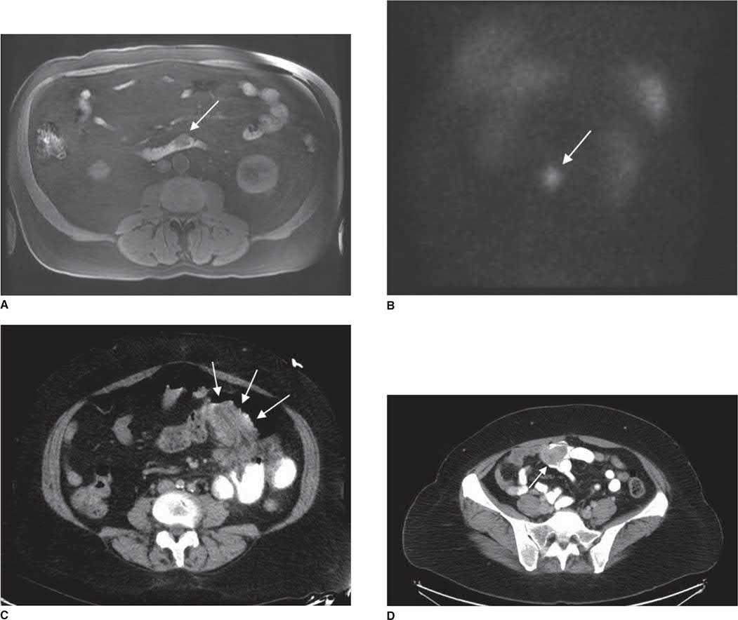
For Meckel's diverticulum, a technetium-99m scan can be obtained where localisation is difficult, the ectopic gastric mucosa taking up the isotope [3].
Jejunal diverticula are frequently an incidental finding at surgery or on radiological imaging [4].
In actinomycosis, pus should be sent for bacteriological examination, which reveals the characteristic sulphur granules [4].
Imaging and localising small-bowel lesions and bleeding
- Enteroclysis detects over 90% of small-bowel tumours and is the test of choice for distal lesions, follow-through only 30–44%, CT 70–80% while also staging spread, angiography or tagged red-cell scans localise bleeding tumours, duodenal lesions are biopsied at gastroscopy with EUS defining the wall layers involved, colonoscopy occasionally reaches the distal ileum, intraoperative enteroscopy visualises lesions beyond endoscopic reach, capsule and double-balloon endoscopy and CT or MR enterography evaluate distal disease, and PET gauges metabolic activity; urinary 5-HIAA and serum chromogranin A are raised in carcinoid syndrome but insensitive, and CEA rises with adenocarcinoma only once liver metastases exist [7].
- For fistulae, CT with enteral contrast is the most useful first test and drains associated abscesses percutaneously, a small-bowel series or enteroclysis defines the origin and excludes distal obstruction, and a fistulogram injected under pressure through a percutaneous catheter may localise a tract the intestinal contrast misses [7].
- Chronic radiation enteritis is evaluated by reviewing total dose, fractionation and treated volume, since lesions localise to high-dose areas; enteroclysis is the most accurate test (sensitivity and specificity over 90%), and CT, though insensitive and non-specific, is needed to exclude recurrent cancer [7].
- Meckel's diverticula are rarely diagnosed before operation except when bleeding; CT sensitivity is too low to be useful, enteroclysis is 75% accurate but impractical acutely, technetium-99m pertechnetate scintigraphy is positive only with ectopic gastric mucosa and about 90% accurate in children but under 50% in adults, and angiography localises acute bleeding [7].
- Duodenal diverticula mimic pseudocysts, biliary cysts and periampullary tumours on ultrasound and CT, are missed by forward-viewing endoscopes and are best shown on upper GI radiographs, while enteroclysis is the most sensitive test for jejunoileal diverticula [7].
- Obscure GI bleeding (no source at gastroscopy and colonoscopy, which reach up to 90% of bleeding lesions) is occult in 20% of cases and arises in the small bowel from angiodysplasia (about 75% in adults), neoplasm (about 10%), Meckel's diverticulum (the commonest cause in children), Crohn's disease, infection, NSAID ulcers, vasculitis, ischaemia, varices, diverticula and intussusception [7].
- Push enteroscopy sees about 60 cm of proximal jejunum with yields of 3–65% and permits cautery, Sonde enteroscopy has been abandoned for capsule endoscopy, which identifies small-bowel pathology in up to 90% and out-yielded contrast study 30% to 7% in a randomised trial without improving rebleeding, transfusion or intervention because it cannot biopsy or treat; after a stopped bleed, push or capsule enteroscopy then enteroclysis (not follow-through) is reasonable, pertechnetate scanning is considered though its yield over 40 is very low, angiography can show angiodysplasia and vascular tumours even without active bleeding and follows a positive tagged red-cell scan in persistent bleeding, and undiagnosed patients who keep bleeding or need transfusion undergo laparoscopy or laparotomy with intraoperative enteroscopy, the endoscope advanced perorally or through an enterotomy by telescoping bowel onto it, the transilluminated bowel inspected with the lights dimmed, lesions marked with serosal sutures for resection, and inspection done on insertion because withdrawal trauma mimics angiodysplasia [7].
- ERCP-related perforation, incidence about 0.5%, is classified by Stapfer as type I free bowel-wall perforation, type II retroperitoneal duodenal perforation from periampullary injury (the commonest), type III duct perforation and type IV retroperitoneal air alone; CT is the most sensitive test, showing pneumoperitoneum, retroperitoneal air, extravasation or paraduodenal fluid, and up to 30% of patients imaged after therapeutic ERCP have asymptomatic retroperitoneal air needing no treatment [7].
- Chylous ascites is diagnosed by paracentesis, cloudy fluid, though clear in fasting postoperative patients, with triglycerides above 110 mg/dL, with CT for nodes, masses and fluid extent and lymphangiography or lymphoscintigraphy to localise leaks for surgical planning; adult intussusception shows a "target sign" on CT, a finding of little significance when seen incidentally without obstructive symptoms; and pneumatosis intestinalis, commonest in the jejunum and idiopathic in 15%, matters to the surgeon for its association with ischaemia and infarction and demands full evaluation [7].
Scoring and Severity
There is no formal grading. The clinically decisive quantities are the length of remnant bowel and the volume of loss.
| Measure | Threshold |
|---|---|
| Resection producing short bowel syndrome | More than 70% of native intestine, as a rule of thumb [1] |
| Widely used length definition in adults | Small intestine under 200 cm [5] |
| Early volume losses after massive resection | May exceed 5 L/day [1] |
| Fistula tract length preventing healing | Short tract, under 2 cm [5] |
| Meckel's neck warranting segmental resection | Neck greater than one-third the diameter of the normal bowel lumen [3] |
Table collects the numerical thresholds cited above. Mortality from enterocutaneous fistula remains high at 10% in recent reports [1].
Treatment and Management
Short bowel syndrome
The first principle is prevention. In Crohn's disease, resection should be limited to segments with a particular complication; in surgery for intestinal ischaemia the smallest possible resection should be performed, with second-look operations to let ischaemic bowel demarcate, so that unnecessary extensive resection is avoided [1].
- After massive resection the course divides into early and late phases.
- Early treatment is directed at controlling diarrhoea, replacing fluid and electrolytes, and prompt institution of total parenteral nutrition in patients who cannot safely tolerate enteral feeding, with vigorous monitoring of intake and output [1].
- Acid hypersecretion is managed with H2 receptor antagonists or a proton pump inhibitor such as omeprazole, and cholestyramine helps where diarrhoea is due to unabsorbed bile salts acting as a cathartic in the colon [1].
For high-volume ostomy losses causing recurrent dehydration, initial management is reduction of gastric secretion with a proton pump inhibitor or H2 receptor antagonist, together with octreotide and loperamide. Small bowel transplantation is not part of initial management [5].
Enterocutaneous fistula in Schwartz's account
Improvement in outcome rests on prevention and, once a fistula occurs, on prompt recognition and intervention, with multidisciplinary care critical [1]. Where a fistula fails to heal on total parenteral nutrition and antisecretory therapy, the cause should be sought among the factors listed above, foreign body, epithelialisation, downstream obstruction, radiation enteritis, infection, malignancy or a short tract [5].
Actinomycosis
Treatment is penicillin or co-trimoxazole, which should be prolonged and in high dosage [4].
HIV-associated intestinal disease
Treatment is directed at the relevant organism, and surgery should be avoided if possible [4].
Fistula management, radiation enteritis and chylous ascites in Schwartz's sequence
- Enterocutaneous fistulae are managed in orderly steps, stabilisation (fluids and electrolytes, usually parenteral nutrition at first, antibiotics and abscess drainage, skin protection with appliances or drains), investigation of the anatomy, decision on options and a timeline, definitive management and rehabilitation, with nutrition and time the keys to spontaneous closure; a trial of oral or enteral feeding suits low-output distal fistulae, octreotide is a useful adjunct for high output and in meta-analysis shortens stay and time to closure without raising closure rates, negative-pressure therapy brought 40% of 91 fistulae to minimal output within a week with 46% spontaneous closure at a mean 90 days, and most surgeons pursue 2–3 months of conservative therapy because 90% of fistulae that will close do so within 5 weeks and later surgery carries lower morbidity [7].
- Factors preventing closure are malnutrition, steroids, malignant, Crohn's or irradiated fistulae, gastric or duodenal site, persistent local sepsis, foreign bodies such as mesh or sutures, an epithelialised or short (<2 cm) tract, high output and distal obstruction, the mnemonic FRIEND (Foreign body, Radiation, Infection/Inflammation, Epithelialisation, Neoplasm, Distal obstruction) [7].
- Acute radiation enteritis is self-limiting and needs only antiemetics and occasionally admission for fluids, rarely forcing a change in radiotherapy; chronic enteritis is treated with antidiarrhoeals and a low-residue diet for obstructive symptoms, and prevention rests on doses below 5000 cGy, multibeam techniques, tilt tables, oral sulfasalazine in small studies, and absorbable mesh slings or other techniques that keep the small bowel out of the pelvis before pelvic radiotherapy [7].
- Bacterial overgrowth in acquired diverticula is treated with antibiotics and asymptomatic diverticula left alone; complications of medial duodenal diverticula penetrating the pancreas are managed endoscopically if possible, bleeding by lateral duodenotomy and oversewing and perforation by wide drainage in emergencies, and diverticulectomy for biliary or pancreatic symptoms is not routinely recommended [7].
- Chylous ascites is treated by addressing the cause, a high-protein low-fat diet with medium-chain triglycerides (absorbed into the portal vein without forming chylomicrons), then fasting with parenteral nutrition and octreotide, paracentesis for respiratory embarrassment, and two-thirds respond conservatively [7].
- Retroperitoneal ERCP perforations without progression or sepsis are managed non-operatively, immediately recognised endoscopic perforations can sometimes be closed endoscopically, and intraperitoneal duodenal perforations need repair with pyloric exclusion and gastrojejunostomy or tube duodenostomy while jejunal and ileal perforations need repair or segmental resection [7].
- For short bowel, initial priorities are the precipitating condition and replacement of the losses of severe diarrhoea, parenteral nutrition for most, gradual enteral feeding once ileus resolves, high-dose H2 antagonists or PPIs against acid hypersecretion, loperamide or diphenoxylate to slow transit and octreotide to cut secretions (though it inhibits adaptation in animals), titrating the two routes over the 1–2-year adaptation period as stool volume falls and absorption improves; parenteral dependence brings catheter sepsis, venous thrombosis, liver and kidney failure and osteoporosis, liver failure often forcing combined liver–intestine transplantation, with 5-year survival of 50–75% on parenteral nutrition, while GLP-2 and glutamine with growth hormone and a high-carbohydrate diet are promising adjuncts [7].
Surgeries
Meckel's diverticulum in Schwartz's account
An incidentally discovered diverticulum is usually left alone, unless gastric mucosa is suspected (the diverticulum feeling thick) or it has a very narrow neck [3]. Diverticulectomy suffices for uncomplicated diverticulitis or bleeding; segmental resection is needed for complicated diverticulitis such as perforation, or where the neck exceeds one-third the diameter of the normal bowel lumen [3].
Jejunal diverticula
- Elective resection of the affected segment can be effective for malabsorption, provided only a limited length of jejunum is involved [4].
- Where perforated jejunal diverticulitis is found at emergency laparotomy, small bowel resection is performed and the choice between primary anastomosis and stoma made on the degree of contamination, physiological stability, and local resources for managing a high-output jejunostomy [4].
- Complications of extensive jejunal diverticulosis can be extremely difficult to treat [4].
Small bowel tumours
Adenomas need resection when identified, often endoscopically [2]. Adenocarcinoma is treated by resection with adenectomy, and by Whipple procedure where the tumour lies in the second part of the duodenum [2].
Resection principles for tumours, diverticula, fistulae and short bowel
- Symptomatic benign tumours are resected or removed endoscopically; duodenal lesions under 1 cm suit endoscopic polypectomy, over 2 cm usually need transduodenal polypectomy or segmental resection (pancreaticoduodenectomy near the ampulla), EUS helps decide for 1–2-cm lesions, endoscopic removal of proven benign periampullary adenomas equals surgery with less morbidity, and adenomas recur so surveillance follows [7].
- FAP patients start screening gastroscopy in the second or third decade, have adenomas removed endoscopically with surveillance at 6 months then yearly, and when surgery is needed generally require a standard rather than pylorus-preserving pancreaticoduodenectomy because the adenomas are multiple, sessile and periampullary, local resection recurs and even the duodenal remnant can recur [7].
- Duodenal adenocarcinoma needs pancreaticoduodenectomy except in the third or fourth part where local resection may serve; jejunal and ileal cancers are widely resected with the mesentery for regional lymphadenectomy as for colon cancer, advanced disease is palliated by resection or bypass, and chemotherapy has no proven adjuvant or palliative efficacy [7].
- Carcinoids are resected with regional lymphadenectomy (nodes are unusual under 1 cm but present in 75–90% over 3 cm) the whole small bowel is examined because 30% are multiple, metastatic disease is debulked for survival and symptom relief, doxorubicin, 5-FU and streptozocin regimens give 30–50% responses without altering the natural history, and octreotide is the most effective drug for the syndrome [7].
- Localised lymphoma is resected with adjacent mesentery and diffuse disease treated primarily by chemotherapy with the value of adjuvant therapy controversial; GISTs are resected segmentally without wide lymphadenectomy because nodal spread is rare, imatinib benefits 80% of unresectable or metastatic patients with 50–60% objective shrinkage and shows promise as neoadjuvant and adjuvant treatment, and sunitinib is used for resistance [7].
- Chronic radiation enteritis surgery is reserved for high-grade obstruction, perforation, haemorrhage, abscess or fistula, aims at limited resection with anastomosis between healthy segments (irradiated bowel is hard to distinguish grossly or on frozen section, and anastomoses between irradiated segments leak in up to 50%), uses bypass when resection is impossible except for bleeding, and carries mortality averaging 10% [7].
- Symptomatic Meckel's diverticula are excised with their bands; bleeding requires segmental ileal resection including the adjacent ulcer, as does a tumour or an inflamed or perforated base; prophylactic removal of incidental diverticula is contested (a meta-analysis found 758 prophylactic diverticulectomies per death prevented) and many adopt a selective policy for patients under 50, band attachments, ectopic tissue or length over 2 cm, without controlled data [7].
- Bleeding or diverticulitis of jejunoileal diverticula is treated by segmental resection and of lateral duodenal diverticula by diverticulectomy [7].
- A fistula that fails 2–3 months of conservative care is treated by resecting the tract with the segment of origin, since simple closure of the bowel opening recurs; in a 23-year review of 153 surgically treated fistulae, mostly small-bowel and iatrogenic after five or more operations, 30-day mortality was about 4%, 1-year mortality 15%, morbidity over 80%, first-attempt success 70% and overall closure 84%, fascial closure reducing refistulisation and death, while another series recorded 30% recurrence linked to high output and to operations without resection [7].
- Chylous leaks after surgery or trauma that fail conservative treatment are localised and repaired with fine non-absorbable sutures or by limited resection if peripheral in the mesentery; peritoneovenous shunts for poor candidates carry sepsis, DIC and high occlusion rates [7].
- Adult intussusception is resected with its lead point for histology, whereas retrograde intussusception after gastric bypass has been reduced without resection [7].
- In short bowel, stomas are reversed whenever possible; transit-slowing operations (segmental reversal, colon interposition, valves, pacing) lack objective benefit and cause obstruction; Bianchi's 1980 longitudinal intestinal lengthening and tailoring separates the dual vasculature and divides the bowel longitudinally for isoperistaltic anastomosis, potentially doubling length in dilated paediatric bowel, and the serial transverse enteroplasty (STEP) lengthens dilated bowel by perpendicular stapler firings without separating the vasculature, achieving enteral autonomy in 47% of 111 registry patients at a median 21 months [7].
- Intestinal transplantation is indicated for life-threatening complications of intestinal failure or parenteral nutrition, impending liver failure, central venous thrombosis, recurrent catheter sepsis and recurrent severe dehydration, as intestine alone (37%), intestine–liver–pancreas (30%) or intestine–liver (24%), multivisceral grafts serving giant desmoids, diffuse dysmotility and splanchnic thrombosis; nearly 80% of survivors have full graft function without parenteral nutrition at the cost of rejection, CMV and post-transplant lymphoproliferative disease [7].
Complications
Extensive resection risks short bowel syndrome, which is why bowel-preserving strategies matter in conditions requiring repeated operations [1]. Mortality from enterocutaneous fistula remains high at 10% [1].
Jejunal diverticula may cause malabsorption from bacterial overgrowth, inflammation, perforation and, rarely, bleeding [4]. Actinomycosis may lead to multiple liver abscesses through portal venous spread, and to secondary fistulation with woody induration [4].
Before the 1960s any cause of intestinal failure was nearly always fatal; numerous treatment strategies have since been developed, though management continues to vary greatly between centres [1].
Prognosis
Outcome in short bowel syndrome depends on remnant length and on whether enteral autonomy can be regained, which is not generally expected once more than 70% of the intestine has been resected [1]. Intestinal failure, once nearly always fatal, is now treatable, although practice varies widely by centre [1].
Enterocutaneous fistula still carries around 10% mortality [1]. In Peutz-Jeghers syndrome the gastrointestinal malignancy risk is small, under 3%, but extraintestinal malignancy is increased, most commonly breast cancer [2].
Survival figures from Schwartz
- Complete resection gives 5-year survival of 50–60% for duodenal and 20–30% for jejunal or ileal adenocarcinoma, 75–95% for localised carcinoid and 19–54% with carcinoid liver metastases, 20–40% for intestinal lymphoma overall and 60% when localised and resectable, and 35–60% for GIST, which recurs in about 35% with size and mitotic index independently prognostic and tumours under 5 cm with fewer than 10 mitoses per high-power field faring excellently [7].
- Acute radiation injury does not predict chronic enteritis [7].
- About 50–70% of short-bowel patients initially on parenteral nutrition eventually achieve independence, more often children than adults; among 124 adults with non-malignant short bowel, survival was 86% at 2 and 45% at 5 years, worse with end-enterostomies or under 50 cm of residual bowel, and graft survival at 1, 5 and 10 years is 80%, 44% and 26% for intestine alone, 62%, 45% and 36% for intestine–liver and 69%, 48% and 33% for intestine–liver–pancreas, with no randomised comparison against chronic parenteral nutrition [7].
References
- Sabiston Textbook of Surgery, 22nd ed., Ch. 91 Small Intestine
- The ABSITE Review, 2022, Small bowel tumors
- The ABSITE Review, 2022, Meckel's diverticulum
- Bailey & Love's Short Practice of Surgery, 28th ed., Ch. 74 The small intestine
- Schwartz's Principles of Surgery: ABSITE and Board Review, Ch. 28 Small Intestine
- Maingot's Abdominal Operations, 13th ed., Ch. 16
- Schwartz's Principles of Surgery, 11th ed., Ch. 28, Small Intestine
- Maingot's Abdominal Operations, 13th ed., Ch. 39