



Hemorrhoids
Summary
- Haemorrhoids are symptomatic enlargements of the normal anal cushions (submucosal vascular tissue that helps maintain continence) commonly presenting with painless bright-red rectal bleeding, prolapse and pruritus [1].
- They are graded first- to fourth-degree according to the extent of prolapse [1].
- Management ranges from dietary and lifestyle measures through outpatient procedures (injection sclerotherapy, rubber band ligation) to surgical haemorrhoidectomy for more advanced disease [1][2].
There is no NICE guideline on haemorrhoids, but NICE has appraised two of the operations, and the two appraisals reach different kinds of conclusion. Stapled haemorrhoidopexy is recommended as an option where surgical intervention is considered appropriate for prolapsed internal haemorrhoids [3]. Haemorrhoidal artery ligation may be used provided normal arrangements are in place for clinical governance, consent and audit, the standard, permissive formula rather than the restrictive "special arrangements" wording [4].
Definition
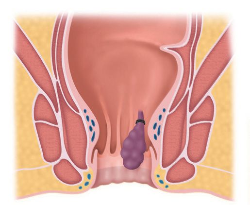
Haemorrhoids are symptomatic enlargements of the internal haemorrhoidal venous plexus (Greek haima, blood, and rhoos, flowing) [1]. Internal haemorrhoids lie proximal to the dentate line and are covered by insensate anorectal mucosa; external haemorrhoids relate to the inferior haemorrhoidal plexus in the skin around the anal verge and are often confused with anal skin tags, which are not true haemorrhoids [1][5].
Pathophysiology
- The internal haemorrhoidal plexus forms the submucosal component of the anal cushions that help seal the anal canal.
- Man's upright posture, absence of valves in the portal venous system, and raised abdominal pressure from pregnancy or straining at defecation contribute to venous engorgement and varicosity formation [1].
- Shearing forces cause mucosal trauma (bleeding) and caudal displacement of the anal cushions (prolapse), impairing venous drainage and causing further engorgement, stasis and transudation of fluid (pruritus); with age, fragmentation of the supporting connective tissue causes loss of elasticity so the cushions no longer retract after defecation [1].
The dentate line explains almost every clinical feature. Because internal haemorrhoids sit proximal to it, in an area devoid of somatic pain fibres, they bleed and prolapse painlessly and can be banded in clinic without anaesthesia; significant pain after rubber band ligation typically results from misplacement of the band below the dentate line and requires immediate band removal [6].
Haemorrhoids as normal anatomy in Schwartz's account
- Haemorrhoids are cushions of submucosal tissue containing venules, arterioles and smooth muscle in the left lateral, right anterior and right posterior positions, thought to form part of the continence mechanism by aiding complete closure of the canal at rest; because they are normal anatomy, treatment is indicated only when they become symptomatic, and excessive straining, raised abdominal pressure and hard stools engorge the plexus and cause the tissue to prolapse, bleed or thrombose [7].
- A skin tag is redundant fibrotic skin at the verge, often the residue of a thrombosed external haemorrhoid, and is frequently confused with symptomatic haemorrhoids; internal haemorrhoids covered by insensate mucosa rarely hurt unless thrombosed and necrotic from severe prolapse, incarceration or strangulation, whereas the richly innervated anoderm means external haemorrhoids must never be ligated or excised without adequate local anaesthesia [7].
- Portal hypertension was long thought to raise haemorrhoidal bleeding through the portal–systemic anastomoses of the plexuses, but haemorrhoidal disease is now known to be no commoner in these patients; rectal varices may occur and bleed, are best treated by lowering portal pressure with suture ligation only for persistent massive bleeding, and haemorrhoidectomy should be avoided for fear of uncontrollable variceal haemorrhage [7].
- Postpartum haemorrhoids follow straining in labour with oedema, thrombosis or strangulation, and haemorrhoidectomy is often the treatment of choice when symptoms were chronic beforehand [7].
Clinical features
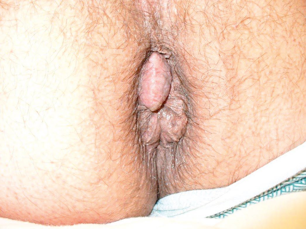
- Bleeding is usually the earliest symptom, characteristically separate from the stool, seen on the paper or as a fresh splash in the pan, and rarely sufficient to cause anaemia, so other causes of bleeding should be excluded [1].
- Pruritus from mucus discharge is common; pain suggests an alternative diagnosis such as anal fissure, unless there is thrombosis [1][8].
- Patients may describe a lump appearing at the anal orifice on defecation [2].
Acute presentations
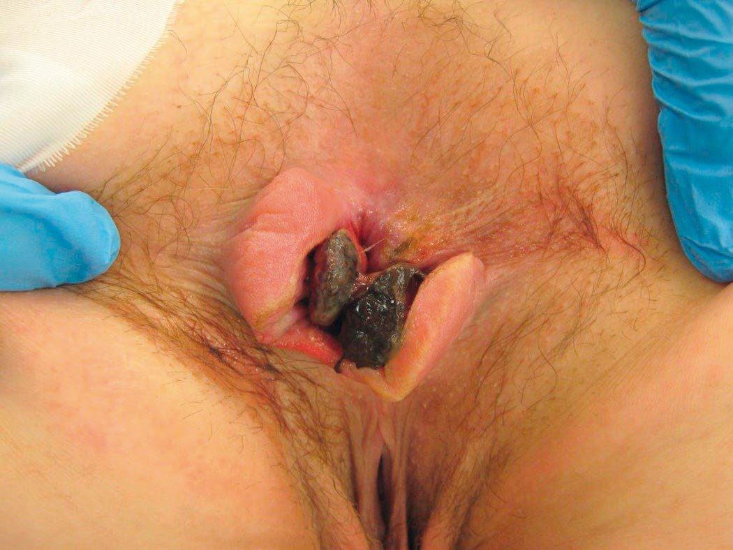
Patients may present acutely with strangulated, ischaemic, gangrenous or acutely thrombosed internal haemorrhoids. These patients generally have a previous history of grade III or IV prolapsing haemorrhoids and present with an acute prolapse that is no longer reducible; if presentation is delayed, incarcerated haemorrhoids may become necrotic and drain bloody or malodorous material [6].
Etiology
Constipation, chronic straining, obesity and previous childbirth are recognised associations [2]. Haemorrhoids rarely start after age 55, and an alternative cause of symptoms should be assumed until proven otherwise in older patients [2].
Diagnosis
Diagnosis is usually made by rigid sigmoidoscopy and proctoscopy; flexible sigmoidoscopy or colonoscopy is used when there is diagnostic uncertainty about the cause of symptoms, particularly bleeding [2]. Colorectal malignancy and other causes of rectal bleeding must be excluded before treatment [1].
Scoring and Severity
Internal haemorrhoids are graded by the degree of prolapse [1][5][6]:
| Grade | Description | Typical management |
|---|---|---|
| First degree | Bleeding only; bulge into the anal canal, no prolapse beyond the anal verge | Medical measures; sclerotherapy, rubber band ligation or infrared coagulation if refractory |
| Second degree | Prolapse through the anus on straining but reduce spontaneously | Rubber band ligation is the most common and effective office procedure |
| Third degree | Prolapse through the anal canal and require manual reduction | Office procedure in selected patients; otherwise excisional haemorrhoidectomy |
| Fourth degree | Permanently prolapsed and irreducible, at risk of strangulation | Excisional haemorrhoidectomy |
Table reformats the grading and its management implications [1][6]. "Mixed" haemorrhoids describes a significant cutaneous (external) component arising from repeated congestion of a prolapsing internal haemorrhoid [1].
Treatment and Management
First-line management addresses bowel and defecatory habits, minimising straining, and adding stool softeners and bulking agents; proprietary creams and phlebotonic suppositories may be used [1].
Office-based procedures
Most patients with grade I and II disease, and selected patients with grade III, who remain symptomatic despite medical management can be offered an office-based procedure, rubber band ligation, sclerotherapy or infrared coagulation [6].
Rubber band ligation (Barron's bander) has largely superseded injection: an elastic band applied above the dentate line strangulates the haemorrhoidal tissue, causing ischaemia and necrosis of the prolapsing mucosa followed by scar fixation to the rectal wall, so it treats both bleeding and prolapse by reducing cushion size and increasing fixation [1][6]. It is the most common and effective office option and has been shown superior to sclerotherapy and infrared coagulation [6]. It should be avoided, or used only with caution, in therapeutically anticoagulated patients or those on antiplatelet drugs, given the risk of serious bleeding from the ulcer when the cushion sloughs, often days after the procedure [6].
Injection sclerotherapy uses 5% phenol in arachis or almond oil, 3–5 mL injected into the pedicle apex rather than into the haemorrhoid itself, to induce fibrosis; injection too superficial causes mucosal ulceration, while too deep can cause prostatitis or pelvic sepsis [1]. Sabiston describes approximately 1 mL of 5% phenol in almond or vegetable oil, or 1% sodium tetradecyl sulfate, injected into the submucosa at the apex of the cushion, and notes that sclerotherapy is appropriately offered to anticoagulated or antiplatelet-treated patients, who are often poor candidates for banding or excision [6]. Infrared coagulation applies infrared light to coagulate protein within the haemorrhoid and is used mostly for grades I and II [6].
The acutely incarcerated haemorrhoid
For incarceration without strangulation or signs of sepsis, efforts focus on reducing the prolapse to resolve the acute crisis and allow a less invasive treatment later. Applying table sugar as an osmotic agent, ice packs, and an anal block can facilitate reduction, and injecting hyaluronidase directly into incarcerated oedematous haemorrhoids can produce prompt and dramatic resolution. Patients who fail a limited trial of non-operative management, or who have strangulation and necrosis, require surgery [6].
Stapled haemorrhoidopexy (using a circular stapler specifically developed for haemorrhoidopexy) is recommended as an option for people in whom surgical intervention is considered appropriate for the treatment of prolapsed internal haemorrhoids [3]. The appraisal examined the HCS33 circular stapler (models PPH01 and PPH03); at the time there was no evidence to make recommendations for the Autosuture stapler with the STRAM kit adaptor [3].
- Haemorrhoidal artery ligation. "Current evidence on haemorrhoidal artery ligation shows that this procedure is an efficacious alternative to conventional haemorrhoidectomy or stapled haemorrhoidopexy in the short and medium term, and that there are no major safety concerns.
- Therefore, this procedure may be used provided that normal arrangements are in place for clinical governance, consent and audit." [4].
- The guidance was published on 19 May 2010 and migrated from interventional procedures guidance IPG342 with recommendations unchanged [4].
- Note the divergence on cost-effectiveness.
- NICE HTG218 endorses haemorrhoidal artery ligation on efficacy and safety grounds without addressing cost-effectiveness.
- The UK HubBLe trial found lower recurrence than rubber band ligation but poorer cost-effectiveness, and Sabiston records that the method was not found to be cost-effective compared with rubber band ligation in terms of incremental cost per quality-adjusted life-year [4].
- A NICE "may be used" is a statement about governance, not a commissioning endorsement, and the two should not be conflated.
Office techniques and their complications in Schwartz's detail
- Many over-the-counter topical preparations are desiccants and relatively ineffective; in rubber band ligation mucosa 1–2 cm above the dentate line is drawn into the applier, generally one or two quadrants are banded per visit, severe pain follows a band placed at or below the dentate line, urinary retention occurs in about 1% (more likely if internal sphincter was caught), necrotising infection is rare but life-threatening, severe pain, fever and urinary retention are its early signs and prompt examination under anaesthesia, debridement, drainage and broad-spectrum antibiotics, and bleeding at 7–10 days when the pedicle sloughs is usually self-limited but may need suture ligation under anaesthesia [7].
- Infrared photocoagulation applied to the apex of each haemorrhoid can treat all three quadrants in one visit but is ineffective for large or significantly prolapsing haemorrhoids; sclerotherapy injects 1–3 mL of phenol in olive oil, sodium morrhuate or quinine urea into the submucosa with few complications beyond occasional infection and fibrosis [7].
- An acutely thrombosed external haemorrhoid causes intense pain and a palpable mass in the first 24–72 hours and is treated by elliptical excision under local anaesthesia in the office (simple incision and drainage rarely works because the clot is loculated) whereas after 72 hours the clot resorbs and pain settles, so sitz baths and analgesia suffice [7].
Surgeries
Indications for haemorrhoidectomy are third- and fourth-degree haemorrhoids; second-degree disease failing non-operative treatment; well-defined mixed haemorrhoids; and bleeding causing anaemia [1]. Surgical excision is very effective, albeit painful, for patients who have not improved or are not candidates for an office-based treatment, and can be offered for symptomatic combined internal and external haemorrhoids with prolapse (grades III–IV) [6].
- Open (Milligan–Morgan) haemorrhoidectomy: the anal mucosa and skin are left open to heal by secondary intention after ligation and excision of each haemorrhoidal pedicle, preserving mucocutaneous bridges to avoid stenosis [1].
- Closed (Ferguson) haemorrhoidectomy: the wound is sutured after excision [1]. This is the most commonly used technique, and is associated with decreased postoperative pain, faster wound healing and reduced risk of postoperative bleeding compared with the open Milligan–Morgan approach [6].
- Stapled haemorrhoidopexy (PPH, procedure for prolapse and haemorrhoids, Longo): a circular stapling device excises a cylinder of mucosa and submucosa above the dentate line and staples the mucosal ends together, also disrupting the feeding arteries and displacing the cushions into the proximal anal canal; less painful with quicker recovery than conventional haemorrhoidectomy, but higher recurrence and risk of serious complications including staple-line dehiscence, rectovaginal fistula and tenesmus [1][6]. It does not address external haemorrhoids, and patients are significantly more likely to have recurrent symptoms requiring additional operative procedures than after excisional haemorrhoidectomy; rectal perforation requiring faecal diversion or low anterior resection has been reported as a rare, severe complication [6].
- Transanal haemorrhoidal (Doppler-guided) artery ligation: ligates the feeding vessels with or without mucopexy; no tissue is excised, but mucosal pexy is required for patients with symptomatic prolapse. The UK HubBLe trial found lower recurrence than rubber band ligation but poorer cost-effectiveness, with better pain scores and complication rates than conventional surgery [1][6].
- Other ablative techniques such as cryotherapy are not commonly used [1].
Technical principles of excisional haemorrhoidectomy
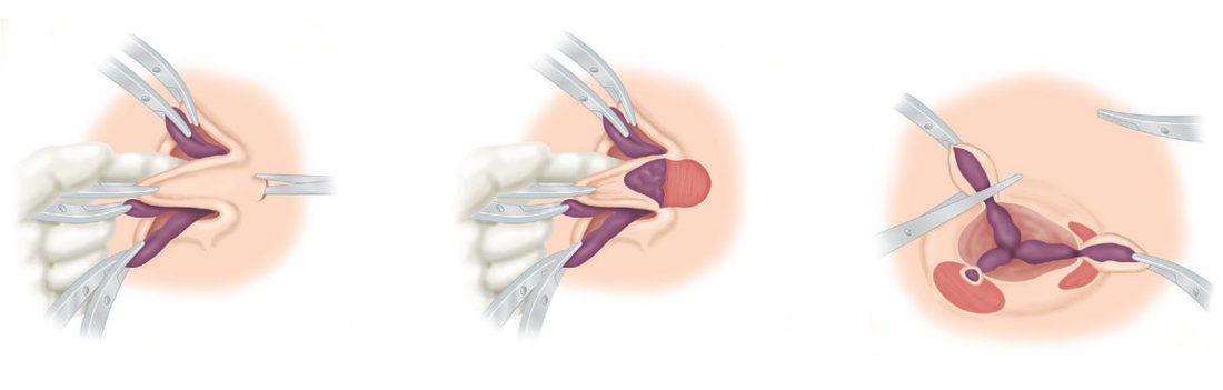
- The principles are removal of only redundant haemorrhoidal tissue and pexy of the haemorrhoidal mucosa to the rectal wall; in most cases removing the largest or most symptomatic haemorrhoid produces the desired relief. Removing all three haemorrhoidal columns creates larger mucosal defects and can cause narrowing or stenosis of the anal canal if adequate intervening mucosa is not preserved, or incontinence if the underlying sphincter is injured [6].
- The haemorrhoid is excised in a diamond shape with the distal apex at the base of the external component on the anoderm, narrowing to the proximal apex at the internal pedicle, and the cushion is dissected off the internal sphincter in a typically avascular plane, the pedicle controlled with a braided absorbable suture ligature [6]. Prophylactic antibiotics are not indicated, the internal sphincter fibres must be identified and preserved regardless of technique, and there is no benefit in packing the anal canal, packing is unlikely to stop postoperative bleeding, and large quantities of blood can accumulate above it, preventing early diagnosis of a bleed [6].
- Postoperative care aims at regular bowel function with liberal stool softeners and laxatives, sitz baths for pain and itching, and a multimodal analgesic regimen to reduce narcotic use [6].
The named operations and their sequelae in Schwartz's account
- The closed submucosal (Parks or Ferguson) haemorrhoidectomy is done prone or in lithotomy under local, regional or general anaesthesia through a Fansler anoscope: an ellipse from just beyond the verge to the anorectal ring is excised, the internal sphincter fibres are identified and brushed away to avoid injury, the apex of the plexus is ligated and the wound closed with a running absorbable suture, all three cushions may be taken but a large area of perianal skin must not be removed lest anal stenosis follow [7].
- The open Milligan–Morgan technique leaves the wounds to heal by secondary intention; Whitehead's circumferential excision above the dentate line with advancement of rectal mucosa to the dentate line has been abandoned by most for the risk of ectropion (Whitehead's deformity) [7].
- Stapled haemorrhoidopexy (PPH) uses an EEA-like stapler to create mucosal and submucosal doughnuts, acting mainly by pexy of redundant tissue, ligation of feeding venules and fixation of mucosa above the dentate line, is best suited to second- and third-degree disease, gives less pain and disability with equivalent complication rates, but its complications include chronic anal pain, bacteraemia, rectovaginal fistula, obstructing stricture and even rectal perforation, and systematic review shows more recurrence than excisional haemorrhoidectomy; Doppler-guided haemorrhoidal artery ligation (transanal dearterialisation) shows early promise with durability unknown [7].
- Urinary retention after haemorrhoidectomy is reported in 10–50% and is reduced by limiting perioperative fluids and giving adequate analgesia; faecal impaction is prevented by preoperative enemas or limited preparation and liberal laxatives; immediate bleeding from an inadequately ligated pedicle mandates urgent return to theatre, and bleeding at 7–10 days from mucosal slough may be observed or need ligation or oversewing under anaesthesia; long-term sequelae are incontinence (usually transient flatus incontinence), anal stenosis from excessive skin excision and ectropion [7].
Complications
Strangulation and thrombosis, ulceration, gangrene, and portal pyaemia are recognised complications of haemorrhoids themselves [1]. Postoperative complications of haemorrhoidectomy are early, pain, acute urinary retention especially in men, and reactionary haemorrhage, which is more common than secondary haemorrhage and may require return to theatre, and late: secondary haemorrhage around day 7–8, anal stricture, anal fissure or submucous abscess, and incontinence from inadvertent internal sphincter damage [1].
Quantitatively, complications after surgical haemorrhoidectomy are relatively uncommon: postoperative haemorrhage is the most common at 1–2%, and acute urinary retention occurs in 1–15% [6]. Pelvic sepsis is a rare but feared complication that can follow either excisional haemorrhoidectomy or an office-based procedure: patients with urinary retention, worsening anal or pelvic pain, or fever must be evaluated emergently, and immediate examination under anaesthesia with debridement of any necrotic tissue is required [6].
Prognosis
- Symptomatic relief is good with appropriately staged treatment; recurrence is more likely after less invasive procedures (injection, banding, haemorrhoidal artery ligation) than after formal haemorrhoidectomy, but formal excision carries higher rates of pain and complication [1].
- The same trade-off holds for stapled haemorrhoidopexy, which gives less pain, pruritus ani and faecal urgency but significantly more recurrent symptoms requiring further operative procedures than excisional haemorrhoidectomy [6].
- No survival or prognostic scoring system applies to this benign condition.
References
- Bailey & Love's Short Practice of Surgery, 28th ed., Ch. 80 The anus and anal canal
- Oxford Handbook of Clinical Surgery, 5th ed., Ch. 12 Colorectal surgery
- NICE Technology Appraisal TA128: Stapled haemorrhoidopexy for the treatment of haemorrhoids (2007), 1 Recommendations; 1.1 www.nice.org.uk
- NICE HealthTech Guidance HTG218: Haemorrhoidal artery ligation (2010, migrated from IPG342), 1.1; Overview www.nice.org.uk
- Schwartz's Principles of Surgery: ABSITE and Board Review, Ch. 29 Colon, Rectum, and Anus
- Sabiston Textbook of Surgery, 22nd ed., Ch. 97 Benign Anorectal Disorders
- Schwartz's Principles of Surgery, 11th ed., Ch. 29, Colon, Rectum, and Anus
- Oxford Handbook of Clinical Surgery, 5th ed., Ch. 26 Emergency surgery topics