



Diverticular Disease
Summary
- Colonic diverticula are acquired outpouchings of mucosa and connective tissue through the colonic wall, found in around 75% of people over 70 in the Western world, overwhelmingly in the sigmoid colon [1][2].
- Most diverticular disease is asymptomatic (diverticulosis), but 10–30% develop symptomatic complications ranging from painful diverticular disease to diverticulitis, abscess, perforation, fistula, stricture and haemorrhage [1].
- Management is chiefly medical, with surgery reserved for complications or recurrent, refractory disease [1][2].
NICE NG147 covers the whole spectrum in one guideline, and it draws a three-way distinction that the textbooks blur: diverticulosis (diverticula present, asymptomatic), diverticular disease (symptomatic but not acutely inflamed) and acute diverticulitis [3]. Each attracts different advice, and three of its recommendations reverse long-standing teaching: seeds and nuts need not be avoided, antibiotics are not offered for diverticular disease, and a no-antibiotic strategy should be considered for acute diverticulitis in a systemically well patient [3].
Definition
- Diverticula are hollow outpouchings of the gastrointestinal tract.
- Congenital diverticula (for example Meckel's) contain all three coats of the bowel wall; acquired diverticula, as in sigmoid diverticular disease, lack a muscularis layer [1].
- Sabiston frames the same distinction as "true" versus "false": true diverticula contain all layers of the bowel wall and are rare and usually congenital, while the vast majority of colonic diverticula are false (pulsion, pseudodiverticula) containing only mucosa and muscularis mucosa [4].
Pathophysiology
- Diverticula form along the lines where penetrating colonic arteries traverse the colonic wall between the taeniae coli, associated with hypertrophy of the surrounding colonic muscle and mucosal thickening, probably driven by high-pressure colonic contractions causing chronic wall pressure [2].
- Epidemiological evidence links the disease to a refined, fibre-deficient Western diet: altered collagen structure with ageing, disordered motility and raised intraluminal pressure (especially in the narrow sigmoid colon) cause mucosal herniation at vessel-penetration points; the rectum, with a complete muscular coat and wider lumen, is rarely affected.
- Diverticular disease is rare in Africa and Asia, where dietary fibre intake is high (Burkitt) [1].
- Diverticulitis represents acute neutrophilic infiltration around an inflamed diverticulum and in the subserosal tissues [2].
Why most diverticulitis stays localised
- Diverticula form classically on the mesenteric side of the colonic wall where the vasa recta traverse the muscular layer; these small vessels are the weak spots.
- When pressure, inflammation, impaired blood supply or trauma acts at these points it can perforate the diverticulum, producing infection and a marked local inflammatory response.
- Because most perforations occur on the mesenteric side, the surrounding mesentery acts as a natural barrier, containing the infection and allowing an abscess to form within the mesenteric tissues rather than spilling into the peritoneal cavity, which is why the majority of diverticulitis is contained.
- If the process outruns that barrier, generalised peritonitis and sepsis follow [4].
The mechanism of diverticulitis itself is thought to be obstruction of the orifice of a diverticulum, with stasis leading to bacterial overgrowth, inflammation and raised intradiverticular pressure, causing ischaemia and microperforation [4].
One anatomical fact has a direct operative consequence. The sigmoid and descending colon are typically affected while the rectum, having an extra layer of muscle, is generally spared, which is why the distal anastomotic margin in operations for diverticulitis should always be within the rectum, to reduce the likelihood of recurrence [4].
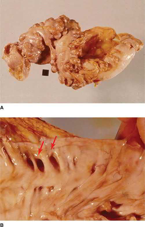
The pulsion mechanism and the segmentation theory in Schwartz's account
- Most colonic diverticula are false diverticula in which mucosa and muscularis mucosa herniate through the colonic wall between the teniae coli, at the points where the nutrient arterial vessels penetrate and presumably weaken the muscle; they are pulsion diverticula produced by high intraluminal pressure, whereas true diverticula containing all wall layers are rare and usually congenital [6].
- The most accepted theory is that a low-fibre diet yields small stool volume, which demands high intraluminal pressure and high wall tension for propulsion; chronic contraction then causes muscular hypertrophy and segmentation, in which the colon behaves as separate compartments rather than a continuous tube, so that pressure is directed radially against the wall instead of into propulsive waves (with an age-related loss of tensile strength and elasticity also proposed) and although none of these theories is proven, a high-fibre diet does appear to lower the incidence of diverticulosis [6].
- Half of the population over 50 in the United States and Europe is estimated to have colonic diverticula, the sigmoid is the commonest site, most cases are asymptomatic, and diverticulitis is estimated to occur in 10–25% of people with diverticulosis, resulting from macroscopic or microscopic perforation of a diverticulum with pericolic contamination [6].
Clinical features
Most diverticular disease is asymptomatic and found incidentally [1]. Recognised presentations include [1][2]:
- Painful diverticular disease: intermittent left iliac fossa pain, distension, flatulence and a sensation of heaviness, which may overlap with irritable bowel syndrome.
- Acute diverticulitis: persistent left iliac fossa pain (occasionally right-sided if a redundant sigmoid loop lies across the midline), fever, tachycardia and localised tenderness or a tender mass; diarrhoea or constipation may accompany it.
- Haemorrhage: typically painless, sudden and profuse, with large-volume dark red or clotted blood (bright red from the sigmoid, darker from the right colon), due to rupture of a peridiverticular submucosal vessel, usually without inflammation.
- Complications: pericolic or paracolic abscess (swinging fever, tender left iliac fossa mass), purulent or faeculent peritonitis from perforation, colovesical fistula (recurrent UTI, pneumaturia) or colovaginal fistula (faeculent vaginal discharge, more common after hysterectomy), and stricture (recurrent colicky pain, distension, bloating from chronic fibrosis).
Fistulae
- Fistulae result from local inflammation and abscess formation decompressing into a neighbouring organ.
- The most common, especially in men, is a colovesical fistula to the dome of the bladder, presenting with recurrent and often polymicrobial urinary tract infections, with pneumaturia and faecaluria; CT can reveal air or contrast in the bladder in the absence of prior instrumentation, and cystoscopy usually discloses inflammation at the fistula site. Colovaginal fistulae occur almost exclusively in women who have had a previous hysterectomy, presenting with vaginal discharge and passage of air per vagina.
- Colocutaneous fistulae usually present at a previous drain site after percutaneous drainage.
- Patients with fistulae usually do not need emergency surgery, because the abscess has already decompressed through the fistula [4].
When to suspect what. Suspect diverticular disease if a person has intermittent abdominal pain in the left lower quadrant with constipation, diarrhoea or occasional large rectal bleeds (the pain may be triggered by eating and relieved by passage of stool or flatus) or tenderness in the left lower quadrant on examination. In a minority of people, and in people of Asian origin, pain and tenderness may be localised in the right lower quadrant, and symptoms may overlap with irritable bowel syndrome, colitis and malignancy [3].
Suspect acute diverticulitis if there is constant abdominal pain, usually severe and localising in the left lower quadrant, with either fever, or a sudden change in bowel habit with significant rectal bleeding or passage of mucus, or left lower quadrant tenderness, a palpable abdominal mass or distension in someone with a previous history of diverticulosis or diverticulitis [3].
Suspect complicated acute diverticulitis and refer for same-day hospital assessment if the person has uncontrolled abdominal pain plus any of the following [3]:
| Symptom or sign | Possible complication |
|---|---|
| Abdominal mass on examination, or perirectal fullness on digital rectal examination | Intra-abdominal abscess |
| Abdominal rigidity and guarding | Bowel perforation and peritonitis |
| Altered mental state, raised respiratory rate, low systolic blood pressure, raised heart rate, low tympanic temperature, no urine output, skin discolouration | Sepsis |
| Faecaluria, pneumaturia, pyuria, or passage of faeces through the vagina | Fistula into the bladder or vagina |
| Colicky abdominal pain, absolute constipation, vomiting or abdominal distension | Intestinal obstruction |
Table reformats the features suggesting complicated acute diverticulitis [3].
Obstruction, fistula frequency and the differential in Schwartz's figures
- Obstructive symptoms occur in approximately 67% of patients with acute diverticulitis and complete obstruction in 10%; approximately 5% of patients with complicated diverticulitis develop a fistula to an adjacent organ, colovesical being commonest, then colovaginal and coloenteric, with colocutaneous fistulae rare [6].
- Diverticular bleeding, from erosion of the peridiverticular arteriole, can be massive but stops spontaneously in 80%; most significant lower gastrointestinal haemorrhage occurs in elderly patients in whom diverticulosis and angiodysplasia coexist, so the exact source may be difficult to identify [6].
- Giant colonic diverticula are extremely rare, usually on the antimesenteric side of the sigmoid, may be asymptomatic or cause vague pain, nausea or constipation, are suggested on plain radiographs and usually diagnosed by barium enema, and can perforate, obstruct or undergo volvulus, so resection of the involved colon with the diverticulum is recommended [6].
- The differential diagnosis of diverticulitis includes malignancy, ischaemic colitis, infectious colitis and inflammatory bowel disease, and for a fistula it includes malignancy, Crohn's disease and radiation injury, in a previously irradiated patient a fistula is recurrent cancer until proved otherwise [6].
Etiology
- Low dietary fibre intake in Westernised populations is the principal implicated factor, combined with age-related collagen change, colonic dysmotility and raised intraluminal pressure [1].
- Right-sided diverticular disease predominates in South East Asia [1].
- Diverticulitis is thought to be mainly a disease of the modern world, coinciding with dietary changes after the Industrial Revolution, and its incidence has risen with increasing life expectancy because it predominantly presents in older individuals [4].
- The prevalence figures are worth holding beside the risk of ever developing diverticulitis.
- Colonic diverticula are found in approximately 40% of individuals aged 50 to 60 and in over 60% of those over 80, yet modern estimates indicate that fewer than 5% of patients with diverticulosis will develop diverticulitis [4].
- Because diverticulosis is so prevalent, however, the absolute burden is large: more than 2.7 million outpatient visits and over 200,000 inpatient admissions annually in the United States, at an estimated cost of more than $2 billion [4].
Diagnosis
- Elective diagnosis is usually by colonoscopy, though this incompletely assesses the number and extent of diverticula and carries perforation risk in a narrowed segment [1].
- Contrast-enhanced CT is the investigation of choice in the acute setting, with excellent sensitivity and specificity for bowel-wall thickening, abscess and extraluminal disease, and is used to guide percutaneous abscess drainage [1][7].
- Endoscopy and contrast studies are generally deferred for six weeks after an acute attack to reduce perforation risk, then used to exclude coexisting carcinoma [1][7].
- Hb, WCC and CRP are checked during acute episodes [1].
A colonic neoplasm can mimic diverticulitis, currently estimated at around 1% to 3% of cases overall and significantly higher against a background of complicated disease, which is why colonoscopy is recommended 4 to 8 weeks after recovery to exclude malignancy [4].
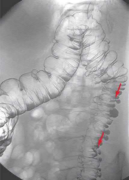
Referral from primary care is deliberately restrictive. Do not routinely refer people with suspected diverticular disease unless routine endoscopic and/or radiological investigations cannot be organised from primary care, or colitis is suspected, or the person meets the criteria for a suspected cancer pathway, in which case refer by that route [3].
- Investigation in secondary care.
- For people with suspected complicated acute diverticulitis referred for same-day assessment, offer a full blood count, urea and electrolytes, and CRP [3]. If inflammatory markers are raised, offer a contrast CT scan within 24 hours of hospital admission to confirm the diagnosis and help plan management; where contrast CT is contraindicated, use non-contrast CT, MRI or ultrasound depending on local expertise [3]. If inflammatory markers are not raised, think about the possibility of alternative diagnoses [3].
- For suspected uncomplicated acute diverticulitis not referred for same-day assessment, reassess in primary care if symptoms persist or worsen, and consider referral to secondary care [3].
Imaging and endoscopy in Schwartz's account
- Plain radiographs detect free intra-abdominal air; CT defines pericolic inflammation, phlegmon or abscess, with uncomplicated disease showing pericolic soft-tissue stranding, wall thickening and/or phlegmon; contrast enema and endoscopy are relatively contraindicated in the acute setting because of the perforation risk [6].
- Because colon carcinoma can present identically, every patient must be evaluated for malignancy after the acute episode resolves, with colonoscopy recommended 4–6 weeks after recovery, and inability to exclude malignancy is itself an indication for resection [6].
- For a fistula the two key tasks are defining its anatomy and excluding other diagnoses: contrast enema and/or small-bowel studies map the tract, CT identifies associated abscesses or masses, and colonoscopy or sigmoidoscopy is usually needed to rule out malignancy [6].
Scoring and Severity
- The Hinchey classification grades the severity of complicated diverticulitis and peritoneal contamination and guides management: Grade I (mesenteric or pericolic abscess; Grade II) pelvic (walled-off) abscess; Grade III (purulent peritonitis; Grade IV) faecal peritonitis [1][7].
- Mortality is markedly higher with perforation than with an inflammatory mass alone (33% versus 3%) [1].
- Complicated diverticulitis is characterised by the presence of an abscess, fistula, obstruction or free perforation [4].
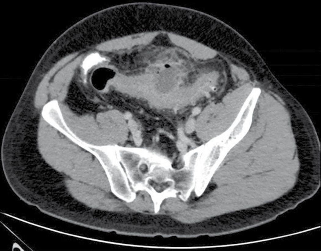
The Hinchey stages as Schwartz defines them
Schwartz states the Hinchey system as follows: stage I is colonic inflammation with a pericolic abscess, stage II colonic inflammation with a retroperitoneal or pelvic abscess, stage III purulent peritonitis and stage IV faecal peritonitis [6].
Treatment and Management
- Medical: a high-fibre diet, bulk-forming laxatives and stool softeners are commonly recommended, though evidence for effectiveness is limited; antispasmodics may help recurrent pain [1].
- Uncomplicated diverticulitis confirmed on CT in an immunocompetent patient without systemic infection may not require antibiotics, as it may be self-limiting; complicated disease with a localised abscess is treated with intravenous antibiotics and image-guided percutaneous drainage [1][8].
- In the Oxford Handbook emergency protocol, uncomplicated diverticulitis is treated with a low-residue diet, high fluid intake and stool softeners for two weeks, with intravenous antibiotics such as co-amoxiclav with or without gentamicin reserved for infective exacerbations [7].
- Recurrent infective episodes may be reduced by cyclical antibiotics and probiotics [2].
Most patients with uncomplicated diverticulitis can be managed as outpatients, with pain medication, short-term dietary alteration (clear liquids then a low-residue diet until inflammation subsides) and, traditionally, antibiotics. A systematic review and meta-analysis found that antibiotics in uncomplicated diverticulitis do not accelerate recovery, prevent complications or prevent subsequent surgery, and some physicians have accordingly stopped prescribing them; outpatient management has been found safe and cost-effective with low readmission rates [4].
- Diverticulosis.
- Tell people the condition is asymptomatic and needs no specific treatment.
- Advise a healthy balanced diet including whole grains, fruit and vegetables, and tell them there is no need to avoid seeds, nuts, popcorn or fruit skins; if they have constipation and a low-fibre diet, increasing fibre gradually may minimise flatulence and bloating, and adequate fluid should accompany any fibre increase.
- Consider bulk-forming laxatives for constipation, and tell people about the benefits of exercise, weight loss if overweight or obese, and stopping smoking in reducing the risk of developing acute diverticulitis and symptomatic disease [3].
- Diverticular disease. Do not offer antibiotics to people with diverticular disease [3]. Advise people to avoid NSAIDs and opioid analgesia if possible, because they may increase the risk of diverticular perforation [3].
- Advise that the benefits of increasing dietary fibre may take several weeks, and that a high-fibre diet should be maintained for life if tolerated [3].
- Consider bulk-forming laxatives if a high-fibre diet is unacceptable or not tolerated or there is persistent constipation or diarrhoea; consider simple analgesia such as paracetamol for ongoing pain, and an antispasmodic for cramping [3].
- If symptoms persist or do not respond, think about alternative causes [3].
- On recurrent diverticular disease, the committee were unable to make recommendations for practice [3].
Acute diverticulitis: antibiotics are stratified by how well the patient is, not by the diagnosis.
| Clinical situation | NICE recommendation |
|---|---|
| Acute diverticulitis, systemically well | Consider a no-antibiotic prescribing strategy; offer simple analgesia such as paracetamol; advise re-presentation if symptoms persist or worsen |
| Systemically unwell, immunosuppressed, or significant comorbidity | Offer an antibiotic prescribing strategy |
| Systemically unwell but not meeting criteria for suspected complicated disease | Offer oral antibiotics |
| Admitted with suspected complicated acute diverticulitis | Offer intravenous antibiotics; review within 48 hours or after scanning if sooner, and consider stepping down to oral |
| CT-confirmed uncomplicated acute diverticulitis | Review the need for antibiotics and discharge depending on coexisting conditions |
Table reformats the antibiotic strategy [3].
The first-choice oral antibiotic for suspected or confirmed uncomplicated acute diverticulitis is co-amoxiclav 500/125 mg three times a day for 5 days; alternatives in penicillin allergy are cefalexin with metronidazole, trimethoprim with metronidazole, or (only when switching from intravenous, with specialist advice) ciprofloxacin with metronidazole [3]. The first-choice intravenous antibiotic for complicated disease is co-amoxiclav 1.2 g three times a day, with cefuroxime plus metronidazole, or amoxicillin plus gentamicin plus metronidazole, as alternatives [3].
Do not offer an aminosalicylate or antibiotics to prevent recurrent acute diverticulitis [3]. This directly contradicts the cyclical-antibiotic approach described in some textbook accounts.
Outpatient and inpatient care, and the case for elective resection in Schwartz's account
- Most uncomplicated diverticulitis responds to outpatient broad-spectrum oral antibiotics for 7–10 days with a low-residue diet; about 10–20% with more severe pain, fever and leukocytosis are admitted for parenteral antibiotics and bowel rest, most improving within 48–72 hours, and failure to improve suggests abscess, for which CT is invaluable and many pericolic abscesses can be drained percutaneously [6].
- Most patients recover without surgery and 50–70% have no further episodes; the traditional advice of elective sigmoid colectomy after the second episode, after the first in very young patients and after any complicated episode has been questioned, since more recent studies suggest the risk of complications and emergency resection does not rise with recurrence, so resection rates have fallen and many surgeons no longer advise colectomy even after two documented episodes if the patient is asymptomatic and carcinoma has been excluded, immunosuppressed patients being the exception, still advised colectomy after a single documented episode [6].
- Abscesses under 2 cm may be treated with parenteral antibiotics alone; larger abscesses are best treated with CT-guided percutaneous drainage and antibiotics, which may allow a one-stage elective procedure or obviate colectomy altogether if recovery is complete [6].
- Incomplete obstruction often responds to fluid resuscitation, nasogastric suction and gentle low-volume water or Gastrografin enemas, allowing full bowel preparation and elective resection, whereas a high-volume oral bowel preparation is contraindicated with obstructive symptoms and obstruction not rapidly relieved mandates laparotomy [6].
- Diverticular haemorrhage is managed by resuscitation and localisation as for lower gastrointestinal bleeding; colonoscopy may occasionally identify a bleeding diverticulum treatable by adrenaline injection or cautery, angiography may be diagnostic and therapeutic, and persistent or recurrent bleeding rarely requires laparotomy and segmental colectomy [6].
Surgeries
- Emergency laparotomy: for generalised peritonitis or failure to respond to optimum medical therapy; carries substantial risk (mortality around 15% overall, approaching 50% with faecal peritonitis) [1].
- Hartmann's procedure: sigmoid resection with formation of a left iliac fossa end-colostomy and closure of the rectal stump; often the safest option with significant contamination or an unstable patient, with a second-stage reversal planned later [1][2].
- Resection with primary anastomosis, with or without defunctioning loop ileostomy: considered selectively in a young, fit patient with limited contamination; evidence suggests outcomes are better than a simple defunctioning stoma without resection [1].
- Laparoscopic lavage: in selected, stable cases in expert hands (Hinchey 2–3) without gross faecal contamination or visible perforation; remains controversial [1][7].
- Elective sigmoid colectomy: considered for recurrent attacks, symptoms affecting quality of life, stricture, or fistula; a laparoscopic approach is favoured where feasible [1]. The aim is to remove the affected segment and perform primary anastomosis of healthy bowel: the proximal margin should be in soft, pliable bowel, and it is not necessary to include all proximal diverticula, but the distal anastomosis should be to the upper rectum, because leaving a segment of distal sigmoid is associated with a higher risk of recurrent diverticulitis. Minimally invasive surgery is safe with faster return of bowel function, less pain and shorter stay, and ureteric stents are often used to mitigate ureteric injury risk by serving as guides amid inflamed tissues [4].
- Percutaneous drainage: for stable patients with a localised abscess (Hinchey I–II), avoiding laparotomy [1].
- Fistula surgery: for colovesical fistula, once malignancy is excluded, the sigmoid is resected off the bladder with catheter drainage for 7–10 days, sometimes with an omental interposition flap; ureteric stents may aid safe dissection [1]. Initial management of any diverticular fistula is broad-spectrum antibiotics to reduce inflammation, followed by colonoscopy and appropriate imaging such as cystoscopy to exclude malignancy and Crohn disease, then resection of the involved colon and fistula tract with primary anastomosis [4].
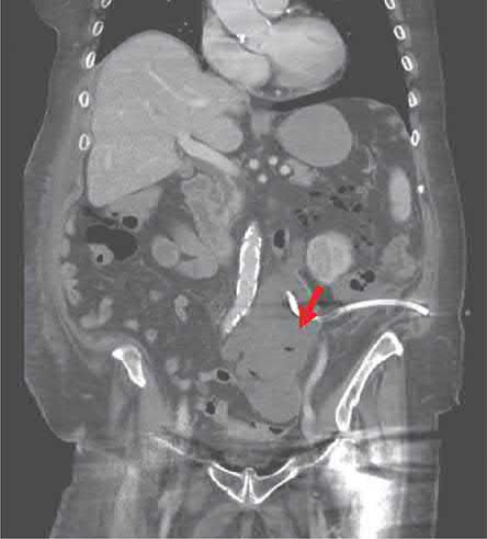
- Abscess management is size-driven, with 3 cm the threshold.
- Offer intravenous antibiotics and a contrast CT to people with acute diverticulitis and a suspected diverticular abscess, and use the scan results to guide treatment based on the abscess's size and location [3]. Consider either percutaneous drainage (if anatomically feasible) or surgery for abscesses greater than 3 cm; for abscesses less than 3 cm, switch to oral antibiotics where possible [3].
- Send pus samples to microbiology so that antibiotic treatment can be tailored to sensitivities, and if the condition does not improve or deteriorates, consider re-imaging [3].
Perforation with generalised peritonitis: lavage and resection are presented as a shared decision, with one hard rule. Offer either laparoscopic lavage or resectional surgery to people with diverticular perforation and generalised peritonitis, after discussing the risks and benefits of both, but if faecal peritonitis is identified intraoperatively, proceed to resectional surgery [3]. This is a more permissive position on laparoscopic lavage than the textbooks take, while retaining the same absolute limit at faecal contamination.
- Choice of resection.
- Offer people with complicated acute diverticulitis having surgery, elective or emergency, either primary anastomosis with or without diverting stoma, or Hartmann's procedure, taking into account the person's age, other conditions and WHO performance status [3]. In people undergoing bowel resection, consider resecting back to the compliant bowel, bowel that is soft, unthickened and unaffected by inflammation [3].
- For those who have recovered from complicated acute diverticulitis but have continuing symptoms, for example stricture or fistula, consider open or laparoscopic resection [3].
Extent of resection and operative choices in Schwartz's account
- In the elective setting sigmoid colectomy with primary anastomosis is the procedure of choice, increasingly laparoscopic; the distal resection must always extend to the rectum because recurrence is high if any sigmoid is retained, the proximal extent must include all thickened or inflamed bowel, but resection of every diverticulum is unnecessary [6].
- Urgent laparotomy is required when an abscess is inaccessible to percutaneous drainage, when the patient deteriorates or fails to improve, or with free air or peritonitis, and in almost all cases the affected segment should be resected: Hinchey I–II patients may be candidates for a one-stage sigmoid colectomy with primary anastomosis, while larger abscesses, peritoneal soiling or peritonitis are most commonly treated by sigmoid colectomy with end colostomy and Hartmann's pouch [6].
- Sigmoid colectomy with primary anastomosis, with or without on-table lavage, protected by a loop ileostomy has also succeeded and suits stable patients, its great advantage being that restoring continuity is simpler than taking down a Hartmann's pouch; the same option applies in obstruction when the patient is stable and proximal and distal bowel look healthy, though sigmoid colectomy with end colostomy remains the safest operation there [6].
- Inflammation and phlegmon raise the risk of ureteric injury during sigmoid mobilisation, so preoperative ureteric catheters can be invaluable; proximal diversion with local drainage alone is reserved for extremely unstable patients or inflammation so severe that resection would harm adjacent organs, and is generally avoided for its high morbidity, mortality and need for multiple operations, while laparoscopic lavage and drainage without resection has been reported safe even with free perforation but carries an expected 20% reoperation rate [6].
- Fistula surgery, once anatomy is defined and other diagnoses excluded, comprises resection of the diverticular segment (usually with primary anastomosis) and simple repair of the secondarily involved organ, with suspicion of carcinoma mandating a wider en bloc resection [6].
- Right-sided diverticula, commoner in younger patients and in people of Asian descent, are usually asymptomatic but when inflamed mimic appendicitis and are often diagnosed only in the operating room; a diverticulectomy may be done for a single large diverticulum with minimal inflammation, but ileocaecal resection is usually preferred [6].
Complications
- Diverticulitis, pericolic or paracolic abscess, purulent or faeculent peritonitis, intestinal obstruction, haemorrhage and fistula formation (colovesical, colovaginal, colocutaneous, or rarely to the retroperitoneum causing a psoas abscess) are recognised complications of diverticular disease [1].
- Patients who present with a diverticular abscess often experience recurring episodes, and both recurrence risk and complication risk are increased in that group; the most recent American Society of Colon and Rectal Surgeons guidelines recommend considering elective resection after successful non-operative treatment of a diverticular abscess, on moderate-quality evidence [4].
- Patients with abscesses not amenable to percutaneous drainage and unresponsive to treatment require urgent surgery [4].
Prognosis
Most diverticulosis remains asymptomatic lifelong. After a first episode of diverticulitis, roughly 25% of patients under 50 will have a further episode, but 75% will not, and the complication rate after elective surgery is low; the risk of complications does not clearly increase with recurrent attacks, which has shifted practice toward more conservative, individualised management [1][8].
The rationale for that shift is worth stating explicitly. After an initial episode of acute uncomplicated diverticulitis only 10% to 35% of individuals have another episode, and although the chance of recurrence rises after further episodes, recurrences generally tend to follow the severity of the initial episode, so the number of attacks of uncomplicated diverticulitis is no longer accepted as a standalone indication for elective resection, as it once was [4].
References
- Bailey & Love's Short Practice of Surgery, 28th ed., Ch. 77 The large intestine
- Oxford Handbook of Clinical Surgery, 5th ed., Ch. 12 Colorectal surgery
- NICE Guideline NG147: Diverticular disease — diagnosis and management (2019), 1.1; 1.1.1; 1.1.2; 1.1.3; 1.1.4; 1.1.5; 1.2; 1.2 Recurrent diverticular disease; 1.2.1; 1.2.2; 1.2.3; 1.2.4; 1.2.5; 1.2.7; 1.2.8; 1.2.9; 1.2.10; 1.2.11; 1.3; 1.3.1; 1.3.2, Table 1; 1.3.3; 1.3.4; 1.3.5; 1.3.6; 1.3.7; 1.3.8; 1.3.9; 1.3.10; 1.3.11; 1.3.12; 1.3.13, Table 2; 1.3.16; 1.3.18; 1.3.20; 1.3.22; 1.3.23; 1.3.24; 1.3.25; 1.3.26; 1.3.27; 1.3.28; 1.3.29; 1.3.30 www.nice.org.uk
- Sabiston Textbook of Surgery, 22nd ed., Ch. 95 Colon and Rectum
- Maingot's Abdominal Operations, 13th ed., Ch. 43
- Schwartz's Principles of Surgery, 11th ed., Ch. 29, Colon, Rectum, and Anus
- Oxford Handbook of Clinical Surgery, 5th ed., Ch. 26
- Schwartz's Principles of Surgery: ABSITE and Board Review, Ch. 29