



Colorectal Cancer
Summary
- Colorectal cancer (CRC) is the second commonest cause of cancer death and the commonest gastrointestinal malignancy in the UK, arising predominantly through an adenoma–carcinoma sequence driven by accumulating genetic mutations [1].
- Around one-third of tumours occur in the rectum and two-thirds in the colon, with a male predominance [1].
- Presentation depends on tumour site, left-sided lesions cause obstructive symptoms and bleeding, right-sided lesions anaemia [1].
- Treatment is stage-directed and multidisciplinary, combining surgical resection with neoadjuvant and adjuvant systemic therapy and, for rectal cancer, radiotherapy [2].
UK colorectal cancer care is defined by four documents that between them cover the whole pathway: the NHS Bowel Cancer Screening Programme for asymptomatic people, NICE HTG690 for the quantitative FIT that now gates symptomatic referral, NICE NG12 for the suspected cancer pathway itself, and NICE NG151 for everything from diagnosis onwards [3][4][5][6].
- Two figures in older textbook accounts are now out of date in the UK.
- Screening is offered to people aged 50 to 74, not 60 to 74, and the screening test is the faecal immunochemical test (FIT), which replaced guaiac faecal occult blood testing in England in June 2019 [3].
- Symptomatic referral is no longer a list of symptom-and-age combinations alone: since 2023 it is gated on a quantitative FIT result of at least 10 micrograms of haemoglobin per gram of faeces [4].
Definition
Colorectal cancer is a malignant neoplasm, almost always an adenocarcinoma, arising from the epithelium of the colon or rectum [7]. It may occur as a polypoid, ulcerating, stenosing or infiltrative tumour mass [1].
Pathophysiology
Most colorectal cancers develop from adenomatous polyps through a sequence of genetic mutations influenced by environmental factors, the adenoma–carcinoma sequence [1]. Mutations of the APC gene occur in two-thirds of colonic adenomas and are thought to arise early in carcinogenesis; K-ras mutations activate cell-signalling pathways and are more common in larger lesions (later events); the p53 gene is frequently mutated in carcinomas but not adenomas, marking the transition to invasion [1].
The three molecular pathways
Sabiston sets out three distinct but potentially overlapping pathways to colorectal cancer, and the distinction matters clinically because it predicts tumour site, patient profile and response to therapy [2]:
| Pathway | Share of CRC | Mechanism | Clinical signature |
|---|---|---|---|
| Chromosomal instability (CIN) | 65–70% | Loss of one allele by insertion, deletion, amplification, aneuploidy or loss of heterozygosity; initiating when it hits a tumour-suppressor or driver gene | The classical adenoma–carcinoma sequence |
| Methylation (CIMP) | Not stated | Promoter hypermethylation silencing gene expression; serrated lesions are the precursor | Older patients, right colon, more common in females; implicated in interval cancers because serrated lesions are flat and subtle |
| Microsatellite instability (MSI) | Not stated | Loss of mismatch repair function through germline variant (Lynch), MLH1 promoter hypermethylation, or double somatic mutation | MSI-high tumours with distinctive pathology including a Crohn-like reaction |
- Table reformats the three CRC pathways [2].
- The four mismatch repair genes are MLH1, MSH2, MSH6 and PMS2; loss of MMR function prevents correction of DNA base-pair mismatches during transcription, and because mismatches accumulate in repeating DNA sequences called microsatellites these tumours are termed microsatellite instability-high [2].
- Some authors suggest that most interval cancers (those found between routine screening examinations) result from failure to detect serrated lesions at colonoscopy [2].
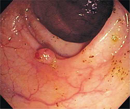
The adenoma–carcinoma sequence and its genes in Schwartz's account
- Colorectal carcinoma is the commonest gastrointestinal malignancy, with over 130,000 new cases and more than 50,000 deaths a year in the United States making it the third most lethal cancer there; incidence is similar in men and women, is falling in the over-50s with national screening, but is rising with worse mortality in people under 50 [9].
- Mutations activate oncogenes (K-ras) and inactivate tumour suppressors (APC, DCC, p53), and APC mutations (first described in FAP) are present in 80% of sporadic cancers; both alleles must be lost to initiate polyp formation, most mutations are premature stop codons giving a truncated protein, and in FAP the site of mutation predicts severity, with 3′ or 5′ mutations producing attenuated disease and central mutations virulent disease [9]. K-ras, a G-protein in the EGFR pathway, is a proto-oncogene because mutating one allele suffices: the mutant cannot hydrolyse GTP and stays permanently active, which is why anti-EGFR agents are ineffective in K-ras-mutant tumours and mutation status guides their use, with BRAF also implicated [9]. MYH on chromosome 1p is a base-excision repair gene whose biallelic loss gives an attenuated-FAP phenotype with autosomal recessive inheritance; p53, crucial for apoptosis in cells with irreparable damage, is mutated in 75% of colorectal cancers; and PTEN deletion underlies juvenile polyposis, Peutz–Jeghers, Cowden's and PTEN hamartoma syndromes as well as MEN IIB, Peutz–Jeghers also carrying STK11 mutations [9].
- Chromosome 18q is deleted in up to 70% of colorectal cancers, removing DCC (a tumour suppressor whose main role is neural differentiation and axonal migration, present in more than 70% of carcinomas and possibly worsening prognosis) and SMAD4 (TGF-β and β-catenin signalling); tumours of the loss-of-heterozygosity pathway sit distally with aneuploidy and worse prognosis, whereas MSI tumours favour the right colon, are diploid and do better [9].
- The pathways are not mutually exclusive, a mismatch-repair gene may itself be silenced by methylation, and the resulting replication errors then inactivate a tumour suppressor [9].
Polyps as Schwartz classifies them
- Polyps are neoplastic (tubular, villous, tubulovillous, serrated), hyperplastic, hamartomatous (juvenile, Peutz–Jeghers, Cronkhite–Canada) or inflammatory (pseudopolyp, benign lymphoid polyp); adenomas occur in up to 25% of Americans over 50, tubular adenomas harbour malignancy in 5%, tubulovillous in 22% and villous in up to 40%, invasive carcinoma is rare below 1 cm and present in 35–50% of polyps larger than 2 cm [9].
- Hyperplastic polyps, usually under 5 mm, are not premalignant but are removed because they cannot be distinguished endoscopically, though large ones over 2 cm may degenerate and hyperplastic polyposis in young adults raises cancer risk; serrated polyps (sessile and traditional serrated adenomas) are flat, hard to see, once thought innocent but now known to progress to invasive cancer and treated like adenomas; and inflammatory pseudopolyps, islands of regenerating mucosa amid mucosal loss in IBD, amoebic, ischaemic or schistosomal colitis, are not premalignant but may mimic FAP when extensive [9].
- Cronkhite–Canada syndrome combines gastrointestinal polyposis with alopecia, cutaneous pigmentation and nail atrophy, with diarrhoea, vomiting, malabsorption and protein-losing enteropathy; most patients die of it despite maximal therapy and surgery is reserved for complications such as obstruction [9].
Clinical features
- Carcinoma of the colon typically occurs after age 50, most commonly in the eighth decade; emergency presentation (20% of cases) carries a substantially worse prognosis even after stage-matching [1].
- Left-sided tumours usually present with change in bowel habit or rectal bleeding; proximal (right-sided) lesions typically present with iron-deficiency anaemia or a palpable mass [1].
- By site [10]:
- Rectal: PR bleeding (deep red, on the surface of stools), change in bowel habit, tenesmus.
- Descending/sigmoid: PR bleeding (dark red, mixed with stool), increased frequency, mucus, bloating.
- Right-sided: iron-deficiency anaemia may be the only presenting feature.
- Emergency presentations (up to 40%): large bowel obstruction, perforation with peritonitis, acute PR bleeding.
Rectal cancer pain is a late symptom, from obstruction of the rectosigmoid or invasion of the sacral plexus, prostate or bladder; weight loss is also late and usually signifies metastatic disease [7]. On digital rectal examination, a tumour within 7–8 cm of the anal verge is felt as an elevated, irregular, hard mass; mobility, tethering or fixity and distance from the anal sphincter should be assessed [7].
Assessment specific to rectal cancer
- The history should elicit nausea, vomiting, bloating or constipation, which give important information about impending large bowel obstruction and the need for more urgent intervention; neuropathic pain in particular indicates a locally invasive tumour involving sacral nerve roots, and patients reporting tenesmus and rectal pain often prove to have low tumours with sphincter invasion [2].
- It is critically important to record the patient's baseline urinary, sexual and bowel function, because imperfect stool continence before surgery predicts poor bowel function and reduced quality of life after low anterior resection with sphincter preservation [2].
- Digital rectal examination supplemented by flexible or rigid proctoscopy performed by the evaluating surgeon is of critical importance: the distance of the tumour from the anal verge and the dentate line should be carefully assessed and documented, and for tumours within the surgical anal canal, potential sphincter or adjacent-structure invasion is determined by assessing mobility in relation to the prostate, vagina and sphincter complex [2].
Etiology
- Predisposing factors include polyposis syndromes (familial adenomatous polyposis, Lynch syndrome, juvenile polyposis), a strong family history, previous polyps or colorectal cancer, chronic ulcerative colitis or colonic Crohn's disease, and a diet low in fruit and vegetables [1].
- Colorectal cancer prevalence correlates with intake of red and processed meat; dietary fibre appears protective, and there is increasing evidence linking colonic microbiota, inflammation and gene methylation to carcinogenesis; smoking and alcohol increase risk, while aspirin and other prostaglandin inhibitors show a protective epidemiological association [1].
- Cholecystectomy may marginally increase right-sided colon cancer risk [1].
Hereditary syndromes
FAP is an autosomal dominant condition caused by mutation of the APC gene on chromosome 5, defined by more than 100 colorectal adenomas, with colorectal cancer risk approaching 100% by age 35–40 if untreated; it is associated with duodenal and gastric polyps, desmoid tumours, osteomas and congenital hypertrophy of the retinal pigment epithelium, the latter combination termed Gardner's syndrome [1][11]. Lynch syndrome (hereditary non-polyposis colorectal cancer) results from mutation of a DNA mismatch-repair gene (MLH1, MSH2, MSH6 or PMS2), producing microsatellite instability and an accelerated adenoma–carcinoma sequence; lifetime colorectal cancer risk is up to 70–80%, with most cancers proximal, and affected women carry a 30–50% lifetime risk of endometrial cancer [1][12]. Diagnosis has historically relied on the Amsterdam II clinical criteria [1][12].
Sabiston covers the less common syndromes in detail, and the surveillance intervals are examinable [2]:
| Syndrome | Gene / inheritance | Key features | Surveillance |
|---|---|---|---|
| MUTYH-associated polyposis | Biallelic MUTYH, autosomal recessive | 10–100 adenomas by the fifth or sixth decade; up to 60% have CRC at presentation; rectal cancer uncommon | Colonoscopy every 1–2 years from diagnosis; baseline upper endoscopy at 30–35 years |
| Juvenile polyposis syndrome | SMAD4 or BMPR1A, autosomal dominant | Hamartomatous polyps; 10–38% lifetime colon cancer risk, average age at diagnosis 34; SMAD4 associated with hereditary haemorrhagic telangiectasia | Upper endoscopy and colonoscopy from 12–15 years, repeated every 1–3 years (every 5 if no polyps) |
| Peutz-Jeghers syndrome | STK11 (LKB1), autosomal dominant | Hamartomatous polyps plus mucocutaneous pigmentation; lifetime risks include breast 32–54%, colon 39%, gastric 29%, pancreas 11–36% | Colonoscopy, upper endoscopy and small bowel imaging from 18 years every 2–3 years; mammography and breast MRI from 30 |
| PTEN hamartoma tumour syndrome (Cowden) | PTEN, autosomal dominant | Trichilemmomas, breast cancer up to 85% lifetime risk, non-medullary thyroid cancer, endometrial cancer 13–30% | Not stated in this table |
Table reformats the non-FAP, non-Lynch hereditary syndromes [2]. About 1–2% of the general population carries a monoallelic MUTYH variant, and fewer than 1% of all CRC diagnoses are due to MUTYH-associated polyposis [2]. Peutz-Jeghers has a de novo rate of 10–20%, so the absence of a family history should not exclude the diagnosis or a referral for genetic testing [2].
Dietary, hormonal and iatrogenic risk factors in Schwartz's account
- More than 90% of cases occur after age 50, the rationale for average-risk screening from 50, and 80% are sporadic while 20% arise with a known family history [9].
- Saturated or polyunsaturated fat raises risk whereas a diet high in oleic acid (olive, coconut, fish oil) does not; vegetable fibre appears protective; alcohol correlates with incidence; calcium, selenium, vitamins A, C and E, carotenoids and plant phenols may reduce risk; and obesity and a sedentary lifestyle dramatically increase cancer mortality [9].
- Cigarette smoking is associated with adenomas, especially after more than 35 years of use; ureterosigmoidostomy raises both adenoma and carcinoma risk; acromegaly, through raised growth hormone and IGF-1, increases risk; and pelvic irradiation may raise rectal cancer risk, notably after radiotherapy for prostate cancer, though whether this is direct radiation damage or an association with another pelvic malignancy is unclear [9].
- Nonsyndromic familial colorectal cancer accounts for 10–15% of cases: lifetime risk is about 6% with no family history, 12% with one affected first-degree relative and 35% with two, diagnosis before 50 raises the risk to relatives, and colonoscopy every 5 years is recommended from age 40 or 10 years before the youngest diagnosis in the pedigree [9].
FAP, attenuated FAP and Lynch syndrome in Schwartz's detail
- FAP accounts for about 1% of colorectal adenocarcinomas, APC testing is positive in 75% of cases and up to 25% present without affected relatives; if a relative tests positive for a known family mutation, annual flexible sigmoidoscopy begins at 10–15 until polyps appear, a negative test allows average-risk screening from 50, and if testing is refused or uninformative annual sigmoidoscopy runs to 24, then every 2 years to 34, every 3 years to 44 and every 3–5 years thereafter, with upper endoscopy every 1–3 years from 25–30 for duodenal and periampullary disease [9].
- Choice of operation rests on age, symptoms, extent of rectal polyposis and the presence of cancer or desmoid: most choose ileal pouch–anal anastomosis unless a distal rectal cancer, a mesenteric desmoid preventing reach or poor sphincter function intervenes, mucosectomy is advocated for the anal transition zone risk but remains controversial, up to 50% experience some incontinence, ileorectal anastomosis demands vigilant rectal surveillance, and COX-2 inhibitors (celecoxib, sulindac) may slow polyp development [9].
- Desmoids arise from fibroblasts, are benign but highly locally invasive in mesentery, pelvis and abdominal wall, may be triggered by surgery and recur after resection, so surgery is avoided if possible; they are often hormone-responsive to tamoxifen, may respond to COX-2 inhibitors, NSAIDs and possibly imatinib; Turcot's syndrome denotes FAP with central nervous system tumours [9].
- Attenuated FAP presents later with 10–100 mainly right-sided polyps, cancer in more than 50% at an average age of 55, duodenal polyposis, APC mutations in only about 30% and MYH mutations in others; colonoscopy starts at 13–15 then every 4 years to 28 and every 3 years thereafter when the mutation is unknown, and total colectomy with ileorectal anastomosis usually suffices because rectal polyps can be snared [9].
- Lynch syndrome (1–3% of colon cancers) causes cancer at an average age of 40–45 in about 70% of carriers, proximal and better-prognosis tumours, a 40% synchronous or metachronous risk and endometrial, ovarian, pancreatic, gastric, small-bowel, biliary and urinary-tract cancers; Amsterdam I requires three relatives with verified large-bowel adenocarcinoma, one a first-degree relative of the others, over two successive generations with one diagnosed before 50, and Amsterdam II admits any Lynch-related malignancy with FAP excluded and pathology reviewed [9]. PMS2 and MSH6 mutations give an attenuated form, MSH6 carrying more endometrial cancer; annual colonoscopy begins at 20–25 or 10 years before the youngest family diagnosis, annual transvaginal ultrasound or endometrial aspiration from 25–35, total colectomy with ileorectal anastomosis is recommended once adenomas or cancer appear because of the 40% second-cancer risk, annual proctoscopy follows, and prophylactic hysterectomy with bilateral salpingo-oophorectomy is considered after childbearing [9].
Diagnosis
- Endoscopy: colonoscopy is the investigation of choice, providing histological diagnosis and detecting synchronous polyps or carcinomas (3–5% of cases), with a small perforation risk (1 in 1,000); flexible sigmoidoscopy detects up to 70% of cancers and almost all causing fresh rectal bleeding [1].
- Rigid sigmoidoscopy or proctoscopy may be used for rectal lesions [10].
- In rectal cancer the reported incidence of synchronous cancers is approximately 3% and of premalignant neoplastic lesions approximately 30%; if an obstructing tumour prevents complete colonoscopy, CT colonography can assess the proximal colon, or a repeat colonoscopy can be attempted after neoadjuvant therapy has downsized the tumour [2].
- Radiology and staging: CT colonography has largely replaced barium enema; CT of thorax, abdomen and pelvis is standard for metastatic staging, while pelvic MRI is required for local staging of rectal cancer, assessing mesorectal fascia involvement [1]. Pelvic MRI is now the preferred modality for locoregional staging of rectal cancer, determining T and N stage and providing critical information on the tumour's relationship to the anterior peritoneal reflection, the circumferential resection margin, and invasion of adjacent structures, endorectal ultrasound, historically important, has been displaced [2].
- Endorectal ultrasound differentiates the five-layer rectal wall, distinguishes T1–2 from T3–4 disease (accuracy 81–94%) and detects perirectal nodes (58–83% accuracy) [12].
- CEA is not useful for diagnosis or screening but is used to monitor for relapse if elevated preoperatively and normalising after resection [1].
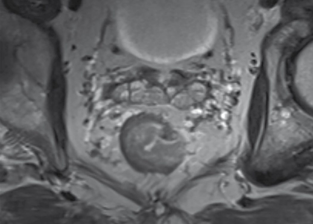
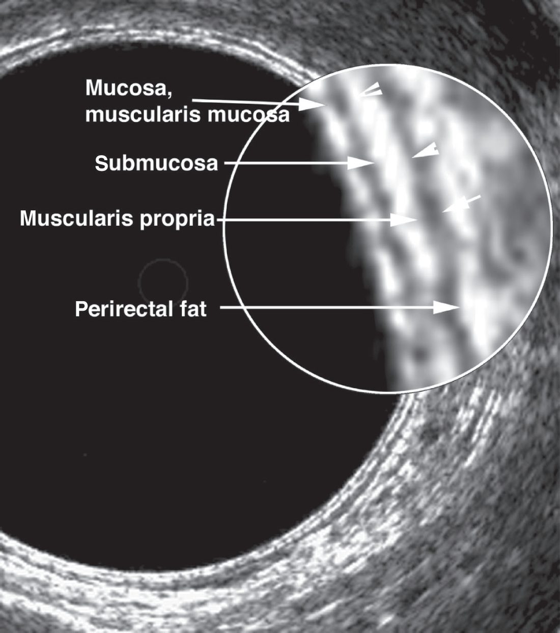
Multidisciplinary review is a central quality metric: all findings should be reviewed by a tumour board of colorectal surgery, medical and radiation oncology, surgical oncology, pathology and radiology, and such review often changes the initial management plan even for experienced practitioners [2].
Symptomatic referral now runs through a number, not a symptom list. Quantitative FIT using HM-JACKarc or OC-Sensor is recommended to guide referral for suspected colorectal cancer in adults with an abdominal mass, a change in bowel habit, or iron-deficiency anaemia; aged 40 and over with unexplained weight loss and abdominal pain; aged under 50 with rectal bleeding and unexplained abdominal pain or weight loss; aged 50 and over with unexplained rectal bleeding, abdominal pain or weight loss; or aged 60 and over with anaemia even in the absence of iron deficiency [4].
Refer using a suspected cancer pathway referral if the FIT result is at least 10 micrograms of haemoglobin per gram of faeces [4]. Two exceptions and one safety net matter:
- FIT should be offered even if the person has previously had a negative FIT result through the NHS bowel cancer screening programme [4].
- People with a rectal mass, an unexplained anal mass or unexplained anal ulceration do not need FIT before referral is considered [4]; NG12 requires referral of those with an unexplained anal mass or unexplained anal ulceration on the suspected cancer pathway [5].
- For people who have not returned a sample, or whose FIT is below 10 µg/g, safety netting processes should be in place, and referral should not be delayed if there is strong clinical concern of cancer because of ongoing unexplained symptoms [4].
Population screening. People aged 50 to 74 are invited every 2 years; a home FIT kit is sent about a week after the invitation letter, results follow 2 weeks after the laboratory receives the kit, and an abnormal result leads to colonoscopy. People aged 75 or over can request a kit every 2 years by telephoning the programme [3].
Molecular testing. Test for RAS and BRAF V600E mutations in all people with metastatic colorectal cancer suitable for systemic anticancer treatment [6].
Screening modalities and their yields in Schwartz's account
- FOBT reduces colorectal cancer mortality by 33% and metastatic disease by 50% yet misses up to 50% of cancers and most adenomas, and 90% of positives do not have cancer; FIT is more sensitive and specific; the multitarget stool DNA test (mutant KRAS, methylated BMP3 and NDRG4 promoter) was 92% sensitive for cancer but 74% specific and 42% sensitive for advanced precancerous lesions, and is endorsed with FIT every 1–3 years [9].
- Flexible sigmoidoscopy every 5 years may cut mortality by 60–70% but misses proximal lesions; FOBT is least effective for rectosigmoid cancers, so Winawer's 12,479-subject study and 11-year Norwegian follow-up support combining annual FOBT with 5-yearly sigmoidoscopy [9].
- Colonoscopy is the most accurate and complete examination, detecting polyps under 1 cm and allowing biopsy, polypectomy, haemostasis and dilatation, with a 0.2–0.3% major complication rate; air-contrast barium enema is 90% sensitive for polyps over 1 cm but compromised by sigmoid diverticulosis; CT colonography matches colonoscopy for cancers and polyps over 1 cm but gives false positives from stool, diverticula, haustra and motion and misses flat adenomas [9].
- Schwartz's Table 29-2 sets surveillance after adenoma at colonoscopy in 3 years, then every 5 if clear (every 3 if polyps, annually for more than 5 adenomas); after cancer at 12 months, 3 years and then 5-yearly; in colitis every 1–2 years after 8 years of pancolitis or 15 of left-sided disease; in FAP annual sigmoidoscopy from 10–12; and in HNPCC colonoscopy every 1–2 years from 20–25 [9].
- Synchronous tumours are present in up to 5%, a Gastrografin enema delineates obstruction and mechanical bowel preparation must be avoided in an obstructed patient, and PET is useful for indeterminate CT lesions and before highly morbid operations such as exenteration or sacrectomy [9].
- Polypectomy sites of sessile lesions should be tattooed, rectal sessile polyps are best excised transanally as one intact specimen because depth of invasion cannot be judged after piecemeal removal, a microperforation in a prepared stable patient may be observed with antibiotics, and post-polypectomy bleeding usually stops spontaneously or is controlled with clips, resnaring, cautery or adrenaline, rarely needing vasopressin infusion or colectomy [9].
Scoring and Severity
Dukes' classification (originally for rectal cancer, later applied to colon cancer): A (confined to the bowel wall (75–90% 5-year survival); B) through the bowel wall, nodes negative (55–70%); C, regional lymph nodes involved (30–60%), subdivided into C1 (pararectal nodes only) and C2 (nodes to the origin of the supplying vessel); D (not part of Dukes' original description) denotes distant metastases (5–10%) [1][10].
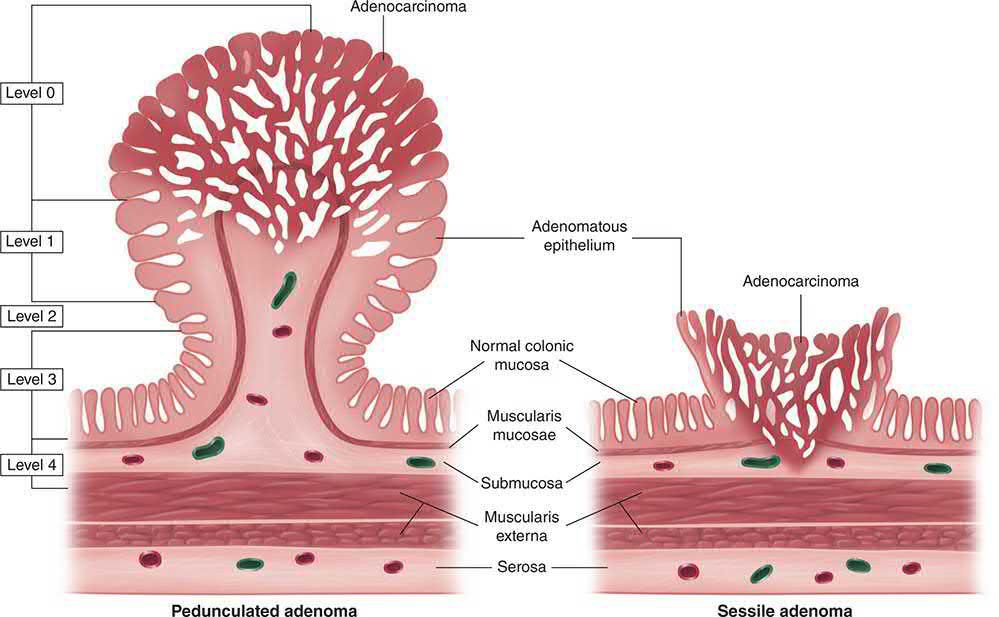
- TNM staging (UICC 8th edition) is now the internationally recognised standard: T1 submucosa, T2 muscularis propria, T3 subserosa/pericolic fat, T4a visceral peritoneum, T4b adjacent organ; N1 (1–3 nodes) or N2 (4 or more nodes); M1a (one organ), M1b (more than one organ), M1c (peritoneum) [1].
- Stage grouping: Stage I = T1–2N0; Stage II = T3–4N0; Stage III = any T with node-positive disease; Stage IV = distant metastasis [12].
- Histological grading and adverse features such as vascular or perineural invasion, an infiltrating margin and tumour budding carry independent prognostic weight; signet-ring adenocarcinoma grows rapidly, metastasises early and carries a poor prognosis [7].
Prognostic factors beyond stage
The AJCC recognises several additional prognostic factors, each independently adverse [2]:
| Factor | Effect |
|---|---|
| Serum CEA greater than 5 ng/mL both before and after resection | Lower disease-free survival |
| Lymphovascular invasion | Increased risk of death; in resected stage II disease, worse disease-free and overall survival |
| Perineural invasion | Independent poor prognostic factor for 5-year disease-free and overall survival |
| Tumour budding (single cells or small clusters in the tumour stroma) | Intermediate and high budding more strongly associated with worse survival than low budding in stage III |
| Mucinous histology | Worse progression-free and overall survival than non-mucinous cancers |
| Signet ring cells | 5-year survival 9–36%, average survival 20–45 months |
Table reformats the AJCC prognostic factors [2].
Nodal risk by T stage and the AJCC 8th-edition subcategories
- T stage is the single strongest predictor of nodal spread: Tis carries no nodal risk, T1–T2 tumours have nodal metastases in 5–20% and T3–T4 in more than 50%, four or more involved nodes (N2) predict a poor prognosis, and the AJCC 8th edition defines N1c as tumour deposits in subserosa, mesentery or pericolic tissue without positive nodes, N2a as four to six and N2b as seven or more nodes, and M1a, M1b and M1c as one site, two or more sites, and peritoneal metastasis respectively [9].
- Upper rectal lymphatics ascend the superior rectal vessels to the inferior mesenteric nodes, lower rectal drainage may follow the middle rectal vessels, and spread along the inferior rectal vessels to internal iliac or groin nodes is rare unless the anal canal is involved or proximal lymphatics are blocked; a mesorectal margin of only 1–2 mm predicts local recurrence and is best assessed on MRI [9].
- A 12-node minimum has been proposed as a quality benchmark, though some investigators find node count does not correlate with staging, chemotherapy use or survival, and negative-node count or lymph-node ratio may refine staging [9].
Treatment and Management
- Management is planned within a multidisciplinary team, taking into account patient fitness, tumour stage and patient preference [7].
- Enhanced recovery after surgery programmes, combining preadmission counselling, carbohydrate loading, avoidance of NG tubes and opiates, short incisions, early mobilisation and early oral intake, reduce length of stay from 10–14 days to as little as 3–5 days [1].
- Preoperative mechanical bowel preparation combined with oral antibiotics reduces surgical site infection, anastomotic leak, ileus and reoperation [1].
Rectal cancer: neoadjuvant therapy and organ preservation
- For rectal cancer, neoadjuvant long-course chemoradiotherapy (5 fractions per week over 6 weeks with concurrent chemotherapy) is used for locally advanced tumours threatening the circumferential resection margin, aiming to downstage and achieve clear margins; short-course radiotherapy (5 days) is used where margins are not threatened but local recurrence risk is high, for example perirectal nodal disease [7].
- Neoadjuvant therapy is usually reserved for locally advanced tumours (T3–4 and/or N1) in the extraperitoneal rectum, because of the historically high local recurrence rates in that group [2].
- The German Rectal Cancer Study compared pre- with postoperative long-course chemoradiotherapy in 823 patients with locally advanced disease and found a significantly lower 5-year local recurrence rate in the neoadjuvant group, 6% versus 13% [2].
- Around 20% achieve a complete clinical response and may be offered non-operative "watch and wait" surveillance (Habr-Gama), with about 30% recurring, most salvageable by surgery [7].
- Habr-Gama's original 2004 observational series of 71 patients with a complete clinical response after neoadjuvant chemoradiotherapy, managed non-operatively with frequent digital examination, proctoscopy and imaging, reported 5-year overall survival of 100% and disease-free survival of 92% [2].
- A complete clinical response requires comprehensive restaging with CT of chest and abdomen, pelvic MRI, and digital examination with flexible proctoscopy by a surgeon experienced in non-operative management; the findings are no mass or nodularity on digital examination; a smooth white or pale scar with or without telangiectasia and no ulceration, mucosal irregularity or stricture on high-definition endoscopy; and no mass or lymph nodes on diffusion-weighted MRI [2].
- Current surveillance protocols call for digital examination with flexible endoscopy and CEA every 3–6 months, pelvic MRI every 6 months, and CT of chest and abdomen every 6–12 months for the first 2 years, with intervals extending thereafter but close monitoring for at least 5 years [2].
Adjuvant therapy
- Adjuvant chemotherapy (fluoropyrimidine-based, with oxaliplatin in high-risk fit patients) is recommended for node-positive (Dukes' C, stage III) colon cancer; benefit in stage II disease is less clear, and MSI-high stage III tumours may not benefit from 5-fluorouracil-based regimens [1][12].
- High-risk features that bring a stage II patient into consideration for adjuvant chemotherapy are poorly differentiated histology, lymphovascular invasion, perineural invasion, bowel obstruction from the tumour, fewer than 12 lymph nodes sampled after resection, localised perforation, a close, indeterminate or positive margin, and high tumour budding [2].
- Duration is stage-dependent: in low-risk stage III disease (T1–3 N1), 3 months of CAPEOX is non-inferior to 6 months with less toxicity, whereas in high-risk stage III disease (T4 N1–2, or any T with N2) 3 months of FOLFOX was inferior to 6 months [2].
- For mismatch-repair-deficient tumours, observation without adjuvant therapy is recommended in stage I and II disease [2].
Palliative options for unresectable disease include chemotherapy, endoluminal stenting of obstructing tumours, or palliative resection [1].
Chemoprevention in Lynch syndrome, a recommendation with no textbook counterpart. Consider aspirin, taken daily for a period of more than 2 years, to reduce the risk of colorectal cancer in people with Lynch syndrome [6]. As of January 2020 this was an off-label use of aspirin, and NICE has produced a patient decision aid to support the discussion [6].
Acute left-sided large bowel obstruction. Consider stenting for people who are going to have treatment with palliative intent; offer either stenting or emergency surgery if potentially curative treatment is suitable [6]. This is a genuine change from the textbook framing of stenting as principally a bridge to surgery.
Preoperative treatment. Do not offer preoperative radiotherapy to people with early rectal cancer (cT1-T2 cN0, M0) unless as part of a clinical trial; offer preoperative radiotherapy or chemoradiotherapy for cT1-T2 cN1-N2 M0 or cT3-T4, any cN, M0 disease [6]. For colon cancer, consider preoperative systemic anticancer therapy for people with cT4 colon cancer [6].
Adjuvant chemotherapy is specified by regimen and duration, and the same recommendation applies to stage 3 colon cancer and to stage 3 rectal cancer treated with short-course radiotherapy or no preoperative treatment: offer CAPOX for 3 months, or if that is unsuitable either FOLFOX for 3 to 6 months or single-agent fluoropyrimidine such as capecitabine for 6 months, basing the choice on histopathology (for example pT1-T3 and pN1 versus pT4 and/or pN2), performance status, personal preferences, comorbidities and age [6]. As of August 2025, CAPOX and the 3-month capecitabine duration were off-label uses, though CAPOX is common in UK clinical practice [6].
Deferral of surgery after a complete response is framed far more cautiously than the watch-and-wait literature. Inform people with a complete clinical and radiological response to neoadjuvant treatment who wish to defer surgery that there is a risk of recurrence, and that there are no prognostic factors to guide selection for deferral of surgery. For those who choose to defer, encourage participation in a clinical trial and ensure that data is collected via a national registry [6]. NICE does not present non-operative management as an established option; it presents it as a patient-initiated deferral requiring registry capture.
Stage-specific decisions in Schwartz's account
- In a malignant polyp, invasion confined to the head of a pedunculated polyp carries under 1% nodal risk and endoscopic excision suffices; in sessile polyps depth of submucosal invasion stratifies risk (Sm1 low, Sm2 and Sm3 intermediate and high), and lymphovascular invasion, poor differentiation, tumour budding or tumour within 1 mm of the margin mandate segmental colectomy [9].
- Adjuvant chemotherapy does not improve survival in stage I, up to 46% of completely resected stage II patients still die of colon cancer so it is suggested for selected high-risk stage II disease though its benefit remains controversial, FOLFOX is routine in stage III, and MSI-high stage III patients in the CRYSTAL subset analysis did not benefit from 5-FU-based chemotherapy [9].
- About 15% of patients with systemic disease have liver-only metastases, 20% of whom are resectable with 20–40% 5-year survival, resection being combined or staged; lung metastases occur in about 20% but only 1–2% of all patients are resectable, with 30–40% long-term survival; HIPEC for carcinomatosis remains unproven with high morbidity; and if unexpected metastases are found at laparotomy, low-volume disease with a straightforward primary favours proceeding while carcinomatosis with a minimally symptomatic primary favours abandoning the operation for early chemotherapy [9].
- For rectal cancer, local excision of T1 lesions is limited to small (<3 cm), well or moderately differentiated tumours or the medically unfit, local recurrence after local excision of uT1N0 and uT2N0 cancers may reach 20% and 40%, and ACOSOG Z6041 (neoadjuvant chemoradiation then transanal excision for T2) gave a 44% pathological complete response and 88% 3-year disease-free survival, though population data show suboptimal survival after local excision [9].
- The German CAO/ARO/AIO-94 trial found equal acute toxicity and complications for pre- and postoperative chemoradiation but postoperative treatment doubled stricture risk and preoperative treatment halved local recurrence (6% versus 12%); the US long course is 5-FU-based chemotherapy with 5–6 weeks of radiation and surgery 6–8 weeks later, the European short course 5 days of radiation and surgery within 1–2 weeks, the two never compared in a randomised trial [9].
- Twenty to 40% of patients relapse after curative surgery, mostly within 2 years; surveillance colonoscopy is at 12 months then every 3–5 years, CEA every 3–6 months for 2 years and annual CT for 5 years despite thin evidence, locally excised rectal tumours need endoscopy every 3–6 months for 3 years then 6-monthly for 2, and intensive surveillance has never been proved to improve survival [9].
Rare colorectal tumours and retrorectal lesions in Schwartz's account
- Up to 25% of gastrointestinal neuroendocrine tumours arise in the rectum, usually as nodules under 1 cm found at colonoscopy with more than 80% overall survival, but more than 60% of tumours over 2 cm have distant metastases; rectal tumours rarely secrete vasoactive substances and carcinoid syndrome is uncommon without hepatic metastases, small lesions are excised transanally while larger, poorly differentiated (small- or large-cell) or muscle-invading tumours need radical surgery, proximal colonic tumours are rarer, bulkier and metastatic in up to two-thirds at diagnosis, and mixed adenoneuroendocrine carcinomas behave like adenocarcinoma and are treated as such [9].
- Submucosal lipomas are resected only when large (over 2 cm) and causing bleeding, obstruction or intussusception, by colonoscopy, colotomy with enucleation or limited colectomy; colorectal lymphoma is about 10% of gastrointestinal lymphoma, favours the caecum by spread from the terminal ileum, and isolated disease is resected with stage-based adjuvant therapy [9].
- Leiomyomas are resected because they cannot be distinguished from leiomyosarcoma, small ones by limited resection and those over 5 cm radically; rectal leiomyosarcoma is usually low grade and diagnosed only after resection with poor prognosis; and 5–10% of GISTs arise in the colorectum, more than 95% express CD117 (KIT), 30–50% are malignant on size and mitotic criteria, resection with microscopically clear margins is the treatment of choice and imatinib shrinks marginally resectable tumours and serves adjuvantly and for metastases [9].
- The retrorectal space lies between the upper two-thirds of the rectum and the sacrum above the rectosacral fascia, bounded by rectum, presacral fascia and the lateral ligaments, and its tumours are heterogeneous embryological remnants: congenital lesions make up almost two-thirds (developmental cysts (dermoid and epidermoid from ectoderm, enterogenous from primitive gut) and anterior meningocele with the pathognomonic "scimitar sign" of a rounded concave sacral border without bony destruction), solid lesions are likelier to be malignant than cystic ones, adult teratomas are malignant in 30%, chordomas from the notochord are the commonest malignancy with characteristic bony destruction, and neurogenic and osseous tumours complete the list [9].
- Most are palpable rectally, pelvic MRI is the most sensitive and specific study, treatment is almost always resection, transabdominally for high lesions, transsacrally for low ones and combined for intermediate ones, and although biopsy was historically avoided for infection and seeding, a recent study refuted needle-tract seeding so most solid lesions should be biopsied to allow neoadjuvant therapy for GIST, sarcoma or metastatic adenocarcinoma, while cystic lesions and especially meningoceles must never be aspirated [9].
Surgeries
- Right/extended right hemicolectomy: for caecal, ascending colon, hepatic flexure and proximal transverse colon tumours; the ileocolic artery is ligated close to its origin ("high tie"); complete mesocolic excision with flush ligation of ileocolic and right colic vessels may improve survival in node-positive disease [1]. Right colectomy includes high ligation of the ileocolic artery, the right colic artery when present, and the right branch of the middle colic artery, with the associated lymph nodes harvested en bloc; the terminal ileum is divided approximately 5 cm from the ileocaecal valve and the transverse colon at its proximal third, and the omentum is resected en bloc with the specimen [2].
- Left hemicolectomy: for descending and sigmoid tumours; the inferior mesenteric artery is ligated close to its origin distal to the left colic branch [1].
- High/low/ultra-low anterior resection: for rectosigmoid, upper, middle and lower rectal tumours, performed with total mesorectal excision for middle and lower-third tumours, sharply dissecting along embryological planes with preservation of pelvic autonomic nerves; TME reduces local recurrence, improves survival, and causes less blood loss and nerve injury than blunt dissection [7][12]. A temporary defunctioning loop ileostomy is usual for anastomoses below the peritoneal reflection given the higher leak risk [1].
- Abdominoperineal excision: for very low rectal or anal-canal tumours not amenable to sphincter preservation, with permanent colostomy [7].
- Local excision (TEMS/TAMIS): full-thickness transanal excision for early (T1, selected T2) low-risk cancers, preserving the rectal reservoir; positive-node risk left behind ranges from about 10% (T1) to 20% (T2) [7].
- Laparoscopic and robotic surgery: equivalent oncological outcomes to open surgery with lower blood loss, wound infection and pain, at the cost of longer operating times and higher expense; robotic assistance may reduce conversion rates in pelvic (rectal) surgery [1].
- Emergency surgery: right-sided obstructing lesions are usually resected with primary anastomosis; left-sided obstruction is managed by Hartmann's procedure, resection with on-table lavage and primary anastomosis, or palliative colonic stenting as a bridge to surgery or definitive treatment [1]. Significant bowel obstruction complicates 8% to 29% of colon cancers and accounts for most emergency presentations; in sigmoid and left colon obstruction a segmental resection of the primary tumour is typical, but if the proximal large bowel has perforated or shows signs of ischaemia, a subtotal colectomy is indicated, and recent evidence supports primary anastomosis in appropriate haemodynamically stable patients rather than the historical distal stump closure with proximal stoma [2].
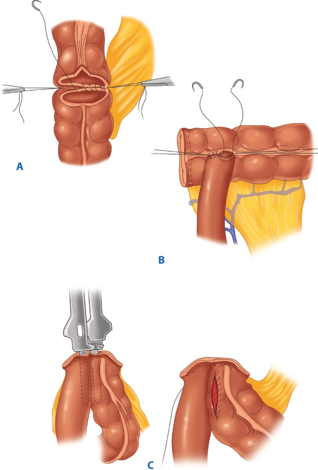
- Early rectal cancer: three options, presented as a shared decision.
- Offer one of transanal excision (including TAMIS and TEMS), endoscopic submucosal dissection, or total mesorectal excision to people with early rectal cancer (cT1-T2, cN0, M0), after discussing the implications of each and reaching a shared decision [6].
- The differences NICE requires to be discussed include that only TME removes lymph nodes and so gives accurate staging; that only TAE and TME allow full-thickness excision; that ESD needs conscious sedation rather than general anaesthesia; that TME may require a stoma and leaves external scarring; and that hospital stay is 1 to 2 days for the endoscopic and transanal options against 5 to 7 days for TME [6].
Surgical technique is constrained in three specific ways. Offer laparoscopic surgery for rectal cancer, considering open surgery where clinically indicated by locally advanced tumours, multiple previous abdominal operations or previous pelvic surgery [6]. Consider robotic surgery only within established programmes that have appropriate audited outcomes [6]. Consider transanal total mesorectal excision only in the context of research [6]. Laparoscopic resection is recommended as an alternative to open resection for both colon and rectal cancer where both are suitable [6].
Volume standards. Hospitals performing major resection for rectal cancer should perform at least 10 of these operations each year, and individual surgeons at least 5 each year [6]. These are explicit numerical thresholds with no textbook equivalent.
Locally advanced and recurrent disease. Consider referring people with locally advanced primary or recurrent rectal cancer that might potentially need multi-visceral or beyond-TME surgery to a specialist centre to discuss exenterative surgery [6].
Information before surgery. People should be told about all treatment options and their possible effects on bowel and sexual function, quality of life and independence; about the reasons a plan might change during care, including conversion from laparoscopic to open surgery or from curative to non-curative treatment, and the likelihood of a stoma, why it might be necessary and for how long; and, where enhanced recovery protocols are used, what these involve and their value [6].
Margins, exenteration and the laparoscopic trials in Schwartz's account
- Most surgeons still seek a 2-cm distal mural margin, and for upper rectal or rectosigmoid tumours a partial mesorectal excision at least 5 cm below the tumour is adequate; TME reduces blood loss and injury to pelvic nerves and presacral plexus compared with blunt dissection [9].
- Pelvic exenteration adds en bloc resection of ureters, bladder and prostate or uterus and vagina to an APR-like dissection, usually with a permanent colostomy and ileal conduit, and sacrectomy is possible up to the S2–S3 junction; pelvic MRI showing sidewall extension, iliac vessel or bilateral sacral nerve involvement or sacral invasion above S2–S3 precludes salvage, intraoperative brachytherapy helps when a clear margin is in doubt, and such operations belong in tertiary centres with colorectal, urological, neurosurgical and plastic surgeons [9].
- The COST, COLOR and MRC CLASICC trials established oncological equivalence of laparoscopic and open colectomy for colon cancer after early fears of port-site recurrence; for rectal cancer ACOSOG Z6051 and ALaCaRT failed to show non-inferiority, across nine randomised trials the CRM was positive in 7.9% laparoscopic versus 6.1% open resections (not significant) but inadequate TME was significantly commoner laparoscopically (13.2% versus 10.4%), COLOR II and COREAN showed equivalent 3-year recurrence and survival, a laparoscopic approach should not be considered for T4 tumours, and ROLARR is awaited for robotic TME quality [9].
- Synchronous cancers, multiple adenomas or a strong family history imply a field defect warranting subtotal or total colectomy, and in a metachronous tumour the surgeon must know which mesenteric vessels were ligated previously because that governs viability of the remaining colon [9].
- For palliation, colonic stents suit obstructing left-sided lesions but cause pain and tenesmus low in the rectum, a diverting stoma with a mucous fistula vents the distal colon, haemorrhage may be embolised angiographically, and exenteration and sacrectomy are generally avoided in stage IV disease [9].
Complications
Anastomotic leak (4–8% for ileocolic and colocolic anastomoses, higher for low pelvic anastomoses, up to 15%), wound infection, postoperative bleeding, ischaemia of the anastomosed bowel, anastomotic stenosis, pouchitis in restorative procedures, and anterior resection syndrome (urgency, incontinence, incomplete evacuation from loss of the rectal reservoir) are recognised complications [1][10]. Stoma complications include ischaemia, retraction, prolapse, stenosis, parastomal hernia and skin irritation [1].
Low anterior resection syndrome is named, defined and given a measurement instrument, treatment of it is a NICE recommendation rather than an afterthought. Give information on LARS to people who will potentially have sphincter-preserving surgery, and advise them to seek help from primary care if they think they have symptoms, specifically increased frequency of stool; urgency with or without incontinence of stool; a feeling of incomplete emptying of the bowels; fragmentation of stool (passing small amounts little and often); and difficulty differentiating between gas and stool [6]. Assess people with symptoms using a validated patient-administered questionnaire, for example the LARS score [6]. Offer treatment in primary care, dietary management, laxatives, anti-bulking agents, anti-diarrhoeal agents or anti-spasmodics, seeking secondary care advice if unsuccessful [6].
Appropriate specialists should discuss possible side effects with everyone who has had surgery for colorectal cancer, including altered bowel, urinary and sexual function [6].
Prognosis
- Prognosis correlates closely with Dukes'/TNM stage: 5-year survival is 75–90% for Dukes' A, 55–70% for B, 30–60% for C, and 5–10% for D (metastatic) disease [1].
- Emergency presentation is an independent marker of worse prognosis even after stage-matching [1].
- MSI-high status predicts a better prognosis stage-for-stage but reduced benefit from 5-fluorouracil-based adjuvant chemotherapy [12].
- The proportion of colorectal cancers occurring in the rectum has risen steadily from 27% in 1995 to 31% in 2019, and the increase is especially notable in people under 50, in whom rectal cancer has risen approximately 2% per year over the past decade [2].
- Follow-up is defined and time-limited.
- For people who have had potentially curative surgical treatment for non-metastatic colorectal cancer, offer follow-up for detection of local recurrence and distant metastases for the first 3 years, including serum CEA and CT scan of the chest, abdomen and pelvis [6].
- This is narrower in both duration and modality than the 5-year clinical-examination-plus-colonoscopy schedules described in the textbooks, and it is worth noting that NICE specifies what follow-up is for (detecting recurrence and metastases) rather than treating it as general oncological aftercare.
References
- Bailey & Love's Short Practice of Surgery, 28th ed., Ch. 77 The large intestine
- Sabiston Textbook of Surgery, 22nd ed., Ch. 96 Cancers of the Colon, Rectum, and Anus
- NHS Bowel Cancer Screening Programme (NHSBCSP): programme overview, NHS England, Target population; The screening test www.gov.uk
- NICE HealthTech Guidance HTG690: Quantitative faecal immunochemical testing to guide colorectal cancer pathway referral in primary care (2023, migrated from DG56), 1.1; 1.2; 1.3 www.nice.org.uk
- NICE Guideline NG12: Suspected cancer: recognition and referral (2015, updated 2026), 1.3.1 www.nice.org.uk
- NICE Guideline NG151: Colorectal cancer (2020, last updated December 2021), 1.1.1; 1.2.1; 1.2.2; 1.2.3; 1.2.4; 1.3.1; 1.3.2; 1.3.3; 1.3.4; 1.3.5; 1.3.7; 1.3.8; 1.3.9; 1.3.10; 1.3.11; 1.3.12; 1.3.13; 1.3.14; 1.3.15; 1.3.16; 1.3.17; 1.3.18; 1.4.1; 1.6.1; 1.6.2; 1.6.3; 1.6.4; Table 1 www.nice.org.uk
- Bailey & Love's Short Practice of Surgery, 28th ed., Ch. 79 The rectum
- Maingot's Abdominal Operations, 13th ed., Ch. 5
- Schwartz's Principles of Surgery, 11th ed., Ch. 29, Colon, Rectum, and Anus
- Oxford Handbook of Clinical Surgery, 5th ed., Ch. 12
- The ABSITE Review, 2022, Ch. Colorectal
- Schwartz's Principles of Surgery: ABSITE and Board Review, Ch. 29
- Maingot's Abdominal Operations, 13th ed., Ch. 54