



Bowel Obstruction
Summary
- Intestinal obstruction is impairment of the normal passage of bowel contents, classified as dynamic (mechanical, peristalsis working against an obstruction) or adynamic (paralytic ileus or pseudo-obstruction, with absent or inadequate peristalsis) [1].
- Adhesions are the commonest cause of small bowel obstruction in Western countries, while colorectal carcinoma is the leading cause of large bowel obstruction [1][2].
- The critical clinical distinction is between simple obstruction, which can usually be managed initially with fluid resuscitation and nasogastric decompression, and strangulating obstruction, a surgical emergency requiring urgent operation [1].
- There is no NICE guideline on bowel obstruction as such, but a patient with obstruction needing a laparotomy falls inside the National Emergency Laparotomy Audit, whose standards are derived from the RCS England report The High-Risk General Surgical Patient: Raising the Standard (2018) and are the closest thing the UK has to a national standard for emergency abdominal surgery [3].
- NELA's own definitions turn on the same clinical distinction the textbooks make: "immediate surgery" includes laparotomy for tender small or large bowel obstruction, strangulated hernia, GI perforation or infarction, generalised peritonitis, uncontrolled haemorrhage or sepsis; "non-immediate surgery" includes non-tender small or large bowel obstruction and infection without sepsis [3].
- NICE NG151 supplies the one disease-specific recommendation, on stenting for malignant left-sided obstruction [4].
Definition
Intestinal obstruction may be dynamic, in which peristalsis works against a mechanical obstruction (acute or chronic), or adynamic, in which there is no mechanical obstruction but peristalsis is absent or inadequate, as in paralytic ileus or pseudo-obstruction [1]. Strangulation denotes obstruction with compromised blood supply to the bowel wall, causing ischaemia [1].
Pathophysiology
- Irrespective of cause, bowel proximal to a mechanical obstruction dilates while distal bowel empties and collapses; proximal peristalsis initially increases to overcome the obstruction, but ultimately becomes flaccid and paralysed if the obstruction persists [1].
- Proximal distension is caused by gas, bacterial overgrowth producing nitrogen and hydrogen sulphide after oxygen and CO2 are reabsorbed, and by fluid, as accumulated digestive secretions of up to about 3.5 L/day from saliva, bile, pancreatic and gastric sources are retained while absorption is retarded.
- The result is dehydration and electrolyte loss from reduced intake, defective absorption, vomiting, luminal sequestration and peritoneal transudation [1].
Strangulation and the closed loop
Strangulation may result from direct pressure on the bowel wall (hernial orifices, adhesive bands), interrupted mesenteric blood flow (volvulus, intussusception), or increased intraluminal pressure in a closed-loop obstruction, one obstructed at both proximal and distal points, for example a malignant colonic stricture with a competent ileocaecal valve. Venous return is compromised before arterial supply, raising capillary pressure and ultimately causing haemorrhagic infarction, bacterial translocation and systemic sepsis [1][5].
The sequence of events in mechanical obstruction, as Schwartz describes it
Gas and fluid accumulate proximal to the obstruction, most of the gas swallowed air, the fluid swallowed liquid plus secretions stimulated by the obstruction itself, and intestinal activity increases to overcome it, accounting for the colicky pain and the diarrhoea some patients experience even with complete obstruction; as distension raises intraluminal and intramural pressure, motility falls, the normally sterile luminal flora changes and bacteria translocate to regional nodes, and when intramural pressure impairs microvascular perfusion the result is ischaemia and necrosis, strangulation [6]. Partial obstruction lets some gas and fluid pass, so the sequence runs more slowly and strangulation is less likely, whereas the closed loop obstructed at both ends, as in volvulus, cannot decompress in either direction and progresses rapidly to strangulation [6].
Clinical features
- Cardinal features are colicky abdominal pain, distension, vomiting and absolute constipation (no flatus), though the pattern varies with the level and cause of obstruction [2].
- In small bowel obstruction, vomiting occurs earlier and distension is less than in large bowel obstruction; in large bowel obstruction, vomiting is a delayed feature because the obstruction is more distal [2].
- Simple obstruction causes discomfort without severe pain, allowing time for investigation; strangulated obstruction causes severe pain, and the patient may be acidotic, very urgent surgery within 6 hours is required to save the bowel and the patient's life [2].
- Closed-loop obstruction classically presents with disproportionate pain and rapid deterioration [1].
Symptoms that grade the level and completeness of obstruction
- Vomiting is more prominent with proximal than distal obstruction and becomes feculent with bacterial overgrowth, signalling an established obstruction; continued passage of flatus or stool beyond 6–12 hours from onset marks partial rather than complete obstruction; distension is most pronounced with distal ileal obstruction and may be absent with proximal obstruction; bowel sounds are hyperactive early and minimal late; and laboratory findings reflect volume depletion with haemoconcentration, electrolyte disturbance and mild leucocytosis [6].
- Pain disproportionate to the abdominal findings, tachycardia, localised tenderness, fever, marked leucocytosis and acidosis suggest strangulation and the need for early operation [6].
- Ileus presents similarly but with diminished or absent rather than hyperactive bowel sounds [6].
Etiology
Small bowel obstruction
- Adhesions, postoperative or from congenital bands, are the commonest Western cause, followed by hernia (acquired, incisional, congenital), tumour, gallstone ileus, foreign body or bolus obstruction (bezoar, food, worms), and inflammatory strictures from Crohn's disease, tuberculosis or previous surgery [1][2].
- The classical distribution is adhesions 40%, hernia 12%, with inflammatory and neoplastic causes making up much of the remainder [1]. Gallstone ileus classically impacts about 60 cm proximal to the ileocaecal valve after erosion of a large gallstone from the gallbladder into the duodenum, producing Rigler's triad on imaging: small bowel obstruction, pneumobilia and an atypical mineral shadow [1].
- Acute intussusception is commonest in infants, peaking at 5–10 months and usually idiopathic, whereas in older children and adults a pathological lead point is more likely [1].
Large bowel obstruction
Colorectal carcinoma, sigmoid or caecal volvulus, pseudo-obstruction, faecal impaction, diverticulitis with fibrotic stenosis, inflammatory bowel disease (Crohn's stricture or ulcerative colitis with toxic megacolon) and anastomotic stricture. Adhesions essentially never cause large bowel obstruction, as the colon is much less mobile than the small bowel [2]. Pseudo-obstruction (Ogilvie's syndrome) occurs without a mechanical cause, typically in critically unwell patients or those with chronic neurological disease, acute medical illness, electrolyte imbalance, orthopaedic trauma or retroperitoneal malignancy [1][2].
Adynamic obstruction
Paralytic ileus may be postoperative (usually self-limiting over 24–72 hours), infective (intra-abdominal sepsis), reflex (spinal or rib fractures, retroperitoneal haemorrhage) or metabolic (uraemia, hypokalaemia) [1].
Adhesions, rarer causes and the causes of ileus
- Obstructing lesions are conceptualised as intraluminal (foreign bodies, gallstones, meconium), intramural (tumours, Crohn's strictures) or extrinsic (adhesions, hernias, carcinomatosis); adhesions from previous surgery account for up to 75% of small-bowel obstruction, over 300,000 Americans undergo surgery for adhesive obstruction each year with no fall over the 20 years to 2007, and small-bowel resection and adhesiolysis are two of the seven procedures accounting for 80% of US emergency operations [6].
- Cancer-related obstruction is usually extrinsic compression or invasion by malignancies from other organs rather than primary small-bowel tumours; intestinal malrotation and midgut volvulus should be remembered in adults without previous surgery, and superior mesenteric artery syndrome (compression of the third part of the duodenum by the artery) in young asthenic patients with chronic proximal obstructive symptoms [6].
- Ileus is provoked by abdominal surgery, infection (sepsis, abscess, peritonitis, pneumonia), electrolyte disturbance (hypokalaemia, hypo- or hypermagnesaemia, hyponatraemia), drugs (anticholinergics, opiates, phenothiazines, calcium-channel blockers, tricyclics), hypothyroidism, ureteric colic, retroperitoneal haemorrhage, spinal cord injury, myocardial infarction and mesenteric ischaemia; chronic intestinal pseudo-obstruction arises from familial or sporadic visceral myopathies and neuropathies, smooth-muscle disorders (scleroderma, muscular dystrophy, amyloid), neurological disease (Chagas, Parkinson's, cord injury), endocrine disease (diabetes, hypothyroidism, hypoparathyroidism), radiation enteritis, drugs and viral infection such as CMV and EBV [6].
Diagnosis
- Plain abdominal and erect chest radiographs are the initial investigation [2].
- For adhesive small bowel obstruction in a stable patient, water-soluble contrast (Gastrografin) given via nasogastric tube or orally with serial abdominal radiographs both predicts outcome (failure to reach the colon by 24 hours predicts non-resolution) and may therapeutically hasten resolution of partial obstruction [2][5].
- CT is preferred in a "virgin" abdomen with no prior surgery, to identify underlying pathology such as tumour or internal hernia (a "mesenteric swirl" sign is typical), and is the investigation of choice for large bowel obstruction and for suspected caecal or sigmoid volvulus [1][2].
- The contrast study has a tighter published threshold than 24 hours.
- In a study of 116 patients given oral water-soluble contrast, the presence of contrast within the colonic lumen within 8 hours of administration had an accuracy of 93% for predicting who would benefit from non-operative therapy.
- Only 19% of patients whose small bowel transit time exceeded 8 hours resolved without operation [7].
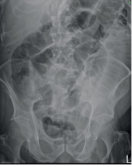
Radiographs, CT and contrast studies
- The diagnostic goals are to distinguish mechanical obstruction from ileus, find the cause, separate partial from complete and simple from strangulating obstruction, with a history of previous operations or intra-abdominal disease and a meticulous search for inguinal and femoral hernias [6].
- The abdominal series (supine and erect abdomen, erect chest) is most specific when it shows the triad of dilated small-bowel loops over 3 cm, air–fluid levels and a paucity of colonic gas; sensitivity is 70–80% and specificity low because ileus and colonic obstruction mimic the findings, with false negatives in proximal obstruction and in fluid-filled loops without gas, the latter typical of closed-loop obstruction [6].
- CT with oral contrast, 80–90% sensitive and 70–90% specific, shows a transition zone with proximal dilatation and distal decompression, contrast held up at the transition and a collapsed colon; a U- or C-shaped loop with mesenteric vessels converging radially on a torsion point suggests a closed loop, and wall thickening, pneumatosis, portal venous gas, mesenteric haziness and poor mural enhancement suggest strangulation [6].
- Water-soluble contrast reaching the colon within 24 hours predicts non-operative resolution with 92% sensitivity and 93% specificity [6].
- CT sensitivity for low-grade or partial obstruction is under 50%, so a small-bowel series (contrast swallowed or via nasogastric tube, water-soluble if perforation is possible) or enteroclysis (200–250 mL barium then 1–2 L methylcellulose solution through a long nasoenteric catheter, giving double-contrast mucosal detail even through overlapping loops) is more sensitive for luminal and mural causes such as primary tumours, and CT enteroclysis is reported superior to plain contrast studies [6].
- In the postoperative patient CT is the test of choice to reveal abscess or peritoneal sepsis and exclude complete mechanical obstruction, and chronic pseudo-obstruction is confirmed by radiographic and manometric studies, sometimes with full-thickness biopsy at laparoscopy or laparotomy [6].
Scoring and Severity
There is no discrete numerical severity score for bowel obstruction. Severity is stratified clinically by the urgency of the surgical indication [1][2]:
| Urgency | Indication |
|---|---|
| Emergency surgery within 6 hours | Obstructed external hernia; clinical features of strangulation; generalised peritonitis; evidence of perforation or ischaemia; complete large bowel obstruction with a competent ileocaecal valve; caecal volvulus |
| Surgery within 24–48 hours | Significant pathology on CT, or failure of a "drip-and-suck" or Gastrografin trial |
| Conservative management up to 72 hours | Adhesive obstruction without signs of strangulation |
Table reformats the urgency stratification [1][2].
- Complete versus partial obstruction is the other severity axis, and it predicts both success of conservative management and risk.
- Non-operative management succeeds in roughly 45% of complete obstructions versus 66% of partial obstructions in one prospective series, and in a second series 42% of complete obstructions
- Comparing complete with partial obstruction managed non-operatively, there was a greater rate of bowel strangulation (10% versus 4%) and need for resection (14% versus 8%) at operation for treatment failure, with mortality 6% versus 0% [7].
NELA's severity framework is procedural rather than anatomical, and it is what a UK emergency general surgeon is audited against. The standards benchmarked are [3]:
| Workstream | Standard | Green threshold |
|---|---|---|
| CT scanning and reporting | For patients requiring immediate surgery, CT undertaken immediately and reported by a senior radiologist (ST3 and above) within one hour, regardless of urgency of request, and communicated to the surgical team before surgery | 85% or above |
| Infection management | Suspected surgical source of infection with NEWS2 of 1–4: antimicrobials within 3 hours; NEWS2 of 5 or more, or 3 or more in any one variable: antimicrobials within 1 hour of the recorded NEWS2 abnormality | 85% or above |
| Timeliness to theatre | Patients with an "RCS immediate" pathology arriving in theatre within 6 hours of arrival at hospital or ED | 85% or above |
| Risk assessment | Risk of morbidity and mortality documented preoperatively and postoperatively, recorded by a senior surgeon (ST3 or above) within four hours of admission using appropriate risk prediction tools | 85% or above |
| Consultant-delivered care | High-risk patients (risk of death 5% or above) with both consultant surgeon and consultant anaesthetist present in theatre | 85% or above |
| Critical care | Patients with an end-of-operation risk of death of 5% or more, by any measure, admitted directly to critical care | 85% or above |
| Elderly care | Patients aged 65 or older and frail, or aged 80 and older, receiving postoperative assessment and management by a perioperative team with expertise in comprehensive geriatric assessment | 85% or above |
Table reformats the NELA principle standards [3]. Amber is 55–84% and red is below 55% for each [3].
- Two of these deserve emphasis because they are frequently missed.
- The 6-hour clock for immediate-pathology patients runs from arrival at hospital, not from the decision to operate [3].
- And the 5% mortality threshold appears twice, once preoperatively, to determine whether consultant surgeon and anaesthetist must both be present in theatre, and again at the end of the operation, to determine direct critical care admission [3].
- For high-risk patients being considered for major surgery there should also be joint preoperative discussion between a senior surgeon (ST3 and above) and a senior radiologist (ST3 and above), in person or by telephone, followed by postoperative comparison of imaging and operative findings, with best care being a discussion between a consultant surgeon and an in-house consultant radiologist [3].
Treatment and Management
Initial resuscitation comprises large-bore intravenous access and crystalloid fluids, nasogastric decompression, urinary catheterisation with fluid-balance monitoring, analgesia, and bloods including FBC, U&Es, CRP, group and save, clotting and lactate [2]. Adhesive small bowel obstruction without signs of strangulation is managed conservatively with intravenous rehydration and nasogastric decompression, generally not prolonged beyond 72 hours, with Gastrografin used both diagnostically and therapeutically; prolonged non-operative delay risks nutritional compromise and worse postoperative recovery, as shown in the NASBO trial [1][2]. Paralytic ileus is managed with nasogastric suction, restricted oral intake until bowel sounds and flatus return, and correction of electrolyte abnormalities [1].
Absolute contraindications to non-operative management are suspected ischaemia, large bowel obstruction, closed-loop obstruction, an acutely incarcerated or strangulated hernia, and perforation; complete small bowel obstruction (dilated small intestine with no distal air) is a relative contraindication [7].
Nasogastric tube technique
The tube should be positioned so that the second of four marks is at the tip of the nares: the first mark is 40 cm from the tip, the normal nares-to-oesophagogastric-junction distance, so if all four marks are outside the nares the tube is probably not in the stomach, and if no marks are visible it is coiled in the stomach or in the duodenum [7]. Oral intake should be nil while a nasogastric tube is in place, and the tube should never be clamped for prolonged periods, because traversing the oesophagogastric junction it renders the lower sphincter incompetent with a risk of aspiration, connecting it to a drainage bag for a brief trial is the appropriate alternative and a test of readiness for removal [7].
For malignant left-sided large bowel obstruction, NICE distinguishes by treatment intent rather than by fitness: consider stenting for people presenting with acute left-sided large bowel obstruction who are going to have treatment with palliative intent, and offer either stenting or emergency surgery if potentially curative treatment is suitable for them [4]. This is a more permissive position on stenting in the curative setting than the traditional "bridge to surgery only in the unfit" framing.
The move from "the sun should never set" to protocolised non-operative care
- Isotonic fluid resuscitation with a bladder catheter is integral, invasive monitoring is reserved for cardiac disease with severe dehydration, antibiotics are not indicated unless ischaemia is suspected and surgery planned, nasogastric decompression reduces nausea, distension, vomiting and aspiration, and long nasoenteric tubes are rarely used because they carry more complications without proven benefit [6].
- The dictum that "the sun should never rise and set on a complete bowel obstruction" rested on the inability of clinical, laboratory or imaging findings to distinguish simple from strangulating obstruction before irreversible ischaemia, but it has been reassessed: in 145 patients with CT-diagnosed high-grade complete obstruction 46% were managed non-operatively, and 66 of the 104 who did not need immediate surgery succeeded without it, so nasogastric decompression and resuscitation are now the usual initial management of non-ischaemic obstruction provided a closed loop is excluded [6].
- Non-operative management succeeds in 65–81% of partial obstructions, only 5–15% of successes fail to improve within 48 hours, so patients not improving by 48 hours are considered for surgery, and National Inpatient Sample data show a 2-day limit of watchful waiting does not raise mortality or morbidity though costs rise [6].
- Water-soluble contrast has therapeutic as well as prognostic value, reducing the need for surgery (odds ratio 0.44), stay by about 2 days and time to resolution by about 28 hours without added morbidity; Schwartz's pathway gives 100 mL through the nasogastric tube, films the abdomen at 8 hours, removes the tube and starts sips if contrast has reached the colon, repeats the film at 24 hours if not, and strongly considers surgery within 72 hours of admission if it still has not [6].
- Early postoperative obstruction (within 30 days) occurs in 0.7–9%, more after pelvic and colorectal surgery, is usually partial and rarely strangulates, so 2–3 weeks of bowel rest, hydration and parenteral nutrition is usually warranted unless complete obstruction or peritonitis demands reoperation; in 180 anterior resections 12.8% developed it at a median day 5 and four needed exploration [6].
- In patients with a cancer history 25–33% of obstructions are adhesive and should not be denied treatment; for recurrent malignancy palliative resection or bypass achieved palliation in over 80% and restored oral intake in over 70% of 81 patients at the cost of 7% fistula or leak and 6% 30-day mortality, and with carcinomatosis bypass avoids a difficult resection or a palliative gastrostomy relieves vomiting [6].
- Ileus is managed by limiting intake, correcting the cause, nasogastric decompression if vomiting or distension is prominent, intravenous fluid and electrolytes, and parenteral nutrition if prolonged; postoperative motility returns first in the small bowel (24 hours), then stomach (48 hours) and colon (2–5 days), so bowel sounds are unreliable and flatus or stool is the useful marker, and prolonged ileus, two or more of nausea or vomiting, intolerance of diet or absence of flatus over 24 hours, distension or radiological confirmation on or after day 4, affects 10–15% after intestinal surgery and costs $750 million to $1 billion a year in the United States [6].
- NSAIDs such as ketorolac with reduced opioids, thoracic epidural local anaesthetic, restricted intra- and postoperative fluids, early feeding, minimal bowel handling, laparoscopy and avoidance of routine nasogastric tubes shorten ileus (the core of ERAS pathways of 15–20 steps), prokinetics have unfavourable efficacy–toxicity profiles, and alvimopan, a peripherally acting μ-opioid antagonist, shortens ileus, stay and readmission in randomised trials though its cost-effectiveness is debated [6].
- Chronic pseudo-obstruction is palliated with fluid, electrolyte and nutritional support avoiding surgery where possible; metoclopramide and erythromycin work poorly, cisapride is restricted to compassionate use for cardiac toxicity, refractory cases need long-term parenteral nutrition, a decompressive gastrostomy or extended resection, and small-bowel transplantation is increasingly applied [6].
Surgeries
At laparotomy, assessment addresses the site and nature of the obstruction and bowel viability [1].
| Feature | Viable bowel | Non-viable bowel |
|---|---|---|
| Colour | Dark but lightens | Remains dark |
| Arterial pulsation | Visible | Absent |
| Surface | Shiny | Dull |
| Consistency | Firm | Flabby and friable |
| Peristalsis | Observable | Absent |
- Table reformats the assessment of bowel viability [1].
- If in doubt, the bowel is wrapped in hot packs for 10 minutes and reassessed [1].
- Adjuncts including Doppler ultrasound and intravenous fluorescein are relatively subjective and are only adjuncts to sound clinical judgement; in patients who would otherwise be left with less than two-thirds of their original bowel length, consideration may be given to resecting only grossly necrotic bowel, preserving questionable segments, and performing an end ostomy or a second-look procedure at 12 to 24 hours [7].
- Adhesiolysis: only the causative adhesion is divided where clear, limiting further dissection to reduce re-adhesion; laparoscopic adhesiolysis is reserved for surgeons with advanced laparoscopic skills, generally for a single band causing obstruction [1][5].
- Resection and anastomosis or stoma formation: for non-viable bowel, tumour, or strictures; small bowel Crohn's strictures are resected with a 2 cm margin from grossly visible disease, frozen-section clearance conferring no benefit [5]. When anastomosing, the surgeon must allow for the discrepancy in diameter and wall thickness between obstructed proximal and decompressed distal bowel: a side-to-side or end-to-side anastomosis may be preferable where the proximal bowel is massively dilated, and a stapled anastomosis may be less safe where there is a large discrepancy in wall thickness or bowel wall oedema, because uniform tissue approximation for a given staple height may not be possible [7].
- Intraoperative decompression: where massive dilatation prevents abdominal closure. Manual retrograde decompression of luminal contents around the ligament of Treitz, through the pylorus and into the stomach for aspiration through the nasogastric tube is the safest and quickest technique, avoiding an enterotomy; a controlled enterotomy with a decompressing tube is strongly discouraged except where distension prevents closure or threatens bowel viability. Distended bowel must be handled gently, since experimental studies show increased bacteraemia after extensive manipulation [7].
- Bypass versus resection: in incurable malignant obstruction where the site cannot be safely dissected, intestinal bypass relieves the obstruction, restores continuity and prevents a closed loop. In carcinomatosis a bypass may be fastest and safest because survival will be short, but patients with chronic inflammatory disease such as Crohn's disease or tuberculosis remain at risk from the bypassed segment and are usually better served by resection [7].
- Right hemicolectomy with primary anastomosis: for a resectable right-sided or proximal transverse obstructing lesion; a proximal stoma or ileotransverse bypass if unresectable [1].
- Hartmann's procedure, or resection with on-table lavage and primary anastomosis: for obstructing left-sided or rectosigmoid lesions; a defunctioning proximal stoma or self-expanding metal stent may bridge to elective or definitive surgery in unfit or palliative patients [1].
- Caecal volvulus: reduction if viable, occasionally requiring needle decompression, with resection or caecopexy (fixation to the right iliac fossa), caecopexy recurs in up to 40% of cases [1].
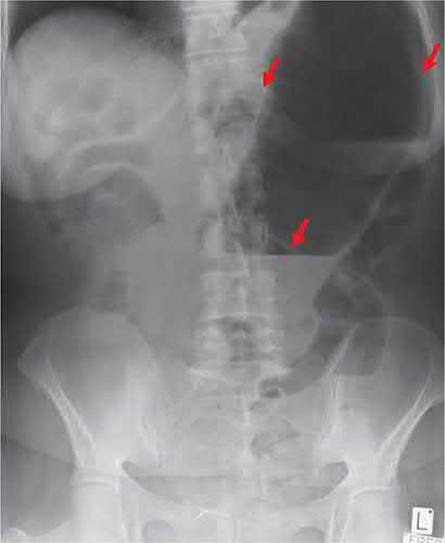
- Sigmoid volvulus: endoscopic (flexible or rigid sigmoidoscopic) detorsion and flatus tube decompression as first-line if there is no gangrene or peritonitis, with elective sigmoid colectomy after stabilisation given the high recurrence risk of up to 40%; immediate surgical exploration is mandated by gangrene, perforation, or necrotic or ulcerated mucosa on endoscopy, generally via a Hartmann's procedure if bowel is non-viable [1][10].
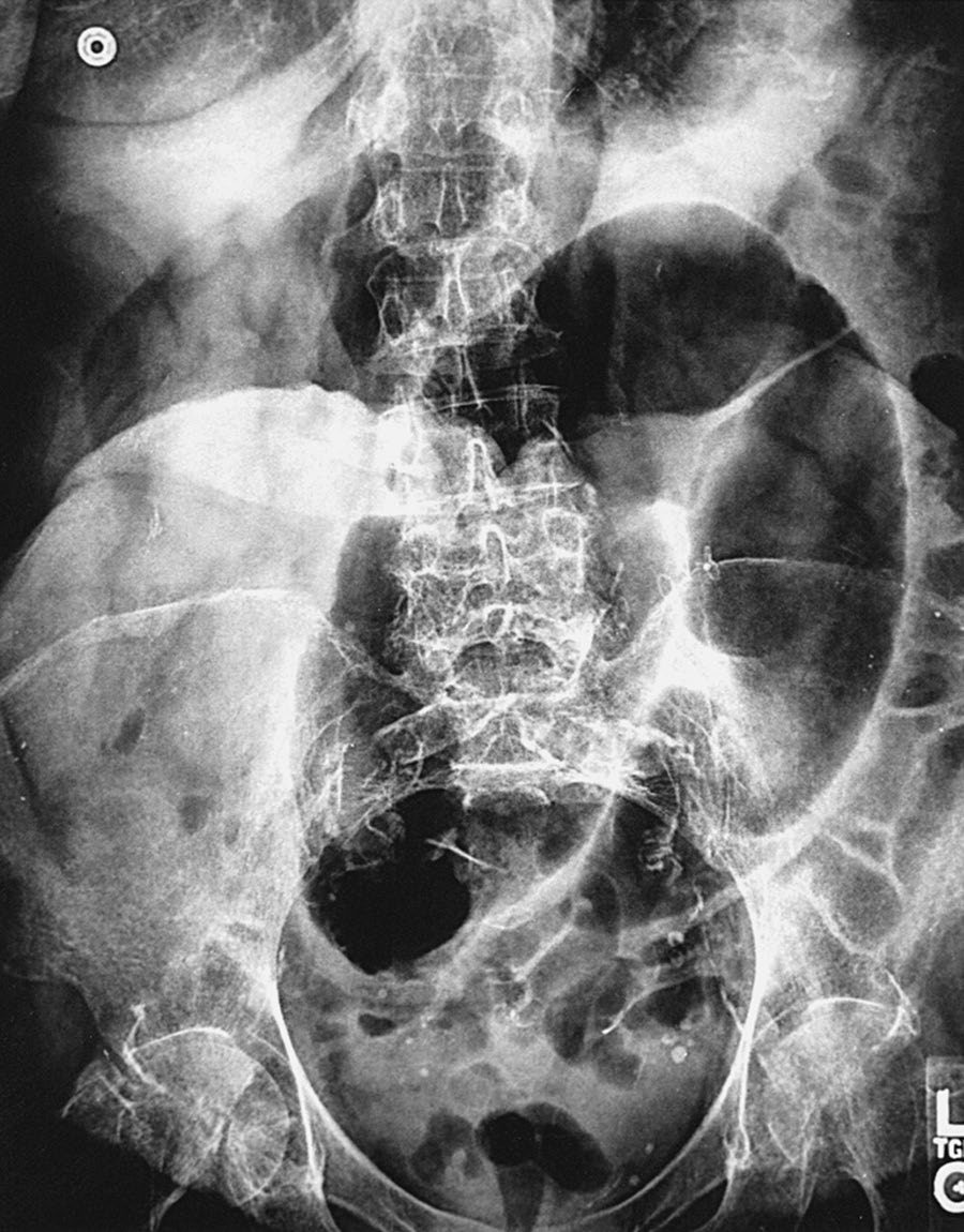
- Intussusception: non-operative reduction by air or barium enema is attempted first in infants, successful in over 70% in experienced units, and is contraindicated with peritonitis, perforation, a known lead point, or profound shock; surgical reduction by gentle compression from distal to proximal is required if radiological reduction fails, with resection for irreducible, infarcted, or lead-point-associated cases [1].
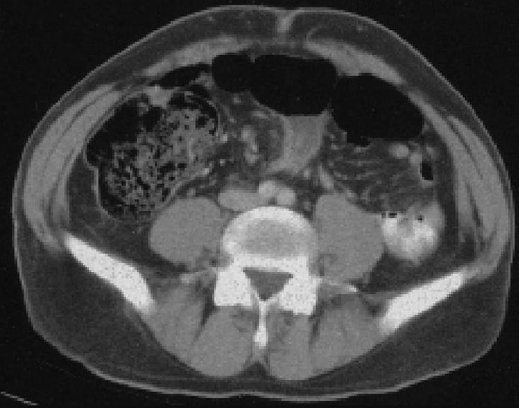
Judging viability, laparoscopy and prevention of adhesions
- Adhesions are lysed, tumours resected and hernias reduced and repaired, but whatever the cause the bowel is examined and non-viable segments resected; normal colour, peristalsis and marginal arterial pulsation indicate viability, visual inspection usually suffices, and Doppler or intravenous fluorescein under ultraviolet light in borderline cases is no better than clinical judgement [6].
- Short doubtful segments are resected with primary anastomosis in the stable patient, but when a large proportion is in question the bowel is left and re-explored at 24–48 hours in a second-look operation [6].
- Laparoscopic adhesiolysis in a propensity-matched study gave fewer complications and surgical-site infections and a shorter stay (4 vs 10 days); it suits early proximal obstruction from a probable single band, distension and multiple adhesions produce conversion rates of 17–33%, and a pooled analysis of 11 comparative studies found no difference in bowel injury or reoperation but longer operating time [6].
- Good technique, gentle handling and minimal foreign material are the cornerstone of adhesion prevention but often inadequate, readmission for adhesive obstruction exceeds 30% over 10 years after colorectal or pelvic surgery, open surgery carries a fourfold risk of obstruction within 5 years compared with laparoscopy in the Swedish register, hyaluronan-based barriers such as Seprafilm clearly reduce adhesions though their effect on obstruction is less well defined, and wrapping an anastomosis in the material may raise leak rates and is discouraged [6].
Colonic volvulus by site in Schwartz's account
- The sigmoid accounts for up to 90% of colonic volvulus, the caecum under 20% and the transverse colon a rarity; chronic constipation producing a redundant colon with a narrow mesenteric base predisposes, fever and leukocytosis herald gangrene or perforation, and some patients report long intermittent obstructive symptoms suggesting chronic recurrent volvulus [12].
- Sigmoid volvulus gives a bent inner tube or coffee-bean appearance with the convexity in the right upper quadrant, opposite the obstruction, and a pathognomonic bird's beak on Gastrografin enema; detorsion is easiest with a rigid proctoscope though flexible endoscopy also works, a rectal tube maintains decompression, and necrotic mucosa, ulceration or dark blood at endoscopy indicate strangulation and mandate operation, with Hartmann's procedure the safest option when dead bowel is found [12].
- Caecal volvulus results from non-fixation of the right colon, usually rotating around the ileocolic vessels with early vascular impairment, though in 10–30% the caecum folds on itself as a caecal bascule; plain films show a kidney-shaped air-filled structure in the left upper quadrant, it can almost never be detorsed endoscopically, exploration is required once diagnosed, and right hemicolectomy with primary ileocolic anastomosis is usually safe and prevents recurrence, whereas detorsion alone or with caecopexy recurs frequently [12].
- Transverse colon volvulus, favoured by non-fixation and megacolon, mimics sigmoid volvulus radiographically but the enema shows a more proximal obstruction, and although colonoscopic detorsion occasionally succeeds most patients need emergency resection [12].
Complications
- Strangulation with bowel infarction and perforation, systemic sepsis from bacterial translocation, dehydration and electrolyte derangement, and postoperative intra-abdominal sepsis are the principal complications [1].
- Recurrent obstruction after adhesiolysis is common, and port-site hernias are a recognised cause of obstruction after laparoscopic surgery [1].
- Recurrent intussusception occurs in up to 10% after non-operative reduction [1].
Prognosis
- Morbidity and mortality from strangulation correlate with the duration and extent of ischaemia; elderly patients and those with comorbidities tolerate strangulation poorly [1].
- The lifetime risk of hospital admission for adhesional small bowel obstruction after abdominal surgery is approximately 4%, with around 2% eventually requiring laparotomy [1].
- Recurrence risk after endoscopic decompression alone is high for both sigmoid volvulus (up to 40%) and caecal volvulus after caecopexy (up to 40%), favouring elective resection in fit patients [1].
Recurrence after adhesive obstruction
Perioperative mortality for non-strangulating obstruction is under 5%, mostly in elderly comorbid patients, and higher for strangulation; fewer than 20% of patients treated conservatively for adhesive obstruction are readmitted with another episode over 5 years, and after operation for adhesive obstruction in 286 patients recurrence was 5.5% at 1 year, 11.3% at 3 and 13.5% at 5 years with reoperation in 3.7%, 4.8% and 5.8%, while a standard hospital-wide policy shortens time to surgery and length of stay [6].
References
- Bailey & Love's Short Practice of Surgery, 28th ed., Ch. 78, Summary boxes 78.13–78.14
- Oxford Handbook of Clinical Surgery, 5th ed., Ch. 26 Emergency surgery topics
- National Emergency Laparotomy Audit: principle standards reported by NELA (February 2025), derived from RCS England The High-Risk General Surgical Patient — Raising the Standard (2018), Principle standards www.nela.org.uk
- NICE Guideline NG151: Colorectal cancer (2020, last updated December 2021), 1.3.1; 1.3.2 www.nice.org.uk
- Schwartz's Principles of Surgery: ABSITE and Board Review, Ch. 28 Small Intestine
- Schwartz's Principles of Surgery, 11th ed., Ch. 28, Tables 28-4 and 28-5
- Maingot's Abdominal Operations, 13th ed., Ch. 38 Small Bowel Obstruction
- Bailey & Love's Short Practice of Surgery, 28th ed., Ch. 8
- Maingot's Abdominal Operations, 13th ed., Ch. 43
- Schwartz's Principles of Surgery: ABSITE and Board Review, Ch. 29
- Sabiston Textbook of Surgery, 22nd ed., Ch. 95
- Schwartz's Principles of Surgery, 11th ed., Ch. 29, Colon, Rectum, and Anus