



Appendicitis
Summary
- Acute appendicitis is the commonest cause of urgent abdominal surgery in the UK, with peak incidence in the teens to early twenties [1][2].
- It classically presents with periumbilical pain migrating to the right iliac fossa, anorexia and vomiting, though the "classic" pattern occurs in only about half of confirmed cases [1].
- Diagnosis is primarily clinical, supported by scoring systems, inflammatory markers and, where uncertain, ultrasound or CT [1].
- Treatment is appendicectomy, laparoscopic or open, although selected uncomplicated cases may be treated with antibiotics alone [1].
- There is no NICE guideline on appendicitis.
- The UK national guidance is the Academy of Medical Royal Colleges / NHS England Evidence-Based Interventions entry on appendicectomy without confirmation of appendicitis, which is about imaging rather than about the operation: its purpose is to reduce the negative appendicectomy rate by defining when a scan is needed and which scan [3].
- It applies to adults and children and carries a hard number the textbooks do not give, a "triple screen" with a negative predictive value above 99% [3].
Definition
Acute appendicitis is acute inflammation of the vermiform appendix [1].
Epidemiology and the two-disease hypothesis
The lifetime incidence of appendicitis is 8.6% in men and 6.7% in women, highest in the second and third decades; the appendicectomy rate in developed countries has fallen over recent decades yet it remains one of the most frequent emergency abdominal operations [4]. Progression to perforation is not predictable and spontaneous resolution is common, so differences in epidemiology and the fact that not all non-perforated disease progresses suggest that non-perforated and perforated appendicitis may be different diseases with different pathogenesis [4].
Pathophysiology
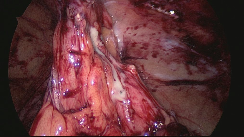
- Obstruction of the appendiceal lumen, by a faecolith or appendicolith, lymphoid hyperplasia, a fibrotic stricture, or occasionally tumour or parasites such as Enterobius vermicularis, appears essential for the development of perforation, although in many early cases the lumen remains patent despite mucosal inflammation and lymphoid hyperplasia [1].
- Once obstructed, continued mucus secretion and inflammatory exudation raise intraluminal pressure, obstructing lymphatic drainage and causing oedema and mucosal ulceration with bacterial translocation to the submucosa.
- Progressive distension causes venous obstruction and ischaemia, permitting bacterial invasion through the muscularis propria and submucosa (acute appendicitis), and finally ischaemic necrosis of the wall (gangrenous appendicitis) with free bacterial peritoneal contamination; alternatively, adherent omentum and small bowel wall off the process to form an appendix mass or abscess [1].
Clinical features
Classically, poorly localised, colicky periumbilical (visceral, midgut) pain precedes anorexia, nausea and one or two episodes of vomiting, before the pain shifts and localises to the right iliac fossa as somatic parietal peritoneum becomes irritated, worsened by coughing or movement [1]. This visceral-to-somatic sequence is present in only around half of confirmed cases; atypical presentation is more common in the elderly [1]. Loss of appetite typically precedes pain by a few hours, and most patients feel nauseated; the pain is referred to the T10 dermatome centrally before localising [6].
Presentation by appendiceal position
Pelvic appendicitis may cause suprapubic discomfort, tenesmus or dysuria rather than anterior abdominal pain, with tenderness elicited only on rectal examination, hence a rectal examination is recommended in every patient with acute lower abdominal pain [1][6]. A retrocaecal appendix may produce a "silent" abdomen with deep tenderness confined to the loin and a positive psoas sign [1].
Signs
Cardinal signs are low-grade pyrexia, localised right iliac fossa tenderness (classically at McBurney's point), muscle guarding and rebound tenderness [1]. Other signs include the pointing sign, Rovsing's sign (palpation of the left iliac fossa causing right iliac fossa pain), the psoas sign, and the obturator sign [1].
Appendicitis in pregnancy
- Appendicitis is the most common non-obstetric emergency in pregnancy and the most frequent reason for general surgical intervention in this group, yet it has a typical clinical presentation in only 50% to 60% of cases [7].
- Several things conspire against the diagnosis: nausea and vomiting are non-specific and common in normal pregnancy; the normal febrile response may be blunted; physical examination is altered because the gravid uterus displaces the appendix to a more cephalad position; and biochemical markers are unreliable, since mild leukocytosis and raised CRP may be physiologically normal in pregnancy [7].
- The surgeon must also consider obstetric emergencies as a cause of abdominal pain, including preterm labour, placental abruption and uterine rupture [7].
The stakes justify routine imaging. With complicated appendicitis the risk of preterm labour is 11% and fetal loss 6%; with uncomplicated appendicitis the rates are lowest at 6% and 2%; and a negative appendicectomy carries rates of 10% and 4%, that is, an unnecessary operation is worse for the fetus than an uncomplicated true positive [7].
Eponymous signs and the weight of individual findings
- Migratory pain (visceral inflammation progressing to the parietal peritoneum) is the classic sign (positive likelihood ratio 2.06); patients lie still, run a low-grade fever of about 38.0 °C and show focal tenderness with guarding, McBurney's point lying one-third of the way from the anterior superior iliac spine to the umbilicus [4].
- The eponymous signs help place the appendix: Rovsing's sign (right lower quadrant pain on release of pressure in the left lower quadrant) for the normal position, Dunphy's sign (pain on coughing) and the iliopsoas sign (pain on hip flexion) for a retrocaecal appendix, the obturator sign (pain on internal rotation of the hip) and pain on rectal or cervical examination for a pelvic appendix [4].
- Andersson's meta-analysis shows that individual findings are weak but combine to high discriminatory power: the strongest positive likelihood ratios are a white cell count of 15 or more (7.20) or 10 or more (4.20), rigidity (2.96), rebound (2.86), guarding (2.48), direct tenderness (2.47), C-reactive protein above 20 mg/L (2.39), indirect tenderness (2.31), pain migration (2.06) and psoas sign (1.99), while symptoms beyond 48 hours actually lower the likelihood (0.49) and anorexia, nausea, vomiting and fever each carry ratios near 1.2–1.6 [4].
- Regional inflammation can also produce ileus, diarrhoea, small-bowel obstruction or haematuria, and the differential of mesenteric adenitis, caecal or Meckel's diverticulitis, acute ileitis, Crohn's disease, pelvic inflammatory disease, torsion of an ovarian cyst or Graafian follicle and gastroenteritis is narrowed by an antecedent viral history, cervical examination and a menstrual history that distinguishes mittelschmerz (mid-cycle pain without fever or leucocytosis) and ectopic pregnancy [4].
- Perforated appendicitis usually presents after 24 hours, although 20% present within 24 hours, and untreated it produces a walled-off right lower quadrant abscess or, less often, psoas or liver abscess, fistula or pylephlebitis [4].
- Chronic appendicitis describes recurrent right lower quadrant pain without fever and an appendicolith or dilated appendix on imaging, usually resolved by appendicectomy, whereas prophylactic appendicectomy without imaging abnormality is not encouraged [4].
Etiology
No single unifying cause is established; decreased dietary fibre and increased refined carbohydrate consumption are implicated, paralleling colonic diverticulitis, with lowest incidence in high-fibre societies [1]. Bacterial proliferation is a mixed aerobic and anaerobic process, usually following luminal obstruction by a faecolith or lymphoid hyperplasia; obstruction by caecal carcinoma is an occasional cause in older patients [1].
Diagnosis
- Diagnosis is essentially clinical; a decision to operate on clinical suspicion alone can still lead to removal of a normal appendix in 15–30% of cases [1].
- Routine investigations include full blood count and urinalysis; selective investigations include pregnancy test, U&Es, CRP, abdominal radiograph, ultrasound and contrast-enhanced CT [1]. Leukocytosis with neutrophilia is present in about 90% of cases, but a normal white cell count is found in 10% and does not exclude the diagnosis [7].
- Ultrasound is particularly useful in children, thin adults and to exclude gynaecological pathology in women of childbearing age (diagnostic accuracy over 90%), though it is not useful in men to diagnose appendicitis; modern CT has around 95% sensitivity and specificity and reduces negative appendicectomy without increasing perforation from delay [1].
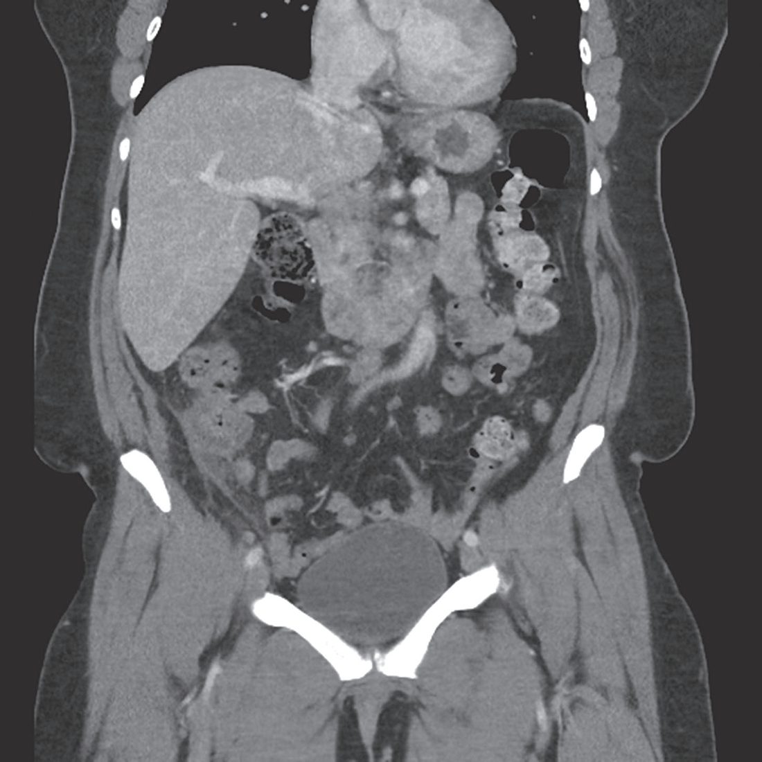
In pregnancy the initial study of choice is ultrasound with graded compression, using the same criteria as in the non-pregnant patient, though sensitivity and specificity fall to about 83% because of the gravid uterus. If ultrasound is equivocal, MRI without gadolinium is the best next study, with excellent preserved accuracy; routine use of MRI in pregnancy reduces the negative appendicectomy rate by 47% without a significant increase in perforation, and is cost-effective. If ultrasound is inconclusive and MRI is not immediately available, CT may be used [7].
- The UK recommendation is to place imaging inside a defined clinical pathway rather than to image everyone or no one. "Consider the imaging of patients with the suspicion of acute appendicitis in a defined clinical pathway.
- Where patients present with a high clinical suspicion of appendicitis, then imaging may not be necessary, but imaging can help identify which patients can be managed conservatively.
- If there is clinical doubt then imaging can reduce the negative appendicectomy rate. Most patients should have an ultrasound as the first-line investigation.
- If the diagnosis remains equivocal, a contrast-enhanced CT (preferably low dose) can be performed to give a definitive diagnosis prior to the patient returning to the surgical unit for a decision on management." [3].
The pathway is explicitly conditional on radiology capacity. It depends on an adequately skilled radiologist (consultant or registrar) or sonographer being available to perform the ultrasound in a timely fashion; if this is not possible, discretion should be used to proceed directly to limited-dose contrast-enhanced CT of the abdomen and pelvis [3].
- The "triple screen" is the single most quotable number here.
- In adults, negative appendicectomy can occur in up to 30% of cases where appendicitis is suspected clinically but imaging is not performed.
- However, the triple screen, CRP under 10, WCC under 10.5 and a neutrophil percentage under 75%, has a negative predictive value above 99% for excluding appendicitis, and imaging is not recommended in that setting [3].
Modality choice by patient group [3]:
| Patient group | Preferred imaging |
|---|---|
| Children | Ultrasound if there is diagnostic uncertainty. CT is not recommended in children given the risks of ionising radiation; MRI can be used in centres with appropriate expertise |
| Young patients, and women where a gynaecological differential is likely | Ultrasound, which is superior to CT in this setting |
| Obese patients | CT may be more appropriate, since ultrasound is more challenging |
| Older patients | CT is usually of more value, because the differential diagnosis is broader |
| Pregnant patients | MRI where available, having accuracy similar to CT without ionising radiation |
Table reformats the EBI guidance on imaging modality [3].
Laboratory markers and imaging performance
- Leucocytosis is usually around 10,000/mm³, higher (about 17,000) with gangrene or perforation; C-reactive protein, bilirubin, IL-6 and procalcitonin have all been proposed to help, particularly in predicting perforation, and the authors regard a white count and CRP, a pregnancy test in women of childbearing age and a urinalysis to exclude stones or pyelonephritis as the appropriate initial tests [4].
- Imaging is used because a negative operation rate is acceptable only below 10% in men and 20% in women, and it is most appropriate when the diagnosis is unclear or operative risk is high, as in pregnancy or multiple comorbidity [4].
- Contrast CT has sensitivity 0.96 and specificity 0.96, showing an enlarged lumen and double-wall thickness over 6 mm, wall thickening over 2 mm, periappendiceal fat stranding or an appendicolith; low-dose protocols deliver 2–4 mSv, little more than the 3.1 mSv of background radiation, and 2-mSv scans do not alter outcomes despite lower resolution, while contrast is omitted for allergy or an eGFR under 30 mL/min/1.73 m² [4].
- Graded-compression ultrasound has sensitivity 0.85 and specificity 0.90, an easily compressible appendix under 5 mm rules out appendicitis, a diameter over 6 mm, pain on compression, an appendicolith, echogenic fat and periappendiceal fluid support it, and is cheaper, more available and radiation-free but operator-dependent, limited in obesity and painful with peritonitis; five meta-analyses found CT more sensitive in five and more specific in four [4].
- MRI has sensitivity 0.95 and specificity 0.92 but is expensive and expertise-dependent, reserved for pregnant or paediatric patients in whom radiation outweighs the convenience of CT [4].
Scoring and Severity
The Alvarado (MANTRELS) score is the most widely used clinical scoring system [1]:
| Component | Feature | Points |
|---|---|---|
| Symptoms | Migratory right iliac fossa pain | 1 |
| Symptoms | Anorexia | 1 |
| Symptoms | Nausea or vomiting | 1 |
| Signs | Right iliac fossa tenderness | 2 |
| Signs | Rebound tenderness | 1 |
| Signs | Elevated temperature | 1 |
| Laboratory | Leukocytosis | 2 |
| Laboratory | Left shift | 1 |
- Table reformats the Alvarado score, total 10 [1]. A score of 7 or more is strongly predictive of appendicitis, and scores of 5–6 warrant further imaging [1].
- More recent consensus guidelines recommend the Appendicitis Inflammatory Response (AIR) score or the Adult Appendicitis Score (AAS) over the Alvarado score, which is useful for ruling appendicitis out but lacks specificity for ruling it in [7].
- Clinically, appendicitis is also broadly classified as mild (localised peritonitis, well patient, can await surgery overnight) or severe (generalised peritonitis, sick patient, requiring very urgent surgery) [2].
Treatment and Management
Non-operative management: a trial of intravenous antibiotics, for example metronidazole with a third-generation cephalosporin, plus bowel rest may be considered in uncomplicated appendicitis with no appendicolith, perforation or abscess, with initial success in around 85%, though 25–33% require surgery within a year for recurrence [1].
- The trial evidence is worth knowing in detail.
- The multicentre randomised CODA trial found antibiotics non-inferior to appendicectomy at 30 days on the EQ-5D, but 29% of patients underwent appendicectomy within 90 days, and patients with an appendicolith treated non-operatively were at higher risk of both appendicectomy and complications [7].
- The smaller single-institution COMMA trial found a similar 25% recurrence rate after non-operative treatment, similar early quality of life, but significantly lower quality of life in the non-operatively treated group at 1 year [7].
- On these data antibiotics are now considered an acceptable first-line treatment for acute uncomplicated appendicitis, and for patients managed non-operatively treatment consists of 10 days of antibiotics covering gastrointestinal flora, the first 24 hours given intravenously [7]. Proven or suspected complicated appendicitis without an abscess or phlegmon should be treated surgically [7].
The appendix mass
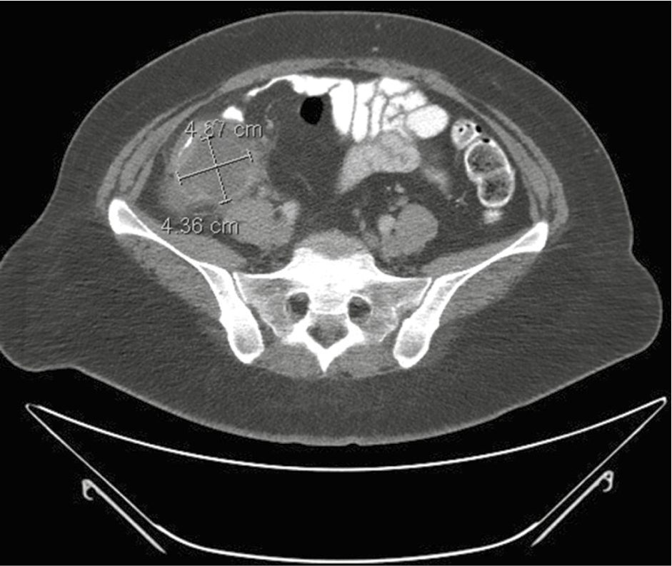
An appendix mass without generalised peritonitis is managed by the conservative Ochsner–Sherren regimen: antibiotics, CT assessment, radiological drainage of any abscess, and 4-hourly observations, with early laparotomy for clinical deterioration, a rising pulse, spreading pain or an enlarging mass. Around 90% resolve without incident, but interval appendicectomy is now debated given a higher-than-expected rate of underlying appendiceal neoplasm, up to 29% in patients over 40 in one trial, so follow-up CT or MRI and colonoscopy are advised in that age group [1].
Antibiotics alone, timing, and the perforated appendix in Schwartz
- Randomised non-inferiority trials of antibiotics (a short intravenous course followed by a fluoroquinolone with metronidazole or oral amoxicillin-clavulanate) against appendicectomy (often open) show in meta-analysis that 26.5% of the antibiotic group need appendicectomy within a year, adverse events are higher (RR 3.18) and recurrences present more often as complicated appendicitis (RR 2.52), so conservative management can be offered through shared decision-making but is not standard except for a significant phobia of surgery [4].
- Delaying surgery under 12 hours after admission antibiotics is acceptable for symptoms shorter than 48 hours and non-perforated, non-gangrenous disease, with no difference in outcome beyond a slightly longer stay [4].
- For long-standing complicated appendicitis management is staged, resuscitation, intravenous antibiotics and image-guided percutaneous drainage, which achieves complete resolution in 79% (more often with lower-grade abscesses, transgluteal and CT-guided drainage), with operation for failure or free perforation, since immediate surgery in the septic patient carries more abscesses and enterocutaneous fistulae from dense adhesions [4].
- About 80% resolve with drainage and antibiotics, and the debate over interval appendicectomy at 6–8 weeks weighs recurrence (7.4–8.8%) and neoplasms found in the specimen (relevant benign 0.7%, malignant 1.3%) against the 91% with no further event at a median 34 months, so shared decision-making is required [4].
- Preoperative antibiotics are given 30–60 minutes before incision, cefoxitin, ampicillin-sulbactam or cefazolin plus metronidazole for uncomplicated disease, clindamycin with a fluoroquinolone, gentamicin or aztreonam for β-lactam allergy, piperacillin-tazobactam or a cephalosporin with metronidazole for perforation, with no postoperative antibiotics for uncomplicated appendicitis and generally under 4 days after source control for perforation (STOP-IT trial), longer with incomplete drainage, persistent catheters or uncertain resolution [4].
- Incidental appendicectomy during other operations is routine in children on chemotherapy, compromised hosts with an unreliable examination, Crohn's patients with a normal caecum, travellers to remote areas and cytoreduction for ovarian cancer, but because adhesions and later complications probably outweigh the risk of future appendicitis it is not otherwise advocated [4].
- Older adults mount less inflammation and present more often with perforation or abscess, so definitive imaging before theatre is prudent [4].
Pregnancy and childhood in Schwartz's account
- Appendicitis complicates 1 in 800 to 1 in 1000 pregnancies, mostly in the first two trimesters, may present with heartburn, bowel irregularity or flatulence, and displaces the point of tenderness; ultrasound is preferred (sensitivity 67–100%, specificity 93–96%, non-visualisation possible), MRI is the alternative (sensitivity 94%, specificity 97%), CT is avoided unless other modalities are inconclusive, fetal loss reaches 36% with perforation so a negative exploration rate as high as 30% is accepted, laparoscopy is safe but carries a reproducibly higher fetal loss (about 7% vs 3%) than open surgery, insufflation pressures of 10–12 mmHg may reduce early labour, and non-operative treatment fails in up to 25% [4].
- Almost 1 in 8 children are worked up for appendicitis; infants and young children perforate in 51–100%, neonates may present with distension, lethargy or irritability, the Pediatric Appendicitis Score (10 points, 2 each for right lower quadrant tenderness and pain with cough, percussion or hopping) predicts appendicitis in 78–96% at 7 or more, and intussusception, gastroenteritis, malrotation, ectopic pregnancy, mesenteric adenitis, omental torsion and ovarian or testicular torsion must be excluded [4].
- Laparoscopic appendicectomy has better outcomes than open in children, urgent operation is advocated for complicated disease without abscess or mass, antibiotics continue at least 3 and preferably 5 days after perforation (APSA guidelines), and non-operative management may be safe with presentation under 48 hours, white count under 18,000, an appendicolith and no rupture on imaging, reducing inflammation in 88–92% but with 22% recurrence at 1 year [4].
Surgeries
Appendicectomy is the standard treatment, performed under general anaesthesia, laparoscopically or open [1]. A single perioperative dose of antibiotics reduces wound infection in the absence of purulent peritonitis; therapeutic intravenous antibiotics covering Gram-negative bacilli and anaerobes are given when peritonitis is suspected [1].
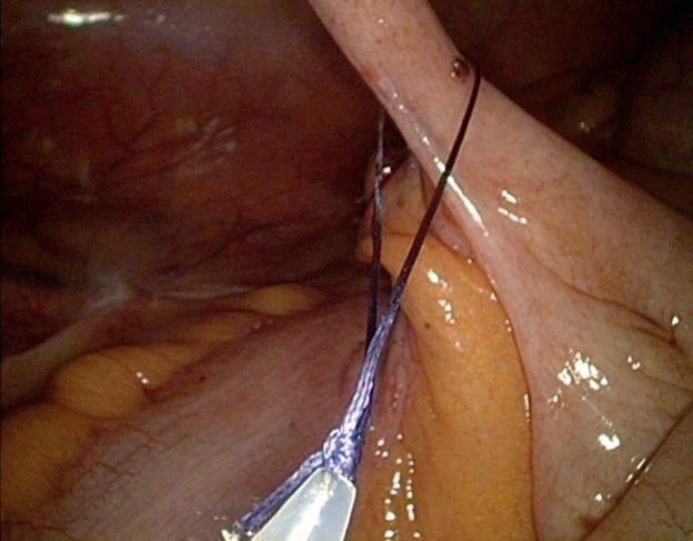
- Laparoscopic appendicectomy: preferred where expertise and equipment allow, particularly in women since it allows assessment of pelvic pathology; associated with lower wound infection and quicker recovery, and now the gold standard approach [1][2]. A three-port technique (umbilical camera port, suprapubic and left-lower-quadrant working ports) is typically used; the appendix is identified at the confluence of the taeniae coli on the caecum, a window is created in the mesoappendix close to the appendiceal base to avoid the appendicular artery, and the base is divided flush with the caecum to avoid stump appendicitis, using a stapler, Endoloop or clips [8]. Where endoloops are used, two loops secure the base and a third is applied distally to avoid spillage of luminal contents, the specimen being divided between them [7].
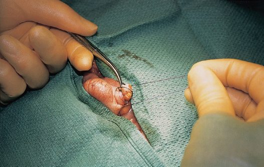
- Open appendicectomy: via a McBurney's (gridiron) incision centred one-third of the way from the anterior superior iliac spine to the umbilicus, or over a palpable mass; a muscle-splitting technique divides external oblique, internal oblique and transversus abdominis along their fibres; the appendix is delivered, its mesoappendix (containing the appendicular artery, an end-artery whose thrombosis causes gangrene) is clamped, ligated and divided, and the base is ligated and divided [1][8]. The classical open sequence continues with placement of a purse-string or Z stitch and inversion of the appendiceal stump [7].
- A normal appendicectomy rate of around 10% is accepted as the price of not missing true appendicitis, confirmed only by histopathology [2].
Incidental appendiceal neoplasms
Appendiceal adenomas and serrated polyps behave like their colonic counterparts and are most often found incidentally on post-appendicectomy pathology review; when confined to the muscularis mucosa and lamina propria without invasion they are adequately managed by simple appendicectomy alone, but they carry an increased risk of synchronous colonic pathology and can be a manifestation of serrated polyposis syndrome, so such patients should be counselled to undergo a full colonoscopy [7]. Pseudomyxoma peritonei occurs when a mucin-producing appendiceal tumour (most commonly a low-grade appendiceal mucinous neoplasm) perforates, spreading tumour cells and mucinous ascites through the abdomen; standard treatment is surgical cytoreduction with hyperthermic intraperitoneal chemotherapy, with outcomes determined primarily by histological subtype, disease burden and completeness of cytoreduction [7].
Adenocarcinoma and neuroendocrine tumours of the appendix
A tumour is found in a small proportion of appendicectomy specimens, and the appendix is one of the commonest sites for gastroenteropancreatic neuroendocrine tumours [9]. Nonmucinous adenocarcinomas of the appendix behave like adenocarcinomas of the colon and rectum and are treated as such: complete staging with cross-sectional imaging and the tumour markers CEA, CA125 and CA19-9, followed by interval right hemicolectomy with sufficient lymph node sampling for staging, and consideration of adjuvant chemotherapy [9]. Some studies suggest that in selected patients with T1, well-differentiated tumours without lymphovascular invasion, a less extensive resection (simple appendicectomy or ileocaecectomy) may suffice, though the rarity of the disease limits prospective study [9]. Goblet cell adenocarcinoma, previously called goblet cell carcinoid, shows both neuroendocrine and mucinous features but is managed like a nonmucinous colonic-type adenocarcinoma, the neuroendocrine component being considered minor in the current WHO classification [9].
Operative technique and the appendiceal critical view
- Open appendicectomy uses an oblique McBurney or transverse Rocky-Davis incision at McBurney's point (a lower midline for a perforation with phlegmon), a muscle-splitting approach in a well-paralysed patient, Trendelenburg with the left side down, tracing the anterior taenia (taenia libera) to find the appendix, early ligation of the mesentery for exposure, simple ligation of a viable base with optional Z-stitch or purse-string imbrication or mucosal fulguration, and medial extension of the incision (Fowler extension) if the appendiceal artery retracts or bleeds; trials show no difference in wound infection between primary and delayed primary closure, and drains have not proved beneficial in complicated or uncomplicated disease [4].
- Laparoscopically the left arm is tucked, access is by periumbilical Hasson or a Veress or optical trocar in the left upper quadrant 3 cm below the costal margin, 5-mm ports go suprapubically and in the left lower quadrant (or right upper quadrant), the appendix is elevated to open the window between mesoappendix and caecum with a Maryland grasper (mobilising the caecum for a retrocaecal or pelvic appendix), the mesoappendix is divided with cautery, clips or bipolar energy, the base with a stapler or endoloop, a staple line across the caecum avoiding the ileocaecal valve if the base is non-viable, or ileocaecectomy if the valve would be compromised, and the specimen is bagged, especially if a neoplasm is suspected [4].
- With a phlegmon the bowel is swept gently with a blunt dissector, friable adjacent loops are separated carefully, the stump is divided first once identified, and conversion is considered for failure to progress [4].
- Single-incision appendicectomy has not improved outcomes or cosmesis in randomised trials and may raise incisional hernia rates, NOTES gave better cosmesis and less pain in a 40-patient meta-analysis but contamination and mucosal closure remain suboptimal, and robotic appendicectomy is ergonomic but expensive with larger ports [4].
- On a negative exploration the peritoneum is examined thoroughly and a normal appendix is often removed to avoid future diagnostic dilemma; ovarian torsion is detorsed with oophoropexy, Crohn's terminal ileitis warrants appendicectomy if the base is uninflamed, Meckel's diverticulitis a segmental resection with primary anastomosis, and an appendiceal mass a laparoscopic appendicectomy or ileocaecectomy without capsular disruption, retrieved in a bag [4].
- The "appendiceal critical view" (appendix at 10 o'clock, taenia libera at 3 o'clock, terminal ileum at 6 o'clock) and identification of where the taeniae merge and disappear locate the true base and prevent stump appendicitis, which arises in a residual stump over 0.5 cm, requires re-excision of the base, and means prior appendicectomy should never be an absolute exclusion of acute appendicitis [4].
Appendiceal neoplasms by type
- Neoplasms occur in about 1% of specimens; almost a third present as acute appendicitis [4].
- Neuroendocrine tumours are rubbery submucosal masses, indolent but capable of nodal or hepatic spread, with carcinoid syndrome in 2.9% when liver metastases exist; the ileocolic nodal basin and liver are inspected, lesions under 1 cm (95%) need only a negative-margin appendicectomy, 2 cm or larger a right hemicolectomy, 1–2 cm have no consensus though right colectomy is often done for mesenteric invasion, enlarged nodes or positive or unclear margins, and serum chromogranin A is measured [4].
- Goblet cell carcinomas (misnamed "goblet cell carcinoids") are adenocarcinoids with both adenocarcinoma and neuroendocrine features, worse than carcinoid but slightly better than adenocarcinoma, with high peritoneal recurrence, so the peritoneum is surveyed and a peritoneal cancer index recorded, and right hemicolectomy is generally appropriate (some reserve it for 2 cm or more); appendiceal lymphomas (1–3% of lymphomas, usually non-Hodgkin's, often with an appendix of 2.5 cm or more) are managed by appendicectomy in most cases [4].
- Primary adenocarcinoma (mucinous, colonic or adenocarcinoid subtypes) most often presents as appendicitis, or with ascites or a mass, is treated by formal right hemicolectomy, perforates early without clearly worsening prognosis, has an overall 5-year survival of 55% and carries a significant risk of synchronous and metachronous neoplasms, about half gastrointestinal [4].
- A mucocele, a mucus-filled appendix from hyperplasia, retention cyst, cystadenoma or cystadenocarcinoma, is usually incidental but presents as appendicitis in a third, appears on imaging as a low-attenuation encapsulated cystic mass whose wall irregularity or soft-tissue thickening suggests neoplasia, and must be excised without capsular disruption because rupture of an adenocarcinoma makes pseudomyxoma peritonei nearly certain; a homogeneous cyst without nodularity may be excised laparoscopically with a stapler across the caecal base and bagged retrieval, appendicectomy with appendiceal lymphadenectomy suffices without mesenteric or peritoneal disease, peritoneal spread is biopsied and scored rather than suboptimally debulked, coexisting colorectal, ovarian and endometrial cancers are sought, and discordant peritoneal histology takes priority [4].
- Under the AJCC 8th edition and PSOGI 2016 consensus, a low-grade appendiceal mucinous neoplasm (LAMN) confined to the appendix is cured by negative-margin appendicectomy; peri-appendiceal acellular mucin carries low recurrence risk and needs resection of the mucin, peri-appendiceal epithelial cells carry high recurrence risk and need peritoneal surveillance by second-look laparoscopy or HIPEC, and distant cells or mucin (low-grade mucinous carcinoma peritonei) need appendicectomy, omentectomy and HIPEC; high-grade mucinous neoplasm is managed identically with slightly worse prognosis; mucinous adenocarcinoma confined to the appendix has a very good prognosis after right hemicolectomy, and with peritoneal dissemination it is treated by cytoreduction and HIPEC with systemic chemotherapy for high-grade histology, poorly differentiated or signet-ring disease having 10-year survival of only 10–20% [4].
- Pseudomyxoma peritonei from appendiceal (or gastric, ovarian, pancreatic and colorectal) primaries is treated as standard by cytoreductive surgery with parietal and visceral peritonectomies and heated (42 °C) intraperitoneal chemotherapy, usually mitomycin; morbidity in high-volume centres now matches other major open GI surgery, early low-volume disease can be treated laparoscopically, and limited disease is far preferable to extensive mucin [4].
Complications
- Perforation (localised or generalised), a right iliac fossa "appendix mass" (appendicitis with a densely adherent caecum, omentum and ileum), right iliac fossa abscess (typically from perforated retrocaecal appendicitis) and pelvic abscess (from perforated pelvic appendicitis) are recognised complications [1].
- Removal of a normal-looking appendix occurs in a proportion of cases despite modern imaging [1].
- Removing a friable, gangrenous or perforated appendix is technically more difficult and requires gentle, meticulous handling, though the technique is the same as for uncomplicated disease [7].
Outcomes in Schwartz's account
Mortality is under 1%; the commonest adverse events are superficial or deep soft-tissue infection, wound infections (usually at the periumbilical port after laparoscopy) are opened and packed without benefit from delayed primary closure, deep abscesses are drained percutaneously with antibiotics, appendicocutaneous or appendicovesical fistulae are managed conservatively first, and bowel obstruction and infertility are infrequent [4]. Meta-analyses of laparoscopic against open appendicectomy show equivalence overall with shorter stay, faster return to work and fewer superficial wound infections after laparoscopy (especially in the obese) against shorter operating time and fewer intra-abdominal infections after open surgery, at similar cost [4].
Prognosis
- Uncomplicated appendicitis treated promptly by appendicectomy has an excellent prognosis.
- Perforation and generalised peritonitis carry increased morbidity, and delayed diagnosis, particularly in infants, the pregnant and the elderly, in whom presentation is often atypical, increases the risk of perforation and postoperative morbidity [1].
- Appendicectomy has been linked epidemiologically to a protective effect against subsequent development of ulcerative colitis [5].
The EBI rationale states the trade-off plainly: in many cases a typical history and physical examination are sufficient to reach a clinical diagnosis, but patients can have a negative appendicectomy, so there is a role for imaging if there is any diagnostic doubt, some reports suggest this is a more cost-effective way of managing suspected appendicitis, and imaging can also identify which patients may be managed conservatively [3]. MRI has comparable accuracy to contrast-enhanced CT but has played a limited role because of scanner access; the lack of ionising radiation makes it a safer option for younger or pregnant patients with an inconclusive ultrasound, where access and expertise allow [3].
References
- Bailey & Love's Short Practice of Surgery, 28th ed., Ch. 76 The vermiform appendix
- Oxford Handbook of Clinical Surgery, 5th ed., Ch. 26 Emergency surgery topics
- Academy of Medical Royal Colleges / NHS England Evidence-Based Interventions Programme: Appendicectomy without confirmation of appendicitis — best practice guidance (published January 2020, last reviewed September 2024), Rationale for recommendation; Recommendation; Summary ebi.aomrc.org.uk
- Schwartz's Principles of Surgery, 11th ed., Ch. 30, The Appendix
- Schwartz's Principles of Surgery: ABSITE and Board Review, Ch. 30
- Browse's Introduction to the Symptoms and Signs of Surgical Disease, 6th ed., Ch. 15 The abdomen
- Sabiston Textbook of Surgery, 22nd ed., Ch. 94 The Appendix
- Maingot's Abdominal Operations, 13th ed., Ch. 41 Appendectomy
- Sabiston Textbook of Surgery, 22nd ed., Ch. 77