



Anal Fissure and Fistula
Summary
Anal fissure (fissure-in-ano) is a longitudinal tear in the anoderm of the distal anal canal causing severe pain on defecation, usually in the posterior midline, and is treated first with sphincter-relaxing agents and, if these fail, lateral internal sphincterotomy [1]. Fistula-in-ano is a chronic abnormal track between the anorectal lumen and the perianal skin, most often arising from cryptoglandular infection, classified by its course relative to the sphincter complex (Parks' classification) and managed with fistulotomy for simple tracks or staged/sphincter-sparing techniques for complex tracks to balance cure against continence [1].
There is no NICE guideline on benign anorectal disease, but NICE has appraised two of the sphincter-sparing fistula procedures and reached opposite conclusions about how freely they may be used. Bioprosthetic plug insertion is supported provided standard arrangements are in place for clinical governance, consent and audit [2]. Radially emitting laser fibre treatment, FiLaC, should only be used with special arrangements for clinical governance, consent, and audit or research [3]. The distinction between "standard" and "special" arrangements is the practical thing to remember: the first is routine practice with audit, the second requires trust governance notification and either audit or enrolment in research.
Definition
An anal fissure is a longitudinal ulcer in the anoderm of the distal anal canal, extending from the anal verge proximally towards, but not beyond, the dentate line [1]. A fistula-in-ano is a chronic abnormal communication extending from the anorectal lumen (internal opening) to an external opening on the perineal or buttock skin, or rarely to the vagina [1].
Pathophysiology
- Anal fissure: the posterior midline is affected in about 90% of cases, possibly related to shearing forces at defecation combined with a less elastic anoderm and increased longitudinal muscle density at that site; anterior fissures are more common in women, sometimes following vaginal delivery [1][4].
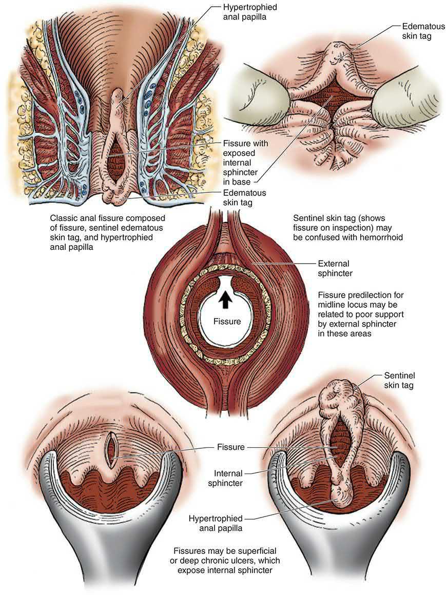
- Affected patients frequently show internal anal sphincter hypertonia, which increases the traumatic effect of hard stool and perpetuates relative ischaemia, reducing mucosal blood supply; pain increases sphincter spasm, which worsens stool hardness and ischaemia in a vicious cycle, producing chronic deep fissures with a hypertrophied anal papilla internally and a sentinel skin tag externally [1].
- Sabiston puts the posterior midline figure at 75%, attributing it to a relatively less robust blood supply there, and notes that the anterior midline is also a frequent site, especially in women [5].
- The sequence is self-perpetuating: a tear from hard stool, diarrhoea, anal receptive intercourse or trauma causes pain, pain causes sphincter spasm, and spasm further reduces blood flow to the anal mucosa, producing relative ischaemia at the fissure site [5]. A fissure found off the midline is atypical and demands a broader differential, Crohn disease, anal cancer, tuberculosis, HIV, syphilis, herpes and leukaemia [5].
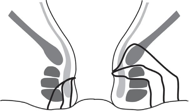
Fistula-in-ano: the majority are idiopathic/cryptoglandular, arising from infection in an intersphincteric anal gland that tracks outward, often first presenting as a perianal abscess (cryptoglandular theory) [1][7]. Anal fistulae may also be associated with Crohn's disease, tuberculosis, lymphogranuloma venereum, actinomycosis, rectal duplication, foreign body, or (rarely) malignancy [1]. Parks' classification, based on the primary track's relation to the external sphincter, defines: intersphincteric (45%, do not cross the external sphincter), trans-sphincteric (40%, cross both sphincters through the ischiorectal fossa, may have secondary/horseshoe extensions), suprasphincteric (10%, curl above puborectalis, often iatrogenic from over-vigorous probing) and extrasphincteric (5%, unrelated to the sphincters, usually from pelvic disease or trauma) [1].
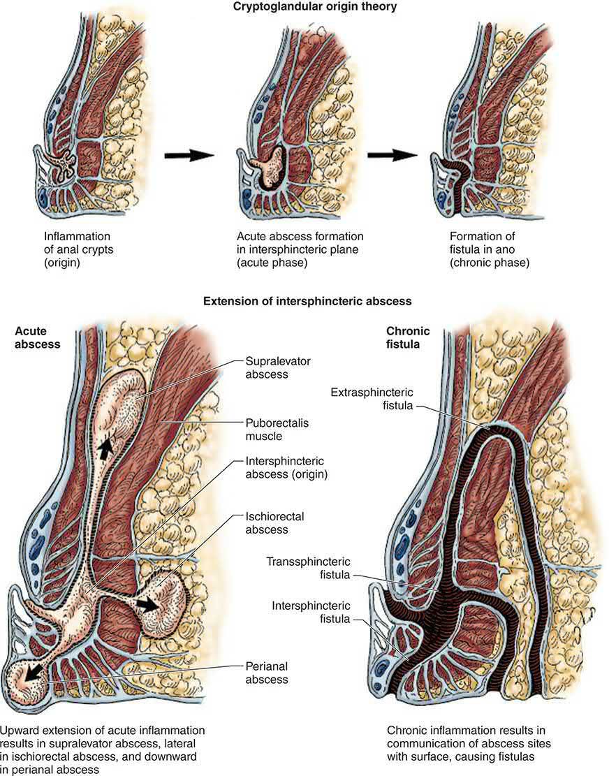
The perianal spaces and the spread of cryptoglandular sepsis in Schwartz's account
- Most anorectal suppuration arises from the anal glands in the intersphincteric plane, whose ducts cross the internal sphincter to empty into the crypts at the dentate line; the perianal space surrounds the anus and is continuous laterally with buttock fat, the intersphincteric space separates the sphincters and runs from the perianal space up into the rectal wall, the ischiorectal space lies lateral and posterior to the anus bounded by external sphincter, ischium, levator ani and the transverse septum and contains the inferior rectal vessels and lymphatics, the two ischiorectal spaces join posteriorly above the anococcygeal ligament but below the levator as the deep postanal space, and the supralevator spaces sit above the levator on either side of the rectum and communicate posteriorly [8].
- A perianal abscess is the commonest manifestation; spread through the external sphincter below the puborectalis produces an ischiorectal abscess, which may be very large yet invisible perianally; intersphincteric abscesses are notoriously hard to diagnose; and supralevator abscesses arise from upward extension of an intersphincteric or ischiorectal abscess or downward extension of intraperitoneal sepsis [8].
- Fissures lie in the posterior midline in the vast majority, anteriorly in 10–15% and off the midline in under 1%, and the tear provokes internal sphincter spasm, pain and anodermal ischaemia in a self-perpetuating cycle; a lateral chronic fissure may signal Crohn's disease, HIV, syphilis, tuberculosis or leukaemia [8].
Clinical features
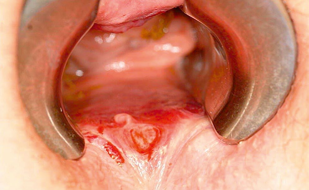
- Anal fissure: acute fissures cause severe anal pain during defecation ("passing glass," "a knife cutting"), recurring at each evacuation, with a trace of fresh blood on the paper; chronic fissures show a hypertrophied anal papilla, sentinel tag, and an indurated ulcer exposing internal sphincter fibres, with itching, discharge, or discharge from an associated intersphincteric fistula [1].
- A fissure that is not midline, or has atypical features, should raise suspicion of Crohn's disease, tuberculosis, syphilis, HIV-related ulceration or squamous cell carcinoma, and warrants examination under anaesthesia with biopsy [1].
- Acute severe "knife-like" pain during defecation with deep throbbing afterward (pelvic floor spasm), and blood on the paper, is the classic acute presentation [9]. A fissure present for more than 6 to 8 weeks is considered chronic, its features being exposed internal sphincter fibres at the base, a hypertrophied anal papilla proximally and a skin tag or sentinel pile distally.
- Pain with defecation tends to be less severe than in an acute fissure but is unrelenting, and in the most severe cases patients dread bowel movements, avoid oral intake and report weight loss [5].
- Patients typically describe the pain as passing "pieces of glass" or "razor blades," followed by throbbing and anal spasm persisting for minutes to hours after defecation [5].
- Fistula-in-ano: intermittent purulent (occasionally bloody) discharge and discomfort relieved temporarily by discharge of pus, often with a preceding history of anorectal sepsis; passage of flatus/faeces through the external opening suggests a rectal rather than anal internal opening [1].
- Presentation may be as an acute perianal abscess (rapid-onset severe pain, swelling, erythema, fever), recurrent perianal sepsis (pressure sensation with intermittent discharge), or chronic low-grade discharge via a punctum [7]. Bilateral external openings should trigger suspicion of a fistula involving the deep postanal space, and multiple external openings (a "watering can perineum") should trigger suspicion of perianal Crohn disease [5].
- Occasionally the external opening is subtle or a considerable distance from the anus; careful inspection with gentle palpation for a cord-like subcutaneous structure helps identify the course, and the patient can often assist by pointing to the site of recurrent pain and drainage [5].
- Approximately 50% of patients with a history of perianal abscess will ultimately develop a fistula [5].
Etiology
- Anal fissures are associated with straining and constipation, and with childbirth (anterior fissures in women) [1].
- Fistula-in-ano most commonly follows cryptoglandular sepsis (often presenting first as a perianal abscess); around 30% of anorectal abscesses eventually develop a fistula-in-ano [4].
- Crohn's disease is an important secondary cause of both conditions, producing atypical, often multiple or laterally sited fissures, and complex fistulae [10].
Diagnosis
Fissure: usually a clinical diagnosis on inspection; if examination cannot be adequately performed in clinic, examination under anaesthesia with biopsy/culture is required to exclude secondary causes [1]. Fistula: clinical assessment aims to determine the internal opening site, external opening site(s), the course of the primary track, secondary extensions, and any complicating conditions; Goodsall's rule predicts the internal opening's likely position from the external opening, though most internal openings are in fact midline [1][4]. Dilute hydrogen peroxide via the external opening, gentle probing, endoanal ultrasound (EAUS) and MRI (the "gold standard," particularly with STIR sequencing) are used to define the tract and detect secondary extensions, informing surgical strategy [1].
Goodsall's rule, stated precisely
Fistulae with an external opening anterior to the transverse bisection of the anus typically track radially, directly into the anal canal, except those more than 3 cm from the anal verge, which may be an anterior extension of a horseshoe fistula originating posteriorly. Fistulae with an external opening posterior to the transverse line often track in a curvilinear fashion to a posterior midline internal opening [5].
Finding the internal opening
- Anoscopy allows direct inspection of the dentate line and may reveal an erythematous crypt or a visible internal opening, and gentle pressure over the tract may induce drainage from the offending crypt.
- In theatre a fistula probe may be passed gently from the external opening through the tract; if the probe does not pass easily, extreme care is needed to avoid creating a false passage and an iatrogenic fistula
- The external opening can be injected with dilute hydrogen peroxide, methylene blue or milk where the internal opening is hard to find, and the anorectal mucosa should be evaluated to exclude a different origin of perianal sepsis such as Crohn disease, atypical ulcers or cancer [5].
- Where the course remains unclear, pelvic MRI with an anal fistula protocol is useful to delineate the anatomy [5].
Goals of fistula treatment
The five goals are to treat any undrained infection, define the fistula anatomy, remove or ablate epithelialised tracts, avoid or minimise the risk of faecal incontinence, and prevent recurrence [5]. Preoperative planning should consider pre-existing continence, stool consistency, any history of sphincter injury or surgery, the amount of sphincter likely to be divided, anterior location in women, and the patient's own attitude to potential imperfections in continence, because there is no simple linear relationship between the extent of operative intervention and continence impairment: some patients report minimal incontinence despite division of a significant portion of sphincter, and others significant incontinence despite division of little or none [5].
Recognising the hidden abscesses in Schwartz's account
- Intersphincteric abscess produces little swelling, deep pain "up inside" worsened by coughing or sneezing, and pain too intense for digital examination, so diagnosis rests on suspicion and examination under anaesthesia; supralevator abscess can mimic intra-abdominal disease and shows as an indurated bulging mass above the anorectal ring on digital examination, and its origin must be identified before drainage; complex or atypical presentations warrant CT or MRI [8].
- Neutropenic patients may harbour serious perianal infection without any cardinal signs of inflammation, so examination under anaesthesia must not be delayed for neutropenia, and any indurated area is incised, drained, biopsied to exclude leukaemic infiltrate and cultured [8].
- Rectovaginal fistulas are classified as low (rectal opening near the dentate line, vaginal opening in the fourchette), middle (between fourchette and cervix) or high (near the cervix); low fistulas usually follow obstetric injury or foreign-body trauma, middle ones severe obstetric injury, resection of a mid-rectal neoplasm, radiation or an undrained abscess, and high ones operative or radiation injury, with Crohn's disease causing fistulas at every level; symptoms range from vaginal flatus to solid stool per vaginam with some incontinence and vaginitis, and small fistulas may be shown by barium enema, vaginogram, endorectal ultrasound or methylene blue instilled rectally with a tampon in the vagina [8].
Scoring and Severity
Fistulae are classified by the Parks' classification (intersphincteric, trans-sphincteric, suprasphincteric, extrasphincteric) as above, condensed clinically by the American Gastroenterology Association classification into: simple fistula (low inter-/trans-sphincteric tract, single external opening) versus complex fistula (high inter-/trans-sphincteric tract, extra-/suprasphincteric tract, presence of abscess/collection, ano-vaginal fistula, or anal stricture), this distinction guides whether a fistula can be managed by straightforward fistulotomy or requires specialist, sphincter-sparing management [1].
| Parks type | Course | Approximate share |
|---|---|---|
| 1 Intersphincteric | Does not cross the external sphincter | 45% |
| 2 Trans-sphincteric | Crosses both sphincters through the ischiorectal fossa; may have secondary or horseshoe extensions | 30–40% |
| 3 Suprasphincteric | Curls above puborectalis; often iatrogenic from over-vigorous probing | 10–20% |
| 4 Extrasphincteric | Unrelated to the sphincters, usually from pelvic disease or trauma | 5% |
Table reformats the Parks classification with its reported distribution [1][5].
Treatment and Management
- Anal fissure: conservative management heals almost all acute and most chronic fissures, dietary fibre, stool softeners, adequate fluids, warm baths, and topical local anaesthetics.
- First-line "chemical sphincterotomy" uses topical glyceryl trinitrate (GTN 0.2%, nitric oxide donor) or diltiazem (2%, calcium-channel antagonist) to relax the internal sphincter, with roughly 50% cure and GTN limited by headache.
- Botulinum toxin injection (10–100 units) is a further option, with temporary incontinence in up to 10% [1][4].
- Incontinence following botulinum injection is a contraindication to subsequent sphincterotomy [4].
The majority of acute fissures resolve with medical management alone, the initial step being increased fluid and fibre to maintain soft formed stools plus sitz baths, which give significant pain relief in over 90% of patients with acute fissures [5]. Chronic fissures are less likely to heal conservatively, and treatment has three aims: address the inciting factors such as constipation or other anal trauma, address the pain and bleeding, and relax and dilate the internal anal sphincter to improve blood flow and allow healing [5].
| Agent | Healing | Practical notes |
|---|---|---|
| Topical nitrates (GTN 0.2%) | Around 50% of chronic fissures | Severe headache is common and leads to cessation of therapy in up to 20% |
| Topical calcium channel blockers (diltiazem 2%, nifedipine) | Similar to nitrates | Preferred first-line topical agent because of a significantly better headache profile |
| Botulinum toxin | Similar to topical therapy as first line; modest improvement as second line | Typical dose 20 to 100 IU, relaxation lasting about 3 months; commonest side effect temporary incontinence to flatus |
| Lateral internal sphincterotomy | 88% to 100% | Superior to nitrates, calcium channel blockers or botulinum toxin; the treatment of choice for chronic anal fissure |
Table reformats the comparative efficacy of treatments for chronic anal fissure [5].
Fistula-in-ano: antibiotics reduce symptoms from sepsis but cannot cure the fistula; medical treatment of underlying Crohn's disease can markedly improve fistulating symptoms [1]. Patients with minimal symptoms may be managed expectantly [1].
Antibiotics, drainage routes and medical fissure therapy in Schwartz's account
- Anorectal abscesses are drained as soon as diagnosed, antibiotics alone are ineffective and are indicated only for extensive cellulitis, immunocompromise, diabetes or valvular heart disease, and delay risks massive necrosis and septicaemia [8].
- A perianal abscess is drained under local anaesthesia by excising a disc of skin to prevent premature closure, without packing, with sitz baths from the next day; horseshoe abscesses need drainage of the deep postanal space through the anococcygeal ligament with counter-incisions over each ischiorectal limb; intersphincteric abscess is drained by a limited, usually posterior, internal sphincterotomy; and a supralevator abscess is drained through the rectum if it arose from an intersphincteric abscess (draining it through the ischiorectal fossa creates a suprasphincteric fistula) but through the ischiorectal fossa if it arose from an ischiorectal abscess (rectal drainage would create an extrasphincteric fistula), while one secondary to intra-abdominal disease is drained by the most direct route with treatment of the primary process [8].
- For fissure, 2% lidocaine jelly adds symptomatic relief to bulk agents, softeners and sitz baths, nitroglycerin often causes severe headache, oral or topical diltiazem and nifedipine may have fewer side effects, arginine (a nitric oxide donor) and topical bethanechol (a muscarinic agonist) are newer options, medical therapy heals most acute but only about 50% of chronic fissures, and botulinum toxin (temporary paralysis by blocking presynaptic acetylcholine release) heals no better than other medical therapy [8].
- Necrotising perineal soft-tissue infection is polymicrobial and synergistic, usually from an undrained cryptoglandular abscess or urogenital source and occasionally after haemorrhoidectomy, shows necrotic skin, bullae or crepitus with systemic toxicity though perineal signs may be minimal, is treated by debridement of all non-viable tissue often over several operations with antibiotics as adjunct, may need colostomy if the sphincter is extensively resected or stool soils the wound, and still carries about 50% mortality [8].
Surgeries
Anal fissure: lateral internal sphincterotomy (division of the internal sphincter away from the fissure itself (open or closed technique)) gives healing rates around 85% but with a risk of altered continence (9% flatus incontinence, 6% soiling, <1% solid incontinence); contraindications include Crohn's disease, ulcerative colitis, women of childbearing age, prior obstetric injury, prior incontinence, or sphincter dysfunction [1][4]. Fissurectomy (excision of the fibrotic edge, sentinel tag and papilla) is an alternative where sphincterotomy is contraindicated, often combined with an anal advancement flap, particularly in postpartum women or those with low resting pressures [1].
Fistula-in-ano:
- Fistulotomy: laying the track open to heal by secondary intention; used for intersphincteric fistulae and low trans-sphincteric fistulae involving less than about 30% of the external sphincter (avoided for anterior fistulae in women); the most important determinant of postoperative continence is the amount of muscle left behind, not divided (a minimum ~2 cm of external sphincter is usually preserved) [1].
- Fistulectomy: coring out the tract with diathermy, giving better anatomical definition than fistulotomy at the cost of longer healing.
- Setons: loose (non-cutting) setons drain sepsis and allow staged fistulotomy or long-term palliation (especially in Crohn's disease); cutting setons gradually divide enclosed muscle ("cheese-wiring") to achieve eradication with less abrupt sphincter division.
- Sphincter-sparing techniques for high/complex fistulae: ligation of the intersphincteric fistula tract (LIFT), and endorectal/anorectal advancement flap; fistula plugs and fibrin glue are generally avoided due to poor results [1][4].
- Perianal/anorectal abscess is treated by prompt incision and drainage, which may be the first presentation of an underlying fistula [4].
What the numbers say
- Reported rates of faecal incontinence after lateral internal sphincterotomy range from 8% to 30%, usually limited to minor episodes of incontinence to flatus and most often in the first 30 days; the procedure should be avoided in patients at higher risk, including those with baseline incontinence [5].
- For fistula surgery, simple low-lying fistulae are commonly defined as those involving less than one-third of the external anal sphincter (intersphincteric or low trans-sphincteric) and may be treated by lay-open fistulotomy.
- Multiple prospective multicentre studies show clinically significant faecal incontinence in under 5% of patients with normal preoperative sphincter function, with a recurrence rate of 2% to 8% [5].
Primary fistulotomy at the time of abscess drainage remains controversial: although it would address the offending crypt and reduce recurrence, inflammation and oedema make accurate assessment of the anatomy difficult and can lead to underestimation of sphincter involvement [5]. Where more sphincter is involved, or where inflammation prevents accurate assessment, initial treatment is a draining seton using a silastic vessel loop or rubber band, which allows a narrow tract to form, prevents the cyclical swelling-pain-discharge pattern, and permits "shortening" of the tract by partial fistulotomy through soft tissue [5].
Minimally invasive tract ablation
- VAAFT (video-assisted anal fistula treatment) uses a fistulascope inserted through the external opening to define the tract and its branches.
- The internal opening is closed by suture ligation or stapling, granulation tissue is debrided, and the tract is ablated under direct vision with an electrode without dividing any sphincter muscle, reported healing 71% to 85% with minor or no incontinence at up to 2 years [5]. FiLaC (fistula laser closure) ablates the tract along its length from the inside using a radially emitting laser probe rather than an electrode; a recent meta-analysis reported healing in 65% at a median of 24 months [5].
- Transplantation of mesenchymal stem cells into the tissue surrounding complex perianal fistulae has shown promise in small single-institution studies and is under randomised investigation [5].
- A note on advancement flaps.
- The classic endorectal advancement flap raises mucosa and submucosa off the internal sphincter.
- Some surgeons include internal sphincter fibres to create a more robust flap with better blood supply, but this has been shown to increase rates of mild-to-moderate incontinence, with reduced manometric resting and squeeze pressures, in up to 35% of patients [5].
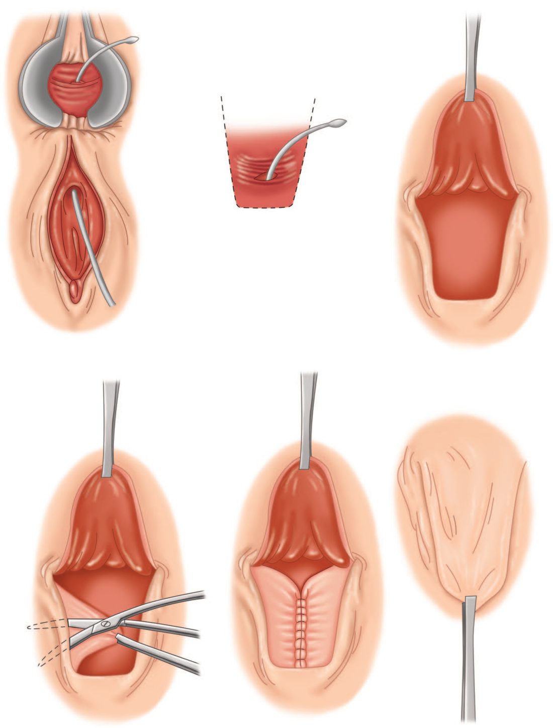
Bioprosthetic plug insertion for anal fistula. "Current evidence on the safety and efficacy of bioprosthetic plug insertion for anal fistula is adequate to support the use of this procedure provided that standard arrangements are in place for clinical governance, consent and audit." [2]. The procedure should only be done by a surgeon experienced in managing anal fistulas [2]. The guidance was published on 25 September 2019, migrated from interventional procedures guidance IPG662 [2].
Note the divergence. Bailey & Love states that fistula plugs and fibrin glue are generally avoided because of poor results; NICE's position is narrower and procedural rather than clinical, it says the evidence is adequate to permit the plug under standard governance, in experienced hands, without asserting that it is the best option [2].
- Radially emitting laser fibre treatment (FiLaC). "Current evidence on the safety and efficacy of radially emitting laser fibre treatment of an anal fistula is limited in quantity and quality
- Therefore, although there are no major safety concerns, this procedure should only be used with special arrangements for clinical governance, consent, and audit or research" [3].
- Clinicians wishing to do it must inform the clinical governance leads in their NHS trusts, ensure patients understand the safety and efficacy and the uncertainties about both with clear written information to support shared decision making, and audit and review clinical outcomes of all patients having the procedure [3]. The procedure should only be done by clinicians experienced in cannulating fistulas who are trained in the use of lasers [3].
- Further research should report patient selection including fistula size, medium- and long-term recurrence rates, and quality-of-life outcomes [3].
Sphincterotomy results, fistula categories and rectovaginal repair in Schwartz's account
- Lateral internal sphincterotomy divides about 30% of the internal sphincter fibres by an open or closed technique, heals more than 95% with immediate pain relief in most, recurs in under 10% and causes incontinence (usually to flatus) in 5–15%; V-Y advancement flaps with or without sphincterotomy are an alternative [8].
- Drainage of an anorectal abscess cures about 50% and the rest develop a fistula whose course follows the anatomy of the original abscess; Goodsall's rule fails when an anterior external opening lies more than 3 cm from the margin, such fistulas tracking to the posterior midline [8].
- An intersphincteric fistula runs through the distal internal sphincter to open near the verge and is laid open by fistulotomy and curettage; a transsphincteric fistula, usually from an ischiorectal abscess, crosses both sphincters and can be treated by sphincterotomy when it involves under 30% of the muscle but needs a seton first when high; a suprasphincteric fistula from the intersphincteric plane loops over the whole external sphincter and is treated with a seton; and an extrasphincteric fistula from the rectal wall exits laterally in the ischiorectal fossa, its extrasphincteric portion opened and drained with any dentate-line tract laid open [8].
- Injecting hydrogen peroxide or dilute methylene blue helps find the internal opening, but care is needed not to create an artificial one and convert a simple fistula into a complex one; complex or non-healing fistulas warrant proctoscopy to assess the rectal mucosa and biopsy of the tract to exclude malignancy, may need many procedures with liberal drains and setons, and may ultimately need faecal diversion [8].
- A cutting seton of suture or rubber band is tightened intermittently in the office to divide the sphincter gradually through fibrosis, whereas a non-cutting soft drain (often a vessel loop) maintains drainage so the tract can later be laid open with less incontinence because scar prevents sphincter retraction, or be left indefinitely; LIFT identifies the tract in the intersphincteric plane with a lacrimal probe, divides it and ligates both ends [8].
- Because up to 50% of obstetric rectovaginal fistulas heal spontaneously, repair waits 3–6 months; low and middle fistulas are best treated by endorectal advancement flap, advancing healthy mucosa, submucosa and circular muscle over the high-pressure rectal opening, with concurrent overlapping sphincteroplasty if the sphincter is injured and diversion rarely needed; high rectovaginal, colovaginal and enterovaginal fistulas are treated transabdominally by resecting the diseased bowel, closing the vagina and interposing omentum or muscle; Crohn's, radiation and malignant fistulas almost never heal spontaneously, a flap is possible in Crohn's only if the rectum is spared, radiation fistulas are not amenable to local flap repair and every radiation fistula must be biopsied to exclude cancer [8].
- Sphincter injury from fourth-degree obstetric tears, infected episiotomy, prolonged labour, midline episiotomy or anal surgery is evaluated by manometry, EMG, pudendal latency and endoanal ultrasound; mild incontinence may respond to diet and biofeedback, the standard repair is wrap-around (overlapping) sphincteroplasty mobilising the divided muscle and reapproximating it without tension, postanal intersphincteric levatorplasty restores the anorectal angle for incontinence from prolapse, and failed or muscle-deficient cases may need gracilis transposition with or without electrostimulation, an artificial anal sphincter (inflatable silastic cuff, pressure-regulating balloon and control pump, prone to infection and erosion), sacral nerve stimulation when the sphincter is intact, or an end stoma [8].
Complications
- Fissure surgery risks fecal incontinence (the most serious complication of sphincterotomy, though the risk is small), haematoma, perianal abscess and recurrent/secondary fistula [1][4].
- The most feared complication of fistula surgery is impaired continence from excessive or inappropriate division of the sphincter complex, which is why fistulotomy is avoided for fistulae involving more than the lower quarter of the external sphincter [4].
- Persistence or recurrence of a fistula usually reflects a missed secondary (often horseshoe) extension [1].
Prognosis
Lateral internal sphincterotomy achieves healing in the great majority (~85%) of chronic fissures, with better long-term results than medical therapy alone, at a small risk to continence [1]. Sabiston puts the healing rate for lateral internal sphincterotomy higher still, at 88% to 100%, and confirms it as superior to topical nitrates, calcium channel blockers or botulinum toxin [5].
- Fistula recurrence and functional outcome depend heavily on fistula complexity and chosen technique, and no unified prognostic scoring system exists.
- The figures that do exist stratify by complexity: simple low-lying fistulae treated by fistulotomy recur in 2% to 8% with clinically significant incontinence in under 5% of those with normal preoperative sphincter function, whereas the sphincter-sparing techniques used for complex fistulae report lower healing, 71% to 85% for VAAFT at up to 2 years and 65% for FiLaC at a median of 24 months [5].
- The trade-off is therefore explicit: the more sphincter that is spared, the lower the cure rate.
References
- Bailey & Love's Short Practice of Surgery, 28th ed., Ch. 80 The anus and anal canal
- NICE HealthTech Guidance HTG528: Bioprosthetic plug insertion for anal fistula (2019, migrated from IPG662), 1.1; 1.2; Overview www.nice.org.uk
- NICE HealthTech Guidance HTG505: Radially emitting laser fibre treatment of an anal fistula (2019, migrated from IPG644), 1.1; 1.2; 1.3; 1.4 www.nice.org.uk
- The ABSITE Review, 2022, Anorectal Disorders
- Sabiston Textbook of Surgery, 22nd ed., Ch. 97 Benign Anorectal Disorders
- Maingot's Abdominal Operations, 13th ed., Ch. 52
- Oxford Handbook of Clinical Surgery, 5th ed., Ch. 12 Colorectal surgery
- Schwartz's Principles of Surgery, 11th ed., Ch. 29, Colon, Rectum, and Anus
- Oxford Handbook of Clinical Surgery, 5th ed., Ch. 26
- Bailey & Love's Short Practice of Surgery, 28th ed., Ch. 75 Inflammatory bowel disease and Ch. 80
- Schwartz's Principles of Surgery: ABSITE and Board Review, Ch. 29