



Mastitis and Breast Abscess
Summary
- Mastitis refers to inflammation of the breast tissue that may or may not be accompanied by infection, and it can occur in both lactating and non-lactating women, the former being more common [1].
- Acute infection is usually caused by Staphylococcus aureus and, if untreated, progresses from cellulitis to suppuration and abscess formation [1].
- Two broad categories exist: lactational infections arising from bacterial entry through the nipple into the duct system, and chronic subareolar infections associated with duct ectasia and periductal mastitis in non-lactating women [2].
- Ultrasound-guided aspiration is now the preferred first-line treatment for a confirmed abscess in most cases, with surgical incision and drainage reserved for those that fail conservative measures [1][2].
There is no NICE guideline on breast infection as a surgical topic. The UK national guidance that bears on it comes from two directions: NICE NG194 on postnatal care, which sets the threshold at which a breastfeeding woman should seek medical advice, and the ABS multi-college diagnostic guidelines, which specify how a suspected abscess is imaged and when the case must be returned to the multidisciplinary meeting rather than discharged [3][4].
Definition
- Mastitis is inflammation of the breast tissue that may or may not be accompanied by infection [1].
- Lactational (puerperal) mastitis occurs in breastfeeding mothers, whereas non-lactational mastitis is inflammation of the breast tissue in a nulliparous woman or occurring at least 6 months after cessation of lactation, and includes periductal mastitis, idiopathic granulomatous mastitis (IGM) and tubercular mastitis [1].
- Duct ectasia and periductal mastitis are described as a common condition of unknown aetiology, characterised pathologically by dilatation of the larger mammary ducts filled with inspissated material containing macrophages and chronic inflammatory debris, with inflammatory complications closely related to smoking [5].
- Periductal mastitis is also termed mammary duct ectasia or plasma cell mastitis [6].
Pathophysiology
Lactational infection
- Bacteria may enter the nipple through a cracked or retracted nipple; in many cases the lactiferous ducts become blocked by epithelial debris, leading to stasis followed by infection, and once within the ampulla of the duct staphylococci cause clotting of the milk and then multiply within the clot [1].
- During breastfeeding, bacteria may gain access to the engorged breast lobules via the nipple and duct system or via the circulation [5].
- Abscess formation is most commonly seen at two stages during lactation: in the first month after childbirth, owing to inexperience or inadequate breastfeeding, and at weaning, owing to engorgement and trauma to the nipple by the baby's teeth [1].
Periductal mastitis
- Periductal mastitis is a chronic non-lactational inflammation around the major milk ducts whose pathogenesis is obscure and thought to be autoimmune in nature, and it is much more common in smokers.
- It may progress to a subareolar inflammatory mass that suppurates, and because thick areolar muscles do not allow the abscess to perforate through the areola, pus follows the path of least resistance, rupturing at the areolar edge to form a mammary or milk duct fistula [1].
- In some cases a chronic indurated mass forms beneath the areola, which mimics a carcinoma, and fibrosis in and around the major milk ducts causes nipple retraction [1].
- In non-lactating women this chronic relapsing subareolar infection is associated with duct ectasia, appears related to smoking and diabetes, and the infections are most often mixed, including aerobic and anaerobic skin flora.
- Repeated infection with resulting inflammatory changes and scarring may lead to nipple retraction or inversion, subareolar masses, and occasionally a chronic fistula from the subareolar ducts to the periareolar skin [2].
Clinical features
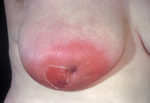
- Initially there is generalised cellulitis, which if untreated progresses to suppuration and abscess formation; an abscess presents as a fluctuant lump (fluctuation may be absent if deep-seated) with pain, signs of inflammation, fever, malaise and difficulty feeding, and may be associated with enlarged tender axillary nodes [1].
- The infected breast will be red, warm, painful and swollen, and the abscess will eventually point and discharge through the skin, with obvious tender ipsilateral axillary lymphadenopathy possible [5].
- The patient develops malaise and fever accompanied by a painful lump in the breast [5]. It is safe for the infant to continue breastfeeding even from the breast containing an abscess, providing the mother can tolerate the process [5].
Lactational mastitis presents with recurrent intermittent breast pain, swelling, tenderness and seropurulent nipple discharge, while breast abscess presents as acute, severe, localised breast pain with swelling, redness and sometimes purulent nipple discharge, commonest in breastfeeding women [7].
Periductal mastitis and duct ectasia
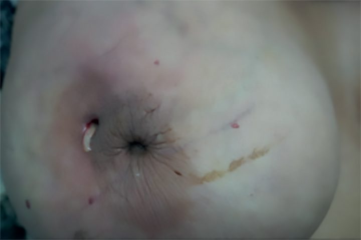
- Periductal mastitis presents with central non-cyclical pain, pus discharge from the nipple and a subareolar tender mass or abscess or a mammary duct fistula, with examination revealing a tender firm subareolar lump or abscess, purulent nipple discharge, thickened tender major milk ducts, and a transverse slit-like nipple retraction resembling a fish's mouth [1].
- Duct ectasia and periductal mastitis present with nipple inversion of this characteristic transverse slit appearance, purulent nipple discharge from dilated ducts, and chronic low-grade periareolar infection with tender thickening that may progress to abscess and fistula formation, typically causing a small scabbed area at the areolar margin that intermittently discharges pus [5].
- Periductal mastitis symptoms include non-cyclical mastodynia, erythema, nipple retraction and creamy nipple discharge, and it can present with a sterile or an infected subareolar abscess [6].
The cancer mimics
- Peau d'orange is a hallmark not only of inflammatory carcinoma but also of acute mastitis [2].
- Idiopathic granulomatous mastitis may present as single or multiple central or peripheral inflammatory breast masses with or without abscess, and may be associated with skin ulceration, nipple retraction, sinus formation, peau d'orange and axillary lymphadenopathy, findings that mimic cancer [1].
- Both duct ectasia and tuberculosis of the breast may present with a painless mass mimicking carcinoma [5].
Etiology
- Most cases of lactational mastitis are caused by S. aureus and, if hospital-acquired, may be due to methicillin-resistant S. aureus [1].
- Infections of the breast are most often caused by Staphylococcus aureus or streptococcal species [2]; these are the organisms most frequently recovered from nipple discharge of an infected breast [8].
- Infectious mastitis is most commonly associated with breastfeeding, with S. aureus the most common organism, streptococcus also implicated, and smoking a risk factor; lactational mastitis arises from blockage of lactiferous ducts, while non-lactational mastitis can be due to chronic inflammatory diseases such as actinomyces or autoimmune disease such as SLE [6].
- Non-lactational mastitis is associated with smoking and due to mixed bacterial growth [9].
- Periductal mastitis risk factors include smoking and nipple piercings, and it usually occurs in peri- or postmenopausal women [6].
- The most common organisms isolated from periductal mastitis and subareolar abscess are staphylococci, enterococci, anaerobic streptococci, and sometimes Bacteroides and mycobacteria [1].
- Mastitis of infants is uncommon and predominantly caused by Staphylococcus aureus [1].
Idiopathic granulomatous mastitis is of unknown aetiology, occurring most commonly in young parous women within the first few years after pregnancy, with an association between IGM and Corynebacterium kroppenstedtii infection postulated [1]; IGM is more common in Hispanic, Middle Eastern, and Southeast Asian populations [2].
Classification, organisms and the rarer infections in Schwartz's account
- Non-lactational infection is intrinsic (from breast abnormality, chiefly periductal mastitis) or extrinsic (from an adjacent structure, chiefly an infected sebaceous cyst); Staphylococcus aureus and streptococci are the usual isolates, staphylococcal infection forming localised, sometimes deep abscesses (subcutaneous, subareolar, periductal or retromammary) whereas streptococcal infection spreads diffusely and superficially and is treated with warm compresses and intravenous penicillins or cephalosporins [10].
- Epidemic puerperal mastitis from virulent MRSA transmitted by the suckling neonate can express pus from the nipple and requires cessation of feeding, antibiotics and surgery, whereas sporadic puerperal mastitis follows nipple fissuring and milk stasis with retrograde infection of interlobular tissue and resolves in over 95% with pump emptying plus antibiotics; Zuska's disease is recurrent retroareolar infection and abscess linked to smoking, managed symptomatically with antibiotics and drainage, wide debridement or terminal duct resection being curative but liable to postoperative infection [10].
- Fungal abscesses (blastomycosis, sporotrichosis) are inoculated by intraoral fungi from the infant near the nipple, discharge blood-stained pus from sinuses and usually respond to antifungals; Candida causes scaly erythema of the inframammary and axillary folds treated by removing maceration and topical nystatin; hidradenitis of the nipple–areola or axilla arises in Montgomery's glands or axillary sebaceous glands in women with acne, mimics Paget's disease or cancer, and needs antibiotics, drainage, excision and sometimes flaps or grafts; and Mondor's "string phlebitis" (1939) of the lateral thoracic, thoracoepigastric or superficial epigastric vein presents as a tender cord with acute lateral breast pain, is benign and self-limiting over 4–6 weeks with anti-inflammatories and warm compresses, biopsied only for an adjacent mass and excised only if refractory [10].
Diagnosis
- Ultrasonography reveals cellulitis as an area of increased echogenicity, and liquefaction necrosis as pus in a hypoechoic collection with floating debris that changes with posture [1].
- Ultrasound evaluation can assist in characterising a breast abscess and help guide needle aspiration [2].
- Ultrasound may help identify a breast abscess if the breast is acutely inflamed, and imaging is rarely necessary otherwise; mammography should be avoided owing to the breast compression required and the pain [7][9].
In periductal mastitis, ultrasonography shows thickened major milk ducts with surrounding inflammation or abscess, and a lump should be biopsied under ultrasound guidance to confirm the diagnosis; pus discharge should be sent for culture and sensitivity and for GeneXpert MTB/RIF testing to rule out tuberculosis [1]. A needle biopsy of a solid mass establishes the diagnosis of IGM, with tissue or aspirate sent for Gram stain and culture, acid-fast bacilli stain and culture, and fungal stain and culture; histologically IGM shows a non-caseating granuloma with chronic inflammation, and the differential diagnoses include tuberculosis, foreign body reaction and sarcoidosis [1].
All breast infections should be followed up to ensure complete resolution and to exclude inflammatory breast cancer, which may present similarly [9]. A non-lactational breast abscess is considered breast cancer until proven otherwise, requiring incision and drainage with antibiotics, plus skin and abscess cavity biopsies for pathology and fluid for cytology; failure to resolve after 2 weeks, or recurrence, requires excisional biopsy including skin to rule out necrotic breast carcinoma [6].
- When to send the patient in.
- NG194 gives the threshold in the postnatal period as a red flag the woman herself is taught to act on: at the first postnatal midwife contact, inform her that worsening reddening and swelling of the breasts persisting for more than 24 hours despite self-management could indicate mastitis, and that she should seek medical advice without delay [3].
- Information given antenatally and postnatally about breastfeeding should cover pain when breastfeeding and when to seek help, and breastfeeding complications such as mastitis or breast abscess and when to seek help [3].
- How a suspected abscess is imaged.
- Under the ABS assessment protocol for breast pain, when there are associated or incidental focal clinical signs in the breast (localised tenderness, nodularity, swelling or a lump) the lump imaging protocol is followed; if infection or abscess is suspected an initial ultrasound scan should be performed and any fluid or pus aspirated and cultured [4].
- Clinical examination in a patient presenting with breast pain should look specifically for signs of infection, pink or red discoloration of the skin with localised swelling and tenderness, and discharge from the nipple or skin, and the history should record current or recent breastfeeding, features of infection, and any recent antibiotic treatment for it [4].
- Breast infection is on the list of cases that must not simply be discharged.
- Breast inflammation, infection, cellulitis or abscess is named explicitly among the presentations requiring further review and/or diagnostic intervention after initial assessment, alongside discordance between elements of triple assessment and equivocal B3/B4 biopsy results [4].
- This is the UK-guideline counterpart of the textbook rule that every breast infection must be followed to complete resolution to exclude inflammatory carcinoma.
Scoring and Severity
There is no formal severity score for breast infection, but two thresholds carry the practical decisions.
Stage of infection determines whether drainage is needed at all: the disease progresses from generalised cellulitis, treated with antibiotics alone, to suppuration and abscess formation, which requires drainage of pus [1].
Abscess size determines how it is drained [1]:
| Ultrasound finding | Drainage method |
|---|---|
| Small abscess | Simple needle aspiration of pus with antibiotic therapy |
| Greater than 3 cm diameter, or containing more than 30 mL of pus | Vacuum suction catheter inserted under ultrasound guidance, with review on alternate days by clinical examination and ultrasonography and aspiration of any residual collection |
| Failure to resolve after aspiration and antibiotics | Surgical incision and drainage, such abscesses are generally multiloculated |
Table reformats the size-based drainage decision [1][2][11].
Two further categorisations govern the antibiotic choice and the index of suspicion for malignancy: lactational versus non-lactational, since lactational infection is usually staphylococcal and non-lactational infection is usually mixed aerobic and anaerobic [1][2]; and, within non-lactational disease, whether the presentation is a straightforward periductal abscess or one of the cancer mimics, IGM, tuberculous mastitis, or inflammatory carcinoma [1][9].
Treatment and Management
Cellulitic stage and lactational mastitis
- During the cellulitic stage, patients should be treated with anti-staphylococcal antibiotics such as cloxacillin, flucloxacillin or erythromycin; breastfeeding from both breasts should be encouraged 2-hourly followed by emptying the breast, and a breast support garment, cold compression and analgesia aid symptomatic relief [1].
- Treatment of lactational mastitis includes advising continued feeding or expressing plus antibiotics such as flucloxacillin; it affects 5% of breastfeeding women and is usually due to staphylococcal infection [9].
- Lactational infectious mastitis is treated with antibiotics alone, continuing breastfeeding, for 2 weeks [6].
- Treatment for breast infections requires antibiotics (depending on the risk of MRSA colonisation) together with regular emptying of the breast [2].
Non-lactational mastitis
- Non-lactational mastitis is treated with antibiotics covering anaerobic bacteria and Gram-negative bacilli, for example amoxicillin plus metronidazole, for 2 weeks; failure to resolve after 2 weeks, or recurrence, requires excisional biopsy including skin to rule out necrotic breast cancer [6][9].
- Subareolar infections may initially manifest as subareolar pain and mild erythema, treatable with warm soaks and oral antibiotics covering aerobic and anaerobic organisms [2].
- Many cases of periductal mastitis resolve with a course of antibiotics combined with needle aspiration of an abscess [1].
- If associated with chronic mastitis, oral antibiotics include metronidazole 400 mg three times daily or co-amoxiclav 750 mg three times daily [7].
Abscess drainage
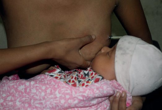
- Contrary to the historical practice of incision and drainage, ultrasound-guided drainage of a breast abscess gives an excellent cosmetic result, does not hamper breastfeeding, and can be done as a day-case procedure with a high success rate; incision and drainage may result in a non-healing milk fistula [1].
- Antibiotics should be continued for 14 days, and the catheter, if used, irrigated with cold normal saline until complete resolution [1].
- Previously almost all breast abscesses were treated by operative incision and drainage, but the initial approach is now antibiotics and repeated aspiration of the abscess, usually ultrasound-guided [8].
- Lactational breast abscess treatment is percutaneous drainage, antibiotics and continued breastfeeding, with incision and drainage reserved for failure to resolve promptly [6].
- Where oral antibiotics are given for a lactational abscess these include flucloxacillin 500 mg three times daily, with aspirational drainage often repeated several times on a daily or alternate-day basis [7].
- Breast abscesses may be effectively aspirated for relief of pressure symptoms under local anaesthetic, and formal incision and drainage is often avoided, especially in lactational abscesses; mini-incision and drainage is used only if aspiration fails or the overlying skin is necrotic [7][9].
Idiopathic granulomatous and tuberculous mastitis
- In IGM, symptomatic patients and those with infection are treated with non-steroidal anti-inflammatory drugs and antibiotics with or without drainage. Anti-tuberculous therapy should not be given as a blanket treatment to all patients with granulomatous mastitis in countries where TB is endemic, it should only be given where there is evidence of TB on imaging, histopathology, microbiological analysis or GeneXpert MTB/RIF [1].
- In persistent or progressive cases, prednisolone (oral or topical) with or without methotrexate has helped in regression [1].
- Antibiotic treatment directed against Corynebacterium species (doxycycline, clindamycin, azithromycin, or levofloxacin, or culture-driven) is a possible first-line treatment; systemic steroids are an additional avenue, with methotrexate for those unable to tolerate steroids, and intralesional steroid injections have shown promising results. Surgical management of IGM is not recommended because it might result in an open, poorly healing wound [2].
- Tubercular mastitis is treated with anti-tuberculous chemotherapy for 6–9 months [1].
Where breastfeeding is stopped or is not started, including where the abscess or its treatment makes feeding impossible, and in the event of the death of a baby, lactation suppression should be discussed, covering how the body produces milk and how long it takes for production to stop; self-help advice including avoiding stimulating the breast, wearing a supportive bra, using ice packs, over-the-counter pain relief, and sparingly expressing milk to ease engorgement; when to seek help; medicines that can be prescribed to suppress lactation and the advantages and disadvantages of each method; and the possibility of becoming a breast milk donor [3].
Note that this UK advice (express sparingly, only to ease engorgement) is the opposite of the management of active lactational mastitis, where the textbooks direct 2-hourly feeding from both breasts followed by emptying the breast. The two situations must not be confused: emptying the breast treats infection, whereas emptying the breast defeats suppression [3].
From aspiration to fistulectomy and total duct excision in Schwartz's account
- Almost all abscesses were once incised, but the initial approach is now antibiotics with repeated, usually ultrasound-guided, aspiration, operative drainage being reserved for failure or for thinned or necrotic skin, with ultrasound delineating the extent, cultures for acid-fast bacilli, anaerobes, aerobes and fungi in chronic or recurrent disease, and biopsy of the cavity wall to exclude cancer when antibiotics and drainage fail [10].
- A tender retroareolar mass is aspirated with a 21-gauge needle on a 10 mL syringe and the fluid cultured in anaerobic transport medium, polymicrobial cover is started pending sensitivities, and a subareolar abscess (unlike a puerperal one) is usually unilocular from a single duct system; in women of childbearing age simple drainage is preferred though anaerobic infection often recurs, and recurrent abscess with fistula is now treated first by fistulectomy with primary closure under antibiotic cover (laying open to granulate being kept for recurrence after fistulectomy), with total duct excision when sepsis is diffuse or fistulas multiple [10].
- Fistulectomy suits a small abscess in one segment recurring in the same segment, mild or no inversion, an unconcerned younger patient, no discharge from other ducts and no prior fistulectomy (the young woman with squamous metaplasia of one duct) whereas total duct excision suits an abscess over half the areolar circumference, recurrence in a different segment, marked inversion the patient wants corrected, purulent discharge from other ducts or recurrence after fistulectomy (the older woman with multiple ectatic ducts) though fistulectomy remains the first operation for localised sepsis at any age and a 2–4-week antibiotic course precedes total duct excision for recurrence after fistulectomy [10].
Surgeries
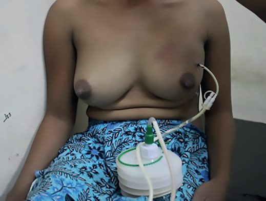
Ultrasound-guided drainage is preferred over incision and drainage for a breast abscess, giving an excellent cosmetic result and not hampering breastfeeding [1]. In abscesses greater than 3 cm in diameter or containing more than 30 mL of pus on ultrasonography, a vacuum suction catheter is inserted under ultrasound guidance to drain the pus; the patient is reviewed on alternate days by clinical examination and ultrasonography, with any residual collection aspirated [1].
- Major milk duct excision, excising a 1.5 to 2 cm length of the ductal cone, is needed in patients with periductal mastitis presenting with a subareolar abscess or sepsis and a mammary duct fistula, and smoking cessation must be encouraged to prevent recurrence [1].
- Excision of chronic abscess cavities is performed in patients with recurrent IGM, and major milk duct excision is indicated in patients with a mammary duct fistula [1].
- Repeated subareolar infections are treated by excision of the entire subareolar duct complex after the acute infection has resolved completely, together with intravenous antibiotic coverage; rarely, patients have recurrent infections requiring excision of the nipple and areola [2].
- Surgical incision and drainage should be reserved for abscesses that do not resolve after needle aspiration and antibiotics, and such abscesses are generally multiloculated [2].
Complications
- Incision and drainage may result in a non-healing milk fistula [1][6].
- Periductal mastitis may progress to a subareolar inflammatory mass that suppurates, with pus rupturing at the areolar edge to form a mammary or milk duct fistula [1].
- A series of infections with resulting inflammatory changes and scarring may lead to retraction or inversion of the nipple, masses in the subareolar area, and occasionally a chronic fistula from the subareolar ducts to the periareolar skin, which may make surveillance for breast cancer more challenging [2].
Recurrent and chronic breast abscess is usually associated with duct ectasia [5]. An antibioma (a hard, oedematous swelling containing sterile pus) may form following treatment of an abscess with long-term antibiotics rather than incision and drainage [12].
Prognosis
Most breast infection resolves without sequelae when treated at the cellulitic stage, and many cases of periductal mastitis resolve with a course of antibiotics combined with needle aspiration of an abscess [1]. Idiopathic granulomatous mastitis is a benign, self-limiting inflammatory breast disease, though persistent or progressive cases need prednisolone with or without methotrexate to achieve regression, and recurrence may require excision of chronic abscess cavities [1].
- The prognosis of the non-lactational group is dominated by recurrence rather than by the acute episode.
- Periductal mastitis is much more common in smokers, and smoking cessation must be encouraged to prevent recurrence after major milk duct excision [1].
- Repeated subareolar infection produces cumulative scarring with nipple retraction and chronic fistula, and rarely requires excision of the nipple and areola [2].
The one prognostic error that matters is diagnostic rather than therapeutic: an inflammatory presentation that does not settle may not be an infection at all. All breast infections should be followed up to ensure complete resolution and exclude inflammatory breast cancer, and failure of a non-lactational infection to resolve after 2 weeks, or its recurrence, mandates excisional biopsy including skin to exclude necrotic carcinoma [6][9].
References
- Bailey & Love's Short Practice of Surgery, 28th ed., Ch. 58 The breast
- Sabiston Textbook of Surgery, 22nd ed., Ch. 68 Diseases of the Breast
- NICE Guideline NG194: Postnatal care. National Institute for Health and Care Excellence, London, UK, 2021., 1.2.4; 1.5 Information and support for breastfeeding; 1.5.21 www.nice.org.uk
- Association of Breast Surgery, Royal College of Radiologists Breast Group, Royal College of Pathologists and partners: Best practice diagnostic guidelines for patients presenting with breast symptoms (November 2010; stated review date November 2012), 2.5 Clinical assessment; 2.5 Imaging; 3.1 The MDM associationofbreastsurgery.org.uk
- Browse's Introduction to the Symptoms and Signs of Surgical Disease, 6th ed., Ch. 13 The breast
- The ABSITE Review, 2022, Ch. 24 Breast – Benign Breast Disease
- Oxford Handbook of Clinical Surgery, 5th ed., Emergency surgery topics
- Schwartz's Principles of Surgery: ABSITE and Board Review, Ch. 17 Breast
- Oxford Handbook of Clinical Surgery, 5th ed., Ch. 6 Breast surgery
- Schwartz's Principles of Surgery, 11th ed., Ch. 17, The Breast
- Bailey & Love's Short Practice of Surgery, 28th ed., Ch. 5 Surgical infection
- Oxford Handbook of Clinical Surgery, 5th ed., Glossary